. 1999 Jun 15;13(12):1601-13.
doi: 10.1101/gad.13.12.1601.

Interaction between FGF and BMP signaling pathways regulates development of metanephric mesenchyme

Affiliations

Interaction between FGF and BMP signaling pathways regulates development of metanephric mesenchyme

A T Dudley et al. Genes Dev. .

Abstract

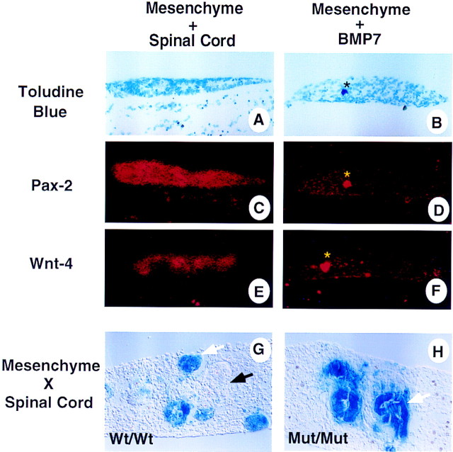
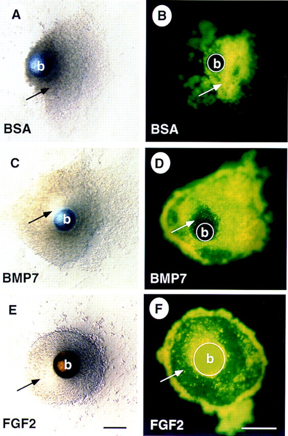
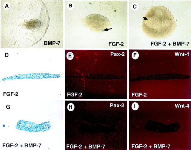
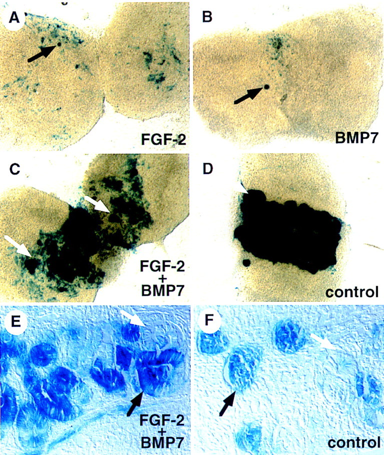
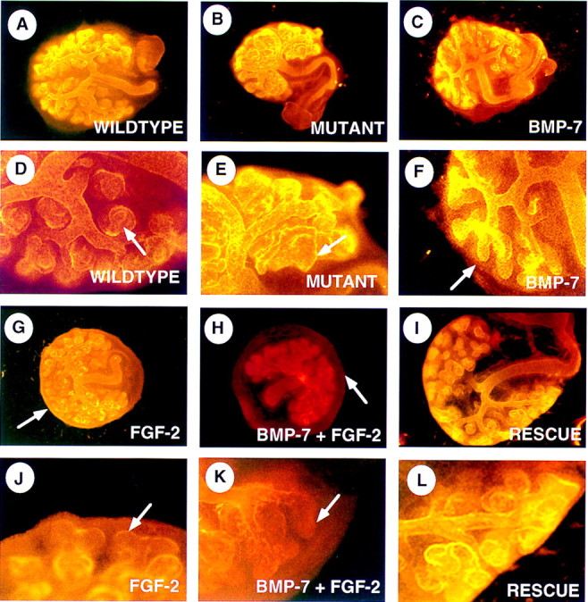
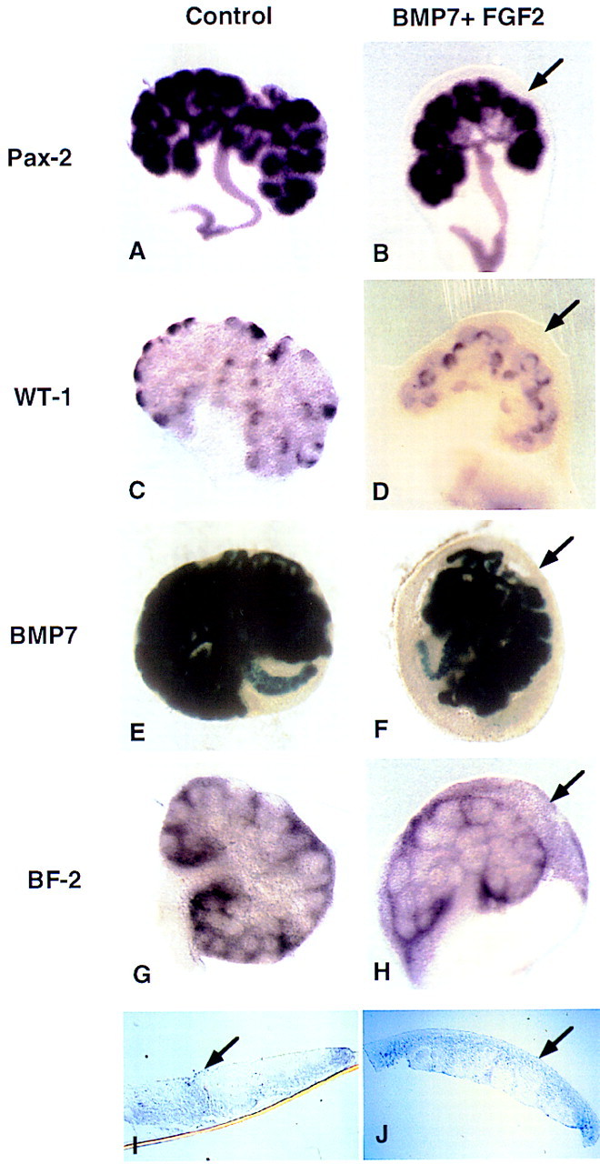
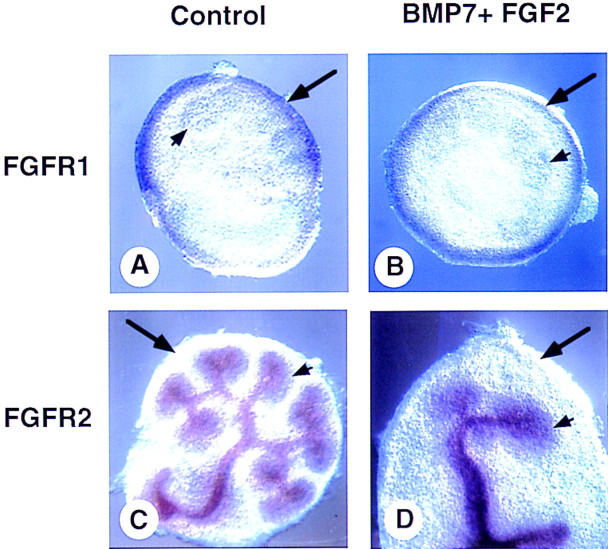
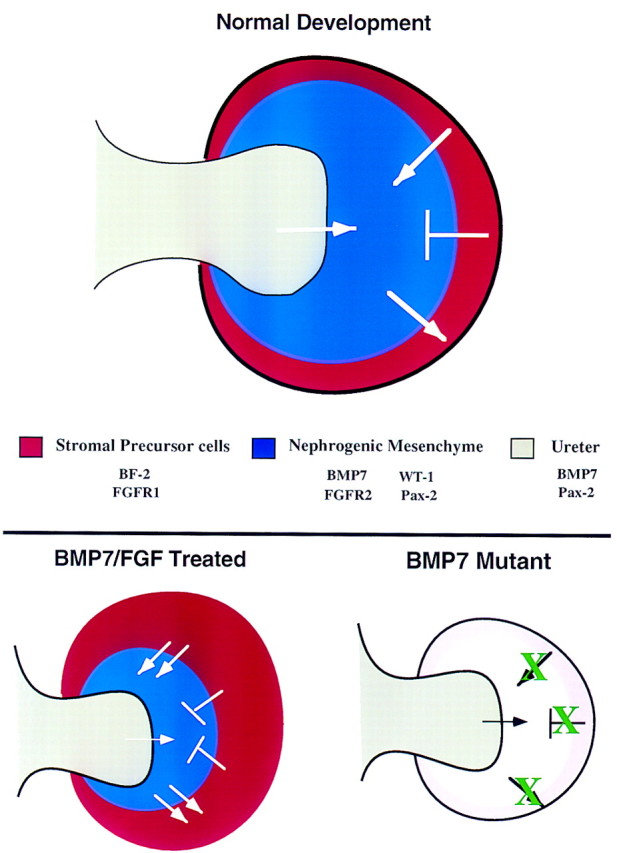
Nephrogenesis in the mouse kidney begins at embryonic day 11 and ends approximately 10 days postpartum. During this period, new nephrons are continually being generated from a stem-cell population-the nephrogenic mesenchyme-in response to signals emanating from the tips of the branching ureter. Relatively little is known about the mechanism by which the nephrogenic mesenchyme cell population is maintained at the tips of the ureter in the presence of signals promoting tubulogenesis. Previous studies have shown that a loss of Bmp7 function leads to kidney defects that are a likely result of progressive loss of nephrogenic mesenchyme by apoptosis. The studies presented here demonstrate that BMP7 signaling can prevent apoptosis in explants of metanephric mesenchyme. The surviving mesenchyme cell population, however, is not competent to respond to signals promoting tubulogenesis. In conjunction with FGF2, BMP7 promotes growth and maintains competence of the mesenchyme in vitro. In addition, FGF2 and BMP7 signaling, both independently and in combination, inhibit tubulogenesis. Interestingly, FGF2 and BMP7 also promote expansion of the stromal progenitor cell population in whole kidney culture. Because BMP7 is not produced by stromal progenitor cells, these data suggest a novel interaction between the nephrogenic mesenchyme and stromal progenitor cell populations. A model for the regulation of nephrogenesis by FGF and BMP signaling is presented.


Figures

Figure 1
Figure 1
BMP7 signaling does not induce nephrogenic mesenchyme. Mesenchyme was isolated from 11.5-dpc embryos and grown in culture for 48 hr in the presence of spinal cord (A,C,E) or recombinant BMP7 protein (B,D,F). After culture, explants were sectioned and processed for in situ hybridization. (A,B) Bright-field and (C–F) dark-field exposures of serial sections. In the presence of spinal cord the mesenchyme condenses, forms immature epithelium, and displays strong expression of Pax2 (C) and Wnt4 (E). In contrast, mesenchyme grown in the presence of BMP7 remains loose and does not express Pax2 (D) or Wnt4 (F). The asterisk in panels B,D, and F indicate an artifact. Recombinant cultures of wild-type spinal cord and Bmp7lacZ/+ mesenchyme display many LacZ+ early tubules (white arrow) as well as nonstaining, more mature tubules (G; black arrow). Cultures of Bmp7 mutant mesenchyme isolated from Bmp7lacZ embryos recombined with mutant spinal cord also display well formed tubules (H).
Figure 2
Figure 2
BMP7 promotes survival of the nephrogenic mesenchyme. Mesenchyme was isolated from 11.5-dpc embryos and grown in culture for 48 hr in the presence of a bead containing BSA (A,B), BMP7 (C,D), or FGF2 (E,F). Explants treated with BSA appear granular and refractory to light. In contrast, there is a well organized, translucent region of mesenchyme in the vicinity of the BMP7-soaked bead (cf. cells indicated by arrow in A and C). Staining with the vital dye TOPRO-1 reveals that cells in the BSA-treated mesenchyme are dying as indicated by fluorescent signal (B), whereas, in the BMP7-treated mesenchyme, cells in the translucent region do not fluoresce, indicating that these cells are viable (cf. cells indicated by arrow in B and D). Interestingly, most of the cells in the explant survive when treated with FGF2 (E,F). Note that the heparin–Sepharose beads in E and F display autofluorescence. (b) Bead. Scale bar, 100μm.
Figure 3
Figure 3
BMP7 and FGF2 function synergistically to promote survival and growth of mesenchyme explants. Mesenchyme was isolated from 11.5-dpc Bmp7lacZ/+ embryos and grown in culture for 48 hr in the presence of BMP7 (A), FGF2 (B,D–F), or both BMP7 and FGF2 (C,G–I). After culture, explants were stained in X-gal or fixed and processed for in situ hybridization. Mesenchyme treated with BMP7 displays limited survival. FGF2 promotes better survival of the mesenchyme. Treatment with both factors results in survival and growth of the isolated mesenchyme. Interestingly, these cultures display translucent regions (C, arrow) similar to that observed around BMP7- or FGF2-soaked beads. Marker analysis reveals LacZ+ cells in cultures treated with FGF2 (B, arrow). In addition, neither FGF2 (D–F) or FGF2 + BMP7-(G–I) treated cultures express Pax2 or Wnt4, demonstrating that induction has not occurred in these explants.
Figure 4
Figure 4
BMP7 and FGF2 signaling maintain nephrogenic mesenchyme in vitro. Mesenchyme was isolated from 11.5-dpc Bmp7 lacZ/+ embryos and grown in culture for 48 hr in the presence of recombinant FGF2 (A), BMP7 (B), or both (C). Mesenchyme was then placed in contact with spinal cord and grown for an additional 48 hr. As a control, freshly isolated mesenchyme was placed immediately in contact with spinal cord (D). After culture, explants were stained in X-gal. Neither FGF2 nor BMP7 alone maintain a population of cells competent to undergo induction, though a few LacZ+ cells and small aggregates of cells are often seen in these cultures (black arrows). In contrast, pretreatment with both factors allows a robust response of the explant to induction (white arrows). Sections through FGF2 + BMP7 (E) and control (F) cultures demonstrate that stained regions contain tubules and forming glomeruli (black arrows) as well as more mature, nonstaining tubules (white arrows). In addition, regions of stained, nonaggregated cells are readily observed in cultures pretreated with FGF2 + BMP7.
Figure 5
Figure 5
BMP7 and FGF2 signaling oppose tubulogenesis. Whole kidneys were isolated at 11.5-dpc and grown on Nuclepore filter rafts. After culture, explants were stained with an anti-laminin antibody via indirect im- munofluorescence. Low magnification (A–C, G–I) and high magnification (D–F, J–L) views of explants are presented. (A,D) Kidney explant grown for 48 hr in the presence of 10% FBS. (B,E) Bmp7 mutant kidney grown for 48 hr in the presence of 10% FBS. Arrows in D and E indicate nephrons that have formed in the periphery. Note how mutant tubules appear larger and less organized than wild-type. Wild- type kidneys grown in the presence of recombinant BMP7 (C,F), FGF2 (G,J), or both factors (H,K). BMP7 seems to have little effect on the branching of the ureter, however, tips of the ureter are often found free of nephrons (F, arrow). FGF2 signaling alone or in combination with BMP7 also reduces nephron formation in the periphery of the explant (arrows in J and K). In contrast to kidneys treated with BMP7 alone, however, FGF2 signaling causes a rind of cells to form in the periphery of the explant (G, arrow). In combination, both factors promote an even greater expansion of this outer cell population (H, arrow). Explants treated with FGF2 + BMP7 for 48 hr, subsequently placed in fresh culture media lacking these factors and grown for an additional 48 hr, display extensive tubulogenesis in the periphery of the kidney (I,L).
Figure 6
Figure 6
BMP7 and FGF2 promote expansion of the stromal progenitor cell population. Whole kidneys were isolated at 11.5 dpc and grown for 48 hr in culture containing 10% FBS in the absence (A,C,E,G,I) or in the presence of both BMP7 and FGF2 (B,D,F,H,J). After culture, explants were processed for whole-mount in situ hybridization or stained in X-gal. (A) Pax2 is expressed in the ureter and in the condensed mesenchyme. (B) A similar expression pattern is observed in the treated kidneys. WT1 is expressed in the condensed mesenchyme associated with the tips of the branching ureter in both control (C) and treated kidneys (D). X-gal staining for the Bmp7lacZ allele also shows a similar expression pattern in both the control (E) and treated (F) explants. In all cases the expanded rind of cells in the periphery of the kidney do not express any of these markers of induced mesenchyme (arrow). The stromal precursor marker BF2 (G), however, is expressed in this expanded population of cells (H). Sections through G and H reveal that BF2 is expressed throughout the expanded region of mesenchyme cells (arrow, I,J). The apparent BF2 signal in the nephrogenic mesenchyme is attributable to the layer of stromal progenitor cells that encompasses the entire explant (G,H).
Figure 7
Figure 7
FGFR expression in BMP7/FGF2-treated kidney explants. Whole kidneys were isolated at 11.5 dpc and grown for 48 hr in culture in 10% FBS (A,C) or in the presence of BMP7 and FGF2 (B,D). After culture, explants were processed for whole-mount in situ hybridization. Fgfr1 is expressed in the most peripheral mesenchyme of both control (A) and treated (B) explants. Fgfr2 is expressed in the ureteric tissue as well as in the condensed mesenchyme of both control (C) and treated (D) explants but is absent from the peripheral mesenchyme. (long arrow) Stromal cells; (short arrow) condensed nephrogenic mesenchyme.
Figure 8
Figure 8
Model of kidney development. This model represents a ureteric bud that has just entered the metanephric mesenchyme and displays the signaling that occurs between the various cell types of the developing kidney based on data in the literature and results presented here. During normal development, signals from the ureteric bud (gray) are required for induction of the nephrogenic mesenchyme (blue). In turn, these cells signal to the adjacent stromal progenitor cells (red) promoting their survival, growth, and differentiation. In a reciprocal manner, the stromal progenitor cells send an antidifferentiation signal to the nephrogenic mesenchyme to control the rate of nephrogenesis. In addition, these cells may also send a survival factor to the nephrogenic mesenchyme. Potential molecules involved in these signaling events are listed. In BMP7/FGF2-treated kidney explants, increased levels of BMP7 and FGF lead to increased survival of the nephrogenic mesenchyme as well as to an expansion of the stromal precursor cells. As a result, there is an increased level of the antidifferentiation signal coming from the stromal precursor cells. Therefore, differentiation of the nephrogenic mesenchyme is reduced. At this time, the source of the FGF signal is unknown. In Bmp7 mutant kidneys, lack of BMP7 signaling (white) leads to loss of proper signaling from the stromal precursor cells (pink), thereby resulting in premature differentiation and perhaps increased cell death in the nephrogenic mesenchyme.

References

    1. Aufderheide E, Chiquet-Ehrismann R, Ekblom P. Epithelial-mesenchymal interactions in the developing kidney lead to expression of tenascin in the mesenchyme. J Cell Biol. 1987;105:599–608. - PMC - PubMed
    1. Barasch J, Qiao J, McWilliams G, Chen D, Oliver JA, Herzlinger D. Ureteric bud cells secrete multiple factors, including bFGF, which rescue renal progenitors from apoptosis. Am J Physiol. 1997;273:F757–767. - PubMed
    1. Cancilla B, Cauchi J, Key B, Nurcombe V, Alcorn D, Bertram J. Immunolocalization of fibroblast growth factor-1 and -2 in the embryonic rat kidney. Nephrology. 1996;2:167–174. - PubMed
    1. Coles HSR, Burne JF, Raff MC. Large-scale normal cell death in developing rat kidney and its reduction by epidermal growth factor. Development. 1993;118:969–981. - PubMed
    1. Davies JA, Garrod DR. Induction of early stages of kidney tubule differentiation by lithium ions. Dev Biol. 1995;167:50–60. - PubMed

Publication types

MeSH terms

Substances

LinkOut - more resources