. 1999 Jun 15;13(12):1614-26.
doi: 10.1101/gad.13.12.1614.

Cdc53/cullin and the essential Hrt1 RING-H2 subunit of SCF define a ubiquitin ligase module that activates the E2 enzyme Cdc34

Affiliations

Cdc53/cullin and the essential Hrt1 RING-H2 subunit of SCF define a ubiquitin ligase module that activates the E2 enzyme Cdc34

J H Seol et al. Genes Dev. .

Abstract

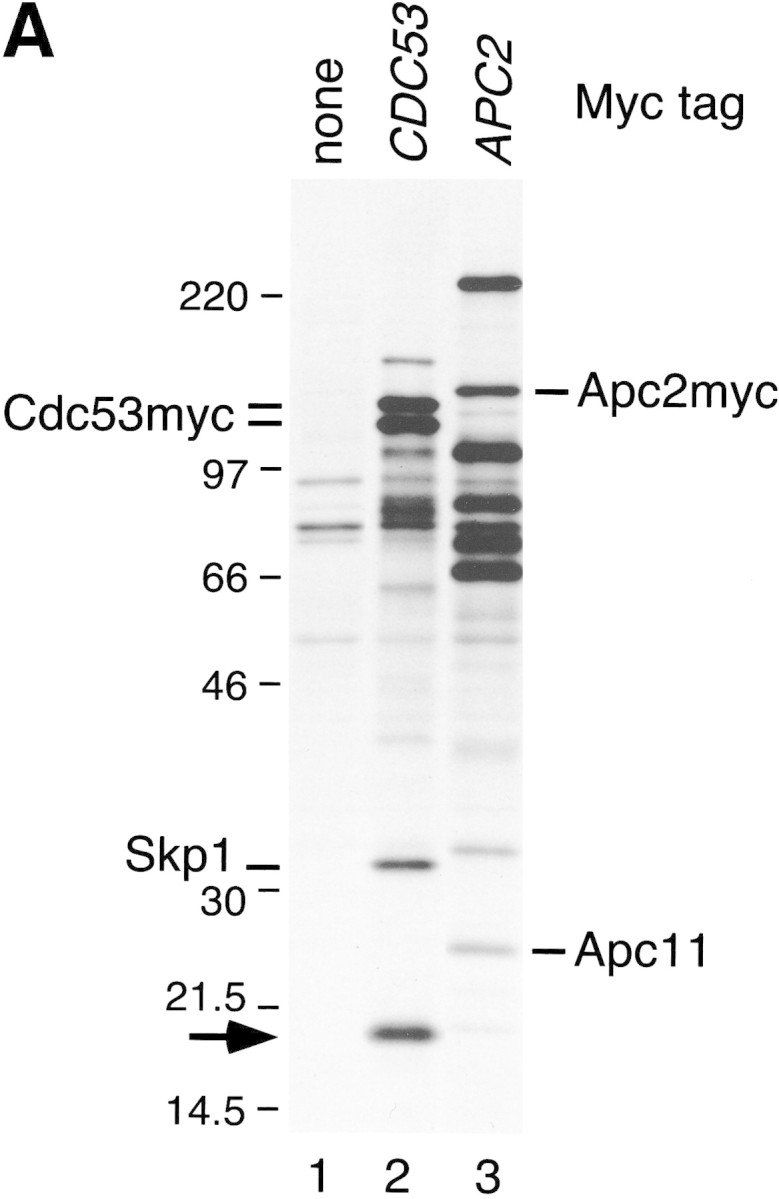
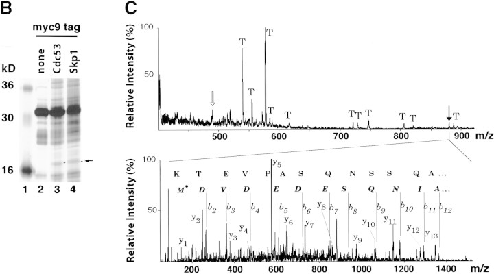
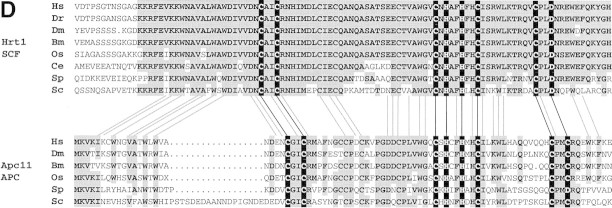
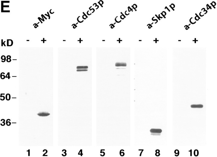
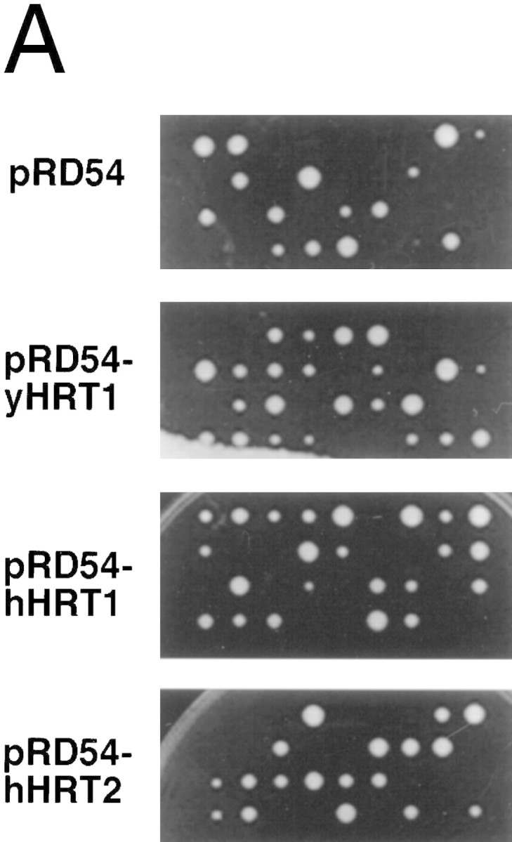
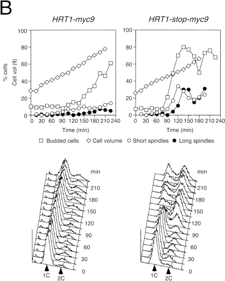
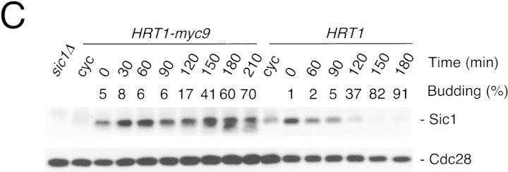
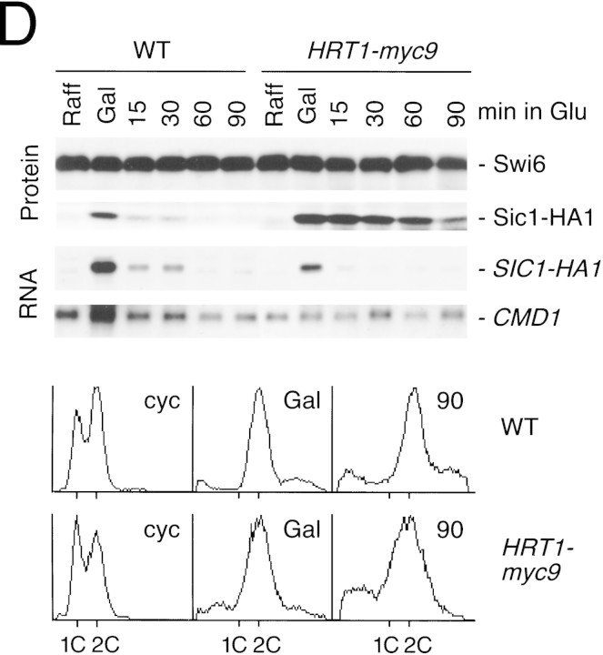
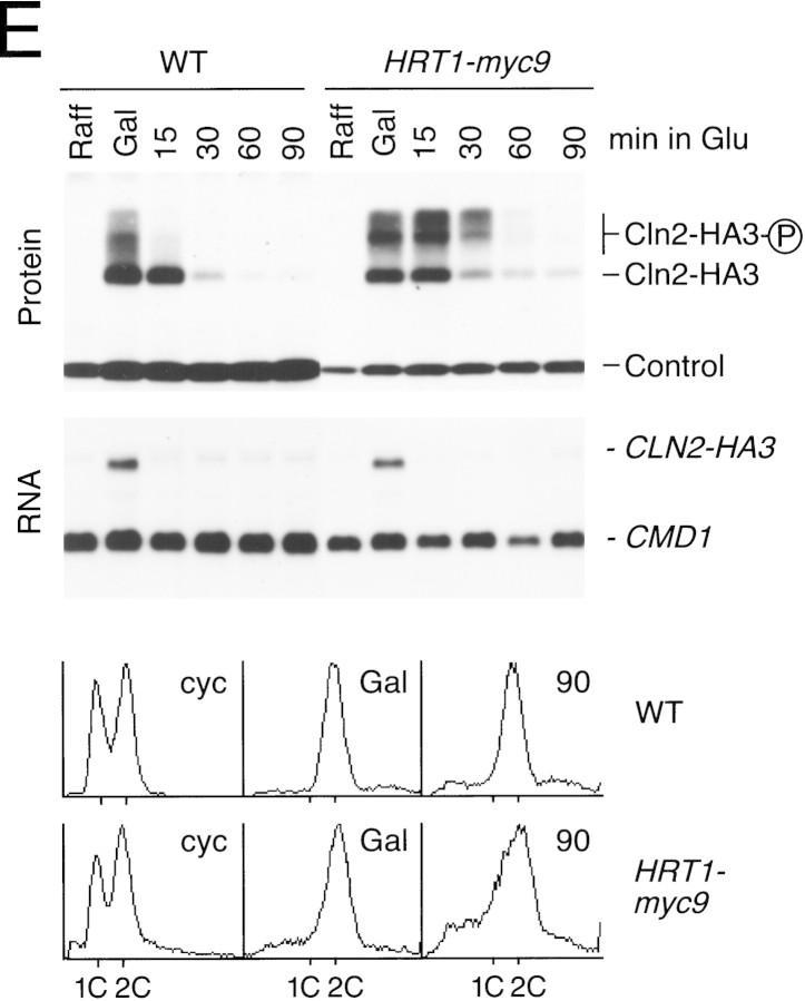
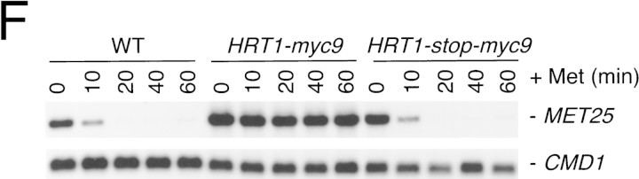
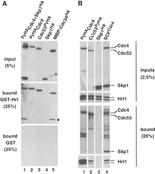
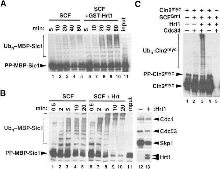
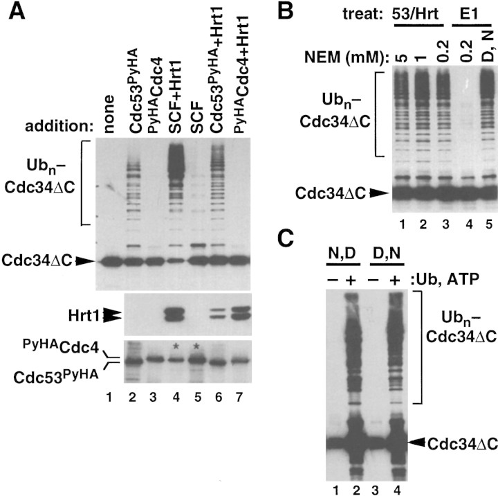
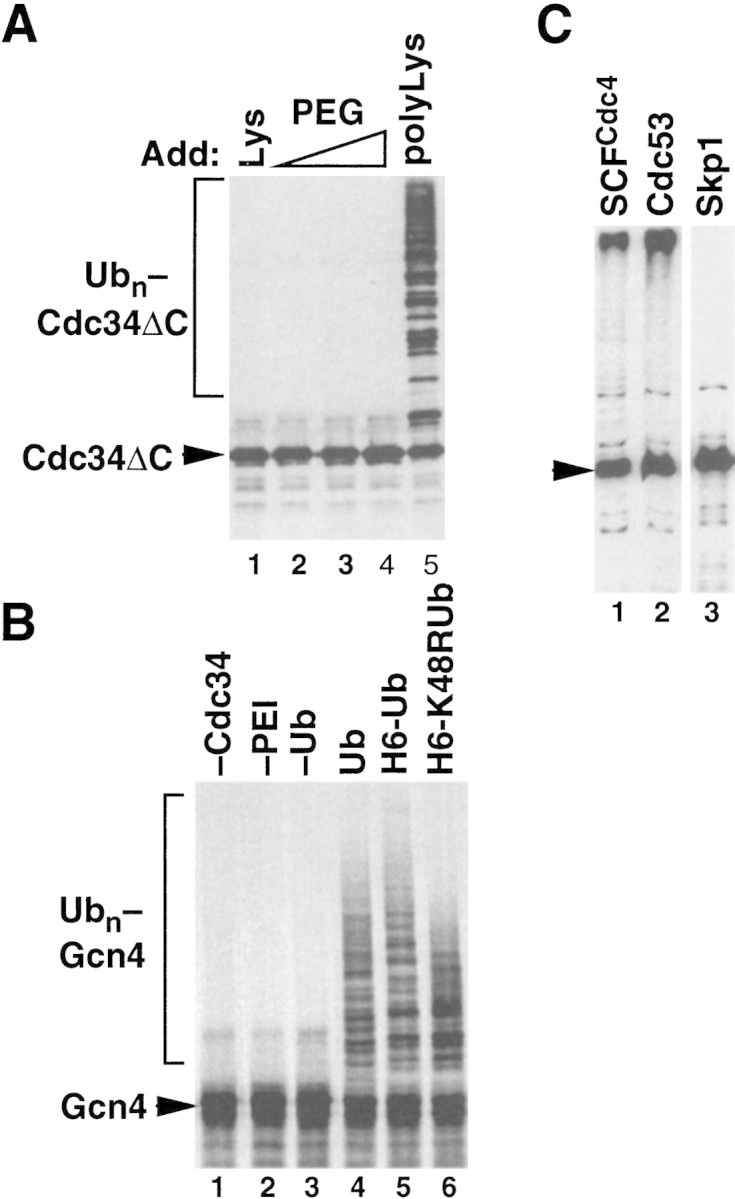
SCFCdc4 (Skp1, Cdc53/cullin, F-box protein) defines a family of modular ubiquitin ligases (E3s) that regulate diverse processes including cell cycle, immune response, and development. Mass spectrometric analysis of proteins copurifying with Cdc53 identified the RING-H2 finger protein Hrt1 as a subunit of SCF. Hrt1 shows striking similarity to the Apc11 subunit of anaphase-promoting complex. Conditional inactivation of hrt1(ts) results in stabilization of the SCFCdc4 substrates Sic1 and Cln2 and cell cycle arrest at G1/S. Hrt1 assembles into recombinant SCF complexes and individually binds Cdc4, Cdc53 and Cdc34, but not Skp1. Hrt1 stimulates the E3 activity of recombinant SCF potently and enables the reconstitution of Cln2 ubiquitination by recombinant SCFGrr1. Surprisingly, SCF and the Cdc53/Hrt1 subcomplex activate autoubiquitination of Cdc34 E2 enzyme by a mechanism that does not appear to require a reactive thiol. The highly conserved human HRT1 complements the lethality of hrt1Delta, and human HRT2 binds CUL-1. We conclude that Cdc53/Hrt1 comprise a highly conserved module that serves as the functional core of a broad variety of heteromeric ubiquitin ligases.


Figures

Figure 1
Figure 1
Identification of Hrt1 as a Cdc53-associated protein. (A) [35S]Methionine-labeled proteins that specifically coimmunoprecipitate with Cdc53myc6 (lane 2) or Apc2myc6 (lane 3) were visualized by SDS-PAGE followed by autoradiography. (B) Proteins coimmunoprecipitating with Cdc53myc9 (lane 3) or Skp1myc9 (lane 4) were separated by SDS-PAGE, and stained with silver. Both preparations consistently contained a 17-kD protein (arrow in A and B). (C) Identifiecation of YOL133w by nanoelectrospray tandem mass spectrometry (Nano ES MS/MS). (Top) Partial mass spectrum of a pool of tryptic peptides recovered after in gel digestion of 17-kD band. (T) Peaks identified as trypsin autolysis products. Tandem mass spectrum acquired from the ion with m/z 488.2 (open arrow) identified peptide AFLIEEQK from ribosomal protein RPL34B (data not shown). Tandem mass spectrum (bottom) of the ion with m/z 880.4 (solid arrow) revealed two nonoverlapping series of fragment y-ions and b-ions (Biemann 1988) that both identified the same Hrt1-derived peptide M*DVDEDESQNIAQSSNQSAPVETK (M* stands for methionine sulfoxide). Carboxy-terminal part of the peptide sequence (...AQSSNQSAPVETK) is covered by continuous series of y-ions. Amino-terminal part of the sequence (M*DVDEDESQNIA) is covered by b-ion series. As every amino acid residue in the peptide sequence can be accounted for, identification of Hrt1 is certain despite the complexity of the spectrum and that only a single peptide was identified. (D) Hrt1 is highly conserved between species and related to the APC subunit Apc11. Budding yeast Hrt1 and Apc11 were each aligned with related sequences from different species. Putative zinc-coordinating residues are highlighted black. Residues identical in at least 4 (Apc11) or 5 (Hrt1) sequences are shaded. Lines connecting the alignments indicate residues conserved between Apc11 and Hrt1. (Hs) Homo sapiens, (Dm) Drosophila melanogaster; (Dr) Danio rerio; (Bm) Bombyx mori; (Os) Oryza sativa; (Ce) Caenorhabditis elegans; (Sp) Schizosaccharomyces pombe; (Sc) Saccharomyces cerevisiae. (E) Hrt1 is an authentic subunit of SCF. Crude extracts from untagged control (lane 1) and HRT1myc9 (lane 2) strains were evaluated in parallel by immunoblotting with 9E10 antibodies. Extracts from untagged (odd-numbered lanes) or HRT1myc9 (even-numbered lanes) cells were immunoprecipitated with 9E10 antibodies fractionated by SDS-PAGE, and immunoblotted with α-Cdc53, α-Cdc4, α-Skp1, or α-Cdc34 as indicated to reveal the corresponding protein.
Figure 1
Figure 1
Identification of Hrt1 as a Cdc53-associated protein. (A) [35S]Methionine-labeled proteins that specifically coimmunoprecipitate with Cdc53myc6 (lane 2) or Apc2myc6 (lane 3) were visualized by SDS-PAGE followed by autoradiography. (B) Proteins coimmunoprecipitating with Cdc53myc9 (lane 3) or Skp1myc9 (lane 4) were separated by SDS-PAGE, and stained with silver. Both preparations consistently contained a 17-kD protein (arrow in A and B). (C) Identifiecation of YOL133w by nanoelectrospray tandem mass spectrometry (Nano ES MS/MS). (Top) Partial mass spectrum of a pool of tryptic peptides recovered after in gel digestion of 17-kD band. (T) Peaks identified as trypsin autolysis products. Tandem mass spectrum acquired from the ion with m/z 488.2 (open arrow) identified peptide AFLIEEQK from ribosomal protein RPL34B (data not shown). Tandem mass spectrum (bottom) of the ion with m/z 880.4 (solid arrow) revealed two nonoverlapping series of fragment y-ions and b-ions (Biemann 1988) that both identified the same Hrt1-derived peptide M*DVDEDESQNIAQSSNQSAPVETK (M* stands for methionine sulfoxide). Carboxy-terminal part of the peptide sequence (...AQSSNQSAPVETK) is covered by continuous series of y-ions. Amino-terminal part of the sequence (M*DVDEDESQNIA) is covered by b-ion series. As every amino acid residue in the peptide sequence can be accounted for, identification of Hrt1 is certain despite the complexity of the spectrum and that only a single peptide was identified. (D) Hrt1 is highly conserved between species and related to the APC subunit Apc11. Budding yeast Hrt1 and Apc11 were each aligned with related sequences from different species. Putative zinc-coordinating residues are highlighted black. Residues identical in at least 4 (Apc11) or 5 (Hrt1) sequences are shaded. Lines connecting the alignments indicate residues conserved between Apc11 and Hrt1. (Hs) Homo sapiens, (Dm) Drosophila melanogaster; (Dr) Danio rerio; (Bm) Bombyx mori; (Os) Oryza sativa; (Ce) Caenorhabditis elegans; (Sp) Schizosaccharomyces pombe; (Sc) Saccharomyces cerevisiae. (E) Hrt1 is an authentic subunit of SCF. Crude extracts from untagged control (lane 1) and HRT1myc9 (lane 2) strains were evaluated in parallel by immunoblotting with 9E10 antibodies. Extracts from untagged (odd-numbered lanes) or HRT1myc9 (even-numbered lanes) cells were immunoprecipitated with 9E10 antibodies fractionated by SDS-PAGE, and immunoblotted with α-Cdc53, α-Cdc4, α-Skp1, or α-Cdc34 as indicated to reveal the corresponding protein.
Figure 2
Figure 2
(A) HRT1 is essential. A diploid hrt1::HIS3/HRT1 strain was transformed with pRD54-based plasmids (pRS316 CEN, URA3) containing either yeast HRT1, human HRT1, or human HRT2 under the control of the galactose-inducible GAL1 promoter. Sporulated diploids were dissected on complete galactose medium, followed by incubation at 24°C for 5 days. (B,C) HRT1 is required for both elimination of Sic1 and for S phase. A myc9 epitope cassette was integrated at HRT1 either immediately upstream (HRT1–myc9, left) or downstream (HRT1–stop–myc9, right) of the stop codon. Small G1 cells were isolated by centrifugal elutriation from cultures grown in YPD at 25°C and inoculated into fresh medium at 37°C at time 0. In B, mean cell volume (fl), budding index, formation of mitotic spindles (top panels), and DNA contents (bottom panels) were assessed at the indicated times. In C, budding index, Sic1 levels, and Cdc28 levels were monitored. (D) HRT1 is required for proteolysis of Sic1. HRT1 cells containing five integrated copies of GAL–SIC1–HA1 and HRT1–myc9 GAL–SIC1–HA1 (one copy) cells were grown in YP–raffinose at 25°C (cyc) and arrested in mitosis with nocodazole (Raff). After shifting the cultures to 32°C, expression from the GAL promoter was induced with galactose (Gal) and then repressed by transferring the cells to YP–glucose. Samples were withdrawn at the indicated time points and assayed for: Swi6 and Sic1-HA1 protein levels (top panels); calmodulin and SIC1–HA1 mRNA levels (middle panels); and cellular DNA content (bottom panels). (E) HRT1 is required for rapid degradation of Cln2. Same as D, except CLN2–HA3 expression was induced with galactose at 35°C, and cells were shifted to 37°C upon transfer to YP–glucose. The band labeled control is an unknown polypeptide recognized by 12CA5. (F) HRT1 is required for methionine-dependent repression of MET25 mRNA. Yeast strains indicated were grown in complete medium containing glucose and lacking methionine at 25°C and were then shifted to 37°C for 2 hr. Methionine was added to the medium and samples were withdrawn at the indicated times. MET25 and CMD1 mRNA levels were assayed by Northern blotting.
Figure 2
Figure 2
(A) HRT1 is essential. A diploid hrt1::HIS3/HRT1 strain was transformed with pRD54-based plasmids (pRS316 CEN, URA3) containing either yeast HRT1, human HRT1, or human HRT2 under the control of the galactose-inducible GAL1 promoter. Sporulated diploids were dissected on complete galactose medium, followed by incubation at 24°C for 5 days. (B,C) HRT1 is required for both elimination of Sic1 and for S phase. A myc9 epitope cassette was integrated at HRT1 either immediately upstream (HRT1–myc9, left) or downstream (HRT1–stop–myc9, right) of the stop codon. Small G1 cells were isolated by centrifugal elutriation from cultures grown in YPD at 25°C and inoculated into fresh medium at 37°C at time 0. In B, mean cell volume (fl), budding index, formation of mitotic spindles (top panels), and DNA contents (bottom panels) were assessed at the indicated times. In C, budding index, Sic1 levels, and Cdc28 levels were monitored. (D) HRT1 is required for proteolysis of Sic1. HRT1 cells containing five integrated copies of GAL–SIC1–HA1 and HRT1–myc9 GAL–SIC1–HA1 (one copy) cells were grown in YP–raffinose at 25°C (cyc) and arrested in mitosis with nocodazole (Raff). After shifting the cultures to 32°C, expression from the GAL promoter was induced with galactose (Gal) and then repressed by transferring the cells to YP–glucose. Samples were withdrawn at the indicated time points and assayed for: Swi6 and Sic1-HA1 protein levels (top panels); calmodulin and SIC1–HA1 mRNA levels (middle panels); and cellular DNA content (bottom panels). (E) HRT1 is required for rapid degradation of Cln2. Same as D, except CLN2–HA3 expression was induced with galactose at 35°C, and cells were shifted to 37°C upon transfer to YP–glucose. The band labeled control is an unknown polypeptide recognized by 12CA5. (F) HRT1 is required for methionine-dependent repression of MET25 mRNA. Yeast strains indicated were grown in complete medium containing glucose and lacking methionine at 25°C and were then shifted to 37°C for 2 hr. Methionine was added to the medium and samples were withdrawn at the indicated times. MET25 and CMD1 mRNA levels were assayed by Northern blotting.
Figure 3
Figure 3
Hrt1 binds Cdc4, Cdc53, and Cdc34. (A) (Lanes 1–4) GST–Hrt1 (middle panel) or GST (bottom panel) purified from E. coli were mixed with crude insect cell lysates (top panel) that contained the proteins indicated above each lane. Proteins retrieved on glutathione-agarose were fractionated by SDS-PAGE and revealed by immunoblotting with α-HA. (Lane 5) Same as lanes 1–4, except MBP–Cdc34ΔCHA purified from E. coli was evaluated for its ability to bind GST-Hrt1. The asterisks denote a proteolytic fragment of MBP–Cdc34ΔCHA. Input lanes contain 5%; bound lanes contain 25% of the material from a single binding reaction. (B) Lysates from Sf9 insect cells that coexpressed Hrt1 plus PyHACdc4 (lane 1), Cdc53PyHA (lane 2), Skp1HA (lane 3), or PyHACdc4/Cdc53/Skp1HA (lane 4) were adsorbed to α-Py beads, and bound proteins were fractionated by SDS-PAGE and revealed by immunoblotting with α-HA (third row of panels) or α-Hrt1 (bottom row of panels) antibodies. Unfractionated insect cell lysates were directly immunoblotted to evaluate relative expression of the individual proteins (top row of panels, α-HA blot; second row of panels, α-Hrt1 blot).
Figure 4
Figure 4
Hrt1 potently stimulates ubiquitin ligase activity of SCFCdc4. (A) Recombinant SCFCdc4 was produced in Sf9 insect cells upon coinfection with baculovirus vectors that express Skp1, Cdc53PyHA, and PyHACdc4, and the complex was purified by adsorption to a protein A resin containing covalently linked antipolyoma monoclonal antibody (α-Py beads). Parallel samples were supplemented with E1 enzyme, Cdc34, ATP, ubiquitin, purified phosphorylated maltose binding protein–Sic1 chimera (PP–MBP–Sic1), and either GST (lanes 1–5) or GST–Hrt1 (lanes 6–10). At the indicated times, samples were evaluated for ubiquitination of phospho–MBP–Sic1 by SDS-PAGE followed by immunoblotting with α-Sic1. (B) (Lanes 1–11) Same as A, except that recombinant SCFCdc4 complexes that either contained (lanes 6–10) or lacked (lanes 1–5) yeast Hrt1 were generated by either triple or quadruple infection of insect Sf9 cells (quadruple infection contained an Hrt1-expressing baculovirus). Input substrate is shown in lane 11. (Lanes 12,13) Levels of SCF subunits in each preparation were evaluated by immunoblotting. (C) Same as B, except that recombinant SCFGrr1 complexes containing (lane 3) or lacking (lane 2) Hrt1 were expressed in insect cells, purified, and mixed with 1/20 volume insect cell lysate that contained recombinant Cln2myc/GST–Cdc28/Cks1 complexes (Cln2myc). In lanes 3 and 4, Cdc34 and substrate, respectively, were omitted from the final reaction. PP–Cln2myc refers to the phosphorylated substrate.
Figure 5
Figure 5
Cdc53/Hrt1 comprise an NEM-resistant ubiquitin ligase module. (A) The proteins indicated were retrieved from baculovirus-infected insect cells on α-Py beads, and mixed with E1, Cdc34ΔC, ubiquitin, and ATP. Following a 60-min incubation at 20°C, reactions were evaluated for Cdc34ΔC autoubiquitination (top panel), levels of Hrt1 (middle panel), and levels of Cdc53PyHA or PyHACdc4 (bottom panel) by SDS-PAGE following by immunoblotting. SCFCdc4 complexes (lanes 4,5) contained PyHACdc4 and untagged Cdc53. Thus, only PyHACdc4 is visible in these lanes in the α-HA blot (bottom panel; see asterisks). (B) Either Cdc53PyHA/Hrt1 (lanes 1–3) or E1 enzyme (lane 4) was pretreated with the indicated amount of NEM for 10 min at 20°C. NEM was quenched with DTT, and complete reactions containing E1, Cdc34ΔC, ubiquitin, ATP, and α-Py bead-bound Cdc53PyHA/Hrt1 were incubated for 60 min at 20°C. For the sample shown in lane 5, DTT was mixed with NEM prior to incubation with E1 to confirm the efficacy of the DTT quench. All reactions were subjected to SDS-PAGE followed by immunoblotting with α-Cdc34. (C) Same as B, except that complete reactions were first assembled and then treated with 5 mm NEM followed by 10 mm DTT (lanes 1,2) or vice versa (lanes 3,4). Afterwards, α-Py bead-bound Cdc53PyHA/Hrt1 was recovered, washed, and supplemented with either buffer (lanes 1,3) or fresh E1, Cdc34ΔC, ubiquitin and ATP (lanes 2,4). The signal in lanes 1 and 3 represents Cdc34ΔC from the first stage incubation that was retained on the Cdc53PyHA/Hrt1 matrix.
Figure 6
Figure 6
Polycations can substitute for SCF. (A) Polylysine activates Cdc34ΔC autoubiquitination. Reactions containing Cdc34ΔC, ATP, ubiquitin, and E1 enzyme were adjusted to either 10 mm lysine (lane 1), 0.01, 0.1, or 1% polyethylene glycol (PEG, lanes 2–4), or 0.01% (∼300 nm) polylysine (lane 5), and incubated at 20°C for 60 min. Reactions were evaluated as described for Fig. 5A. (B) Polyethyleneimine is an E3 for Gcn4. 35S labeled Gcn4 produced by in vitro translation in reticulocyte lysate and enriched by DEAE chromatography was mixed with E1, Cdc34, ATP, the indicated ubiquitin derivatives (lanes 4–6; H6 stands for His6) and 0.0005% polyethyleneimine (PEI), and incubated at 25°C for 60 min. Reactions conducted in the absence of either Cdc34, PEI, or ubiquitin are depicted in lanes 1–3. Gcn4 was visualized by SDS-PAGE followed by autoradiography. (C) Fusion of Sic1 to Cdc34 does not activate its ubiquitination. Cdc34ΔC–Sic1 (arrowhead) was mixed with E1, ubiquitin, and ATP in the presence of immobilized SCFCdc4 (lane 1), Cdc53PyHA (lane 2), or (as a negative control) Skp1HA (lane 3). Samples were incubated and processed as described for Fig. 5A.

Similar articles

Cited by

References

    1. Altschul S, Madden T, Schaffer A, Zhang J, Zhang Z, Miller W, Lipman D. Gapped BLAST and PSI-BLAST: A new generation of protein database search programs. Nucleic Acids Res. 1997;25:3389–3402. - PMC - PubMed
    1. Bai C, Sen P, Hofmann K, Ma L, Goebl M, Harper JW, Elledge SJ. SKP1 connects cell cycle regulators to the ubiquitin proteolysis machinery through a novel motif, the F-box. Cell. 1996;86:263–274. - PubMed
    1. Banerjee A, Gregori L, Xu Y, Chau V. The bacterially expressed yeast CDC34gene product can undergo autoubiquitination to form a multiubiquitin chain-linked protein. J Biol Chem. 1993;268:5668–5675. - PubMed
    1. Barral Y, Jentsch S, Mann C. G(1) cyclin turnover and nutrient-uptake are controlled by a common pathway in yeast. Genes & Dev. 1995;9:399–409. - PubMed
    1. Bartel B, Wunning I, Varshavsky A. The recognition component of the N-end rule pathway. EMBO J. 1990;9:3179–3189. - PMC - PubMed

Publication types

MeSH terms

Substances