. 1999 Jul 2;290(1):9-20.
doi: 10.1006/jmbi.1999.2860.

Display of epitopes on the surface of tobacco mosaic virus: impact of charge and isoelectric point of the epitope on virus-host interactions

Affiliations

Display of epitopes on the surface of tobacco mosaic virus: impact of charge and isoelectric point of the epitope on virus-host interactions

M Bendahmane et al. J Mol Biol. .

Abstract

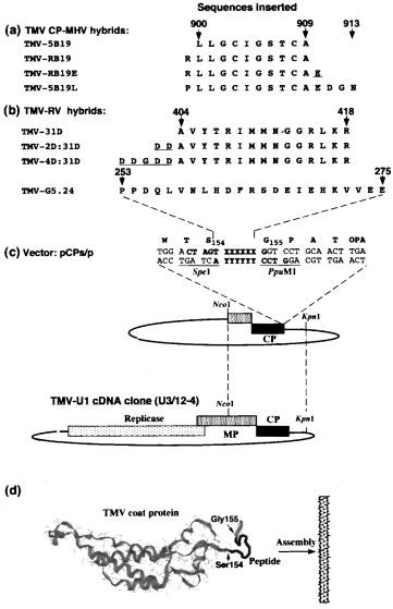
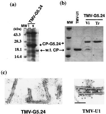
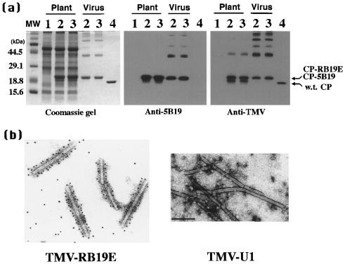
The biophysical properties of the tobacco mosaic tobamovirus (TMV) coat protein (CP) make it possible to display foreign peptides on the surface of TMV. The immunogenic epitopes G5-24 from the rabies virus (RV) glycoprotein, and 5B19 from murine hepatitis virus (MHV) S-glycoprotein were successfully displayed on the surface of TMV, and viruses accumulated to high levels in infected leaves of Nicotiana tabacum Xanthi-nn. The peptide RB19, which contains an arginine residue plus the 5B19 epitope fused to the CP (TMV-RB19), resulted in the induction of necrotic local lesions on inoculated leaves of N. tabacum Xanthi-nn and cell death of infected BY2 protoplasts. RNA dot blot assays confirmed that expression of the acidic and basic pathogenesis-related PR2 genes were induced in infected Xanthi-nn leaf tissue. TMV that carried epitope 31D from the RV nucleoprotein did not accumulate in inoculated tobacco leaves. Analysis of hybrid CPs predicted that the isoelectric points (pI):charge value was 5.31:-2 for wild-type CP, 5.64:-1 for CP-RB19, and 9.14:+2 for CP-31D. When acidic amino acids were inserted in CP-RB19 and CP-31D to bring their pI:charge to near that of wild-type CP, the resulting viruses TMV-RB19E and TMV-4D:31D infected N. tabacum Xanthi-nn plants and BY2 protoplasts without causing cell death. These data show the importance of the pI of the epitope and its effects on the hybrid CP pI:charge value for successful epitope display as well as the lack of tolerance to positively charged epitopes on the surface of TMV.


Figures

Figure 1
Figure 1
Virus hybrids and amino acid sequence of the peptides used in this study. (a) Peptides from the MHV S-glycoprotein. Numbers above the arrows represent amino acids in S-glycoprotein Buchmeier et al 1984, Luytjes et al 1989. (b) Peptides from rabies virus glycoprotein and nucleoprotein. The amino acid coordinates in the nucleoprotein (for the peptide 31D) and in the glycoprotein (for the peptide G5-24) are indicated (Dietzschold et al., 1990). Amino acid residues inserted to neutralize the positive charge of the peptides are underlined. (c) Site of insertion of the peptides in TMV CP. The amino acid coordinates for w.t. CP are indicated (Goelet et al., 1982). The map of the vector pCPs/p developed in this study is shown. (d) Computer-assisted graphic representation of TMV CP structure. The position of the insertion of the different peptides at the C terminus of the CP is indicated.
Figure 2
Figure 2
(a) Coomassie blue-stained gel showing the accumulation of hybrid CP-G5.24 in N. tabacum Xanthi-nn plants infected with TMV-G5.24. i, inoculated leaf; s, systemically infected leaf; w.t. CP, wild-type TMV coat protein; Mw, protein molecular mass standards, in kDa. (b) Coomassie blue-stained gel showing the patterns of the hybrid CP-G5.24 in virus purified from plant inoculated with in vitro-transcribed viral RNA (lane Tr), or from plant inoculated with TMV-G5.24 virus particles (lane Vi). (c) Immunogold labeling of TMV-G5.24 and TMV-U1 using the monoclonal MAb5 antibody raised against the G5-24 peptide. Electron microscopy was performed on Phillips CM100 electron microscope at magnification of 39,000 ×. The scale bar represents 200 nm.
Figure 3
Figure 3
(a) Coomassie blue-stained gel of extracts of tobacco plants inoculated with two independent infectious clones of TMV-4D:31D, clones 13 and 16. The position of the CP-4D:31D is indicated. i, inoculated leaf; s, upper non-inoculated leaf; w.t. CP, wild-type TMV coat protein; Healthy, non infected tobacco plant; Mw, protein molecular mass standards, in kDa. (b) Western blot analysis of the accumulation of the CP-31D, CP-2D:31D and CP-4D:31D in infected BY2 protoplasts using anti-TMV antibody. The positions of molecular mass protein markers are shown in kDa.
Figure 4
Figure 4
(a) Symptoms of TMV-U1, TMV-RB19 and TMV-RB19E on inoculated leaves of N. tabacum Xanthi-nn and Xanthi-NN plants at 5 d.p.i. Plants were inoculated with in vitro transcribed viral RNAs and held at 26 °C. (b) Change in diameters of necrotic local lesions produced by TMV-RB19 on inoculated N. tabacum Xanthi-nn and by TMV-U1 or TMV-RB19 on N. tabacum Xanthi-NN. (c) and (d) Viability of BY2 protoplasts following infection with TMV-U1, TMV-5B19, TMV-RB19 or TMV-RB19E. Protoplasts were inoculated with viral RNA and then incubated at 24 or 31 °C. (c) Light microscopy (top) and fluorescence microscopy (bottom) showing the auto-fluorescence of dead cells at 36 h.p.i. (d) Percentage of viable cells determined microscopically after staining for live cells with FDA.
Figure 5
Figure 5
(a) Characterization of CP-5B19 and CP-RB19E. Proteins were extracted from infected leaves or from purified virus, separated by SDS-PAGE, and stained with Coomassie blue or electroblotted to nitrocellulose membrane and reacted with anti-5B19 or anti-TMV antibodies. Samples are from plants inoculated with TMV-5B19 (sample 2), TMV-RB19E (sample 3), TMV-U1 (sample 4) or from healthy plant (sample 1). Mw, protein molecular mass in kDa. (b) Immunogold labeling of TMV-RB19E and TMV-U1 (negative control) using monoclonal antibody raised against the 5B19 peptide. Electron microscopy was performed on Phillips CM100 electron microscope at magnification of 39,000 ×. The scale bar represents 200 nm.
Figure 6
Figure 6
Accumulation of (a) basic and (b) acidic PR2 mRNA in N. tabacum Xanthi-nn and Xanthi-NN plants following inoculation with TMV-U1 or TMV-RB19 at 4 d.p.i. RNA dot blots were performed using similar amounts of total RNA and then hybridized with sequences to detect (a) basic or (b) acidic PR2 gene expression. The signal intensity of the isotope label was measured using phosphoimager followed by Multi-Analyst computer program analyses (Bio-Rad, Hercules, CA), and the relative values are displayed as graphs.

References

    1. Abbink T.E.M., Tjernberg P.A., Bol J.F., Linthorst H.J.M. Tobacco mosaic virus helicase domain induces necrosis in N gene-carrying tobacco in the absence of virus replication. Mol. Plant Microbe Interact. 1998;11:1242–1246.
    1. Asselin A., Zaitlin M. Characterization of a second protein associated with virions of tobacco mosaic virus. Virology. 1978;91:173–181. - PubMed
    1. Beachy R.N., Fitchen J.H., Hein M.B. Use of plant viruses for delivery of vaccine epitopes. Ann. NY Acad. Sci. 1996;792:43–49. - PMC - PubMed
    1. Bendahmane M., Fitchen J.H., Zhang G., Beachy R.N. Studies of coat protein mediated resistance to TMV: correlation between assembly of mutant coat proteins and resistance. J. Virol. 1997;71:7942–7950. - PMC - PubMed
    1. Brederode F.T., Linthorst H.J.M., Bol J.F. Differential induction of acquired resistance and PR gene expression in tobacco by infection, ethephon treatment, UV light and wounding. Plant Mol. Biol. 1991;17:1117–1125. - PubMed

Publication types