. 1999 Jul;65(7):3121-8.
doi: 10.1128/AEM.65.7.3121-3128.1999.

Identification of a new gene family expressed during the onset of sexual reproduction in the centric diatom Thalassiosira weissflogii

Affiliations

Identification of a new gene family expressed during the onset of sexual reproduction in the centric diatom Thalassiosira weissflogii

E V Armbrust. Appl Environ Microbiol. 1999 Jul.

Abstract

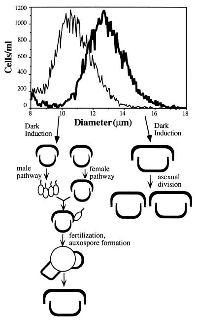
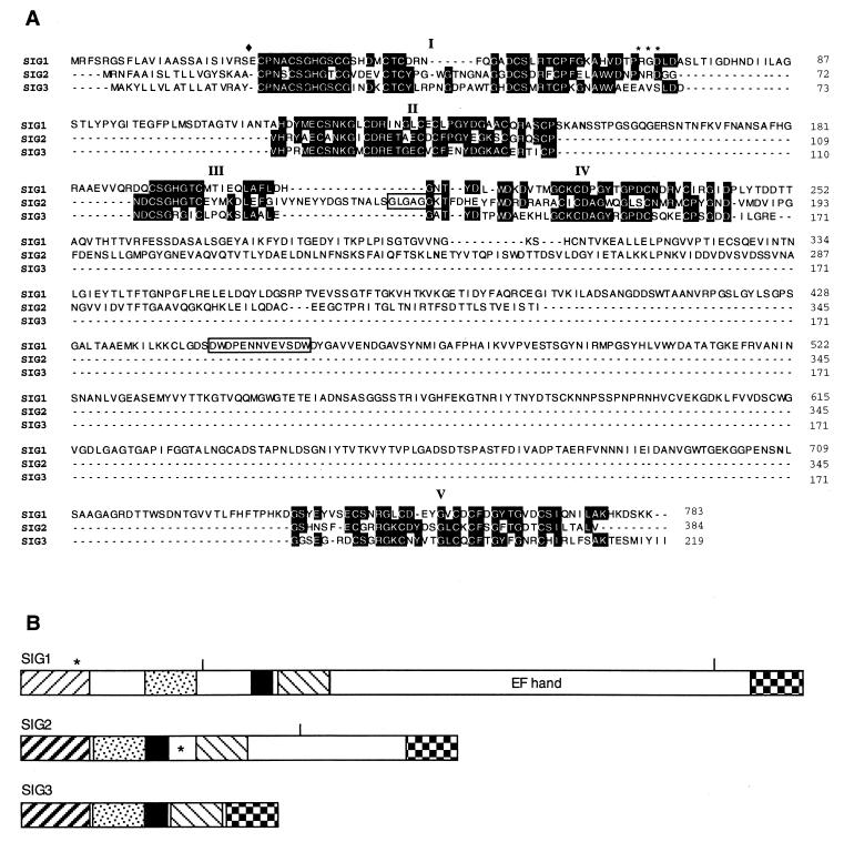
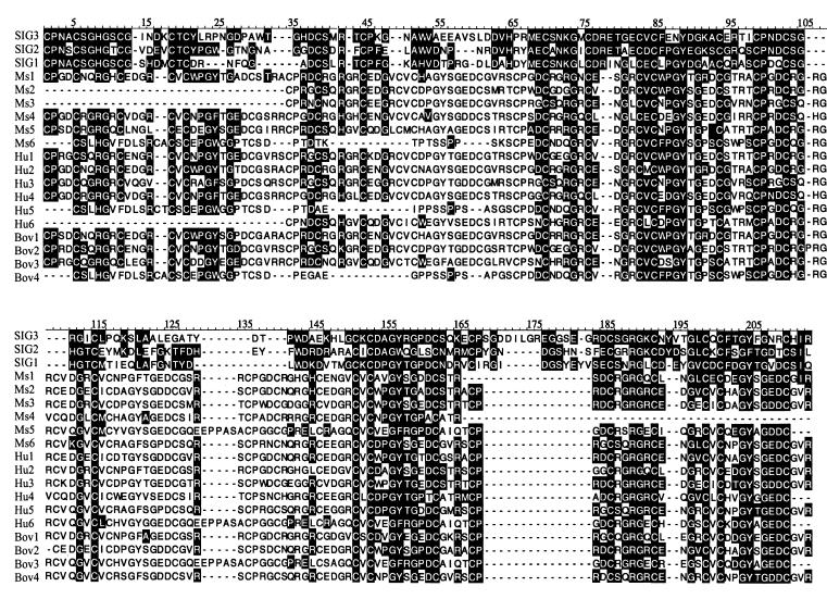
An intriguing feature of the diatom life cycle is that sexual reproduction and the generation of genetic diversity are coupled to the control of cell size. A PCR-based cDNA subtraction technique was used to identify genes that are expressed as small cells of the centric diatom Thalassiosira weissflogii initiate gametogenesis. Ten genes that are up-regulated during the early stages of sexual reproduction have been identified thus far. Three of the sexually induced genes, Sig1, Sig2, and Sig3, were sequenced to completion and are members of a novel gene family. The three polypeptides encoded by these genes possess different molecular masses and charges but display many features in common: they share five highly conserved domains; they each contain three or more cysteine-rich epithelial growth factor (EGF)-like repeats; and they each display homology to the EGF-like region of the vertebrate extracellular matrix glycoprotein tenascin X. Interestingly, the five conserved domains appear in the same order in each polypeptide but are separated by variable numbers of nonconserved amino acids. SIG1 and SIG2 display putative regulatory domains within the nonconserved regions. A calcium-binding, EF-hand motif is found in SIG1, and an ATP/GTP binding motif is present in SIG2. The striking similarity between the SIG polypeptides and extracellular matrix components commonly involved in cell-cell interactions suggests that the SIG polypeptides may play a role in sperm-egg recognition. The SIG polypeptides are thus important molecular targets for determining when and where sexual reproduction occurs in the field.


Figures

FIG. 1
FIG. 1
Coulter size distributions of the responsive (thin line) and unresponsive (thick line) isolates used for sexual induction and a simplified schematic of the resulting life cycle features of the responsive (left side) and unresponsive (right side) cells. The mean cell diameter of the responsive culture was 10.8 μm, and the mean cell diameter of the unresponsive culture was 12.9 μm.
FIG. 2
FIG. 2
Comparison of the steady-state levels of transcription of 10 differentially expressed cDNA clones. Spots of approximately 0.5 μg of the total cDNAs isolated from the unresponsive (U) and responsive (R) cultures 5 h after the dark-induced cultures were returned to continuous light are shown.
FIG. 3
FIG. 3
Structure and expression of Sig1, Sig2, and Sig2. (A) Schematic of the genomic structures of the Sig1, Sig2, and Sig3 transcription units. Rectangles, exons; lines, introns; solid rectangles, untranslated RNA. Arrows indicate approximate locations of the PCR primers used for RT-PCR. (B) Two hundred nanograms of poly (A)-selected RNA isolated from the responsive (R) and unresponsive (U) cultures 5 h after the dark-induced cultures were returned to continuous light were reverse transcribed. Total genomic DNA (G) or equal amounts of the first strand-cDNAs (R or U) were PCR amplified with the primers specific to Sig1, Sig2, and Sig3 shown in panel A. Molecular weight markers (in kilobases) are shown on the left. PCR primers specific to carbonic anhydrase (Cal) were used to PCR amplify equal amounts of the first-strand cDNAs, and PCR products were analyzed on a second gel; molecular weight markers for this gel are shown on the right.
FIG. 4
FIG. 4
Alignment and structure of the predicted amino acid sequences of SIG1, SIG2, and SIG3. (A) Alignment of the predicted amino acid sequences; amino acid numbering, beginning with the initiator methionine, is shown to the right of each line. Dashes, alignment gaps; solid diamond, potential cleavage site of the signal sequences; boldfaced N’s, potential N-linked glycosylation sites; areas highlighted in black, amino acid identity domains I through V. The location of RGD in SIG1 is indicated by asterisks. The EF hand in SIG1 and the ATP/GTP binding site in SIG2 are boxed. (B) Schematic of the structure and orientation of domains I through V within the SIG polypeptides. The five different patterns represent the five different domains, arranged from left to right with domain I leftmost. Only domain I of SIG2 and domain III in each polypeptide do not contain an EGF-like motif. Open rectangles, nonconserved regions. In SIG1, the asterisk above domain I indicates the location of the RGD motif. In SIG2, the asterisk indicates the location of the ATP/GTP binding motif. The short vertical lines above SIG1 and SIG2 indicate the locations of potential N-linked glycosylation sites.
FIG. 5
FIG. 5
Alignments of composites of the predicted amino acid sequence from domains I through V for SIG1, SIG2, and SIG3 with the EGF-like domains of tenascin X from mice (GenBank accession no. 2564958), humans (4), or cows (13). Alignment gaps are indicated by dashes. Identical and similar amino acids (similarly charged acidic [D and E] or basic [R, K, and H] residues or uncharged [M, L, V and A] residues) are highlighted in black. Amino acids 19 to 206 of SIG3 are included. Amino acids 391 to 575 (Ms1), 298 to 451 (Ms2), 236 to 389 (Ms3), 484 to 637 (Ms4), 546 to 732 (Ms5), and 132 to 296 (Ms6) of the mouse sequence, amino acids 283 to 467 (Hu1), 407 to 591 (Hu2), 221 to 405 (Hu3), 500 to 681 (Hu4), 146 to 312 (Hu5), and 593 to 748 (Hu6) of the human sequence, and amino acids 403 to 619 (Bov1), 279 to 463 (Bov2), 558 to 744 (Bov3), and 142 to 308 (Bov4) of the bovine sequence are included.

Similar articles

Cited by

References

    1. Armbrust E V, Chisholm S W. Patterns of cell size change in a marine diatom: variability evolving from clonal isolates. J Phycol. 1992;28:146–156.
    1. Armbrust E V, Olson R J, Chisholm S W. Role of light and the cell cycle on the induction of spermatogenesis in a centric diatom. J Phycol. 1990;26:470–478.
    1. Baylor College of Medicine. 3 May 1999, revision date. [Online.] Clustal W. Human Genome Sequencing Center, Baylor College of Medicine, Houston, Tex. http://dot.imgen.bcm.tmc.edu:9331/multi-align/multi-align.html. [20 February 1999, last date accessed.]
    1. Braarud T. Microspores in diatoms. Nature. 1939;143:899.
    1. Bristow J, Tee M K, Gitelman S E, Mellon S H, Miller W L. Tenascin-X: a novel extracellular matrix protein encoded by the human XB gene overlapping P450c21B. J Cell Biol. 1993;122:265–278. - PMC - PubMed

Publication types

Substances

Associated data

LinkOut - more resources