. 1999 Jun;5(6):1393-400.

Antinuclear antibodies as potential markers of lung cancer

Affiliations

Antinuclear antibodies as potential markers of lung cancer

F Fernández-Madrid et al. Clin Cancer Res. 1999 Jun.

Abstract

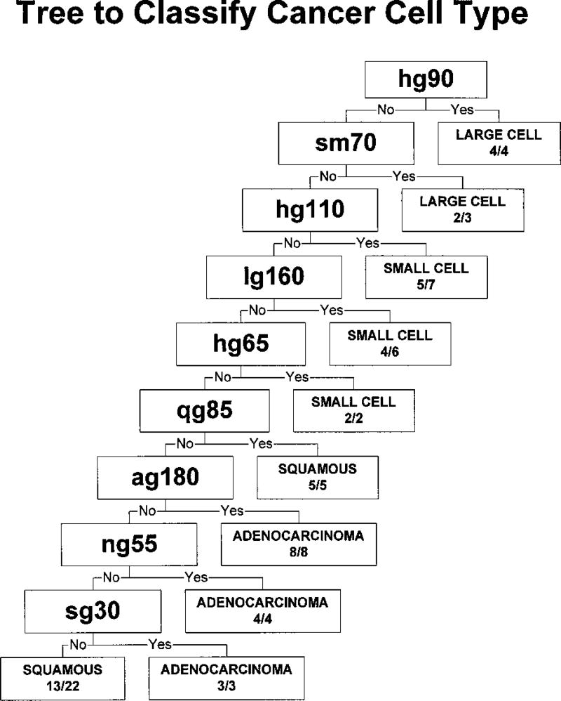
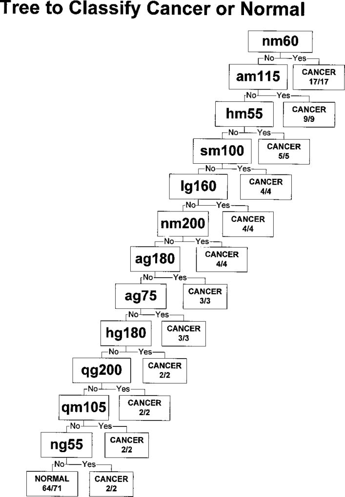
There are multiple case reports of antinuclear antibodies (ANAs) in patients with malignancies, yet to date there has not been a systematic survey of ANAs in lung cancer. We have previously reported that autoantibodies to collagen antigens resembling those found in the connective tissue diseases are consistently detected in the sera from lung cancer patients. In this work, we looked for the presence of ANAs in the sera from these same patients. Sera from 64 patients with lung cancer and 64 subjects without a history of cancer were retrospectively tested for reactivity on immunoblots of nuclear extracts of HeLa, small cell carcinoma, squamous cell carcinoma, adenocarcinoma, large cell carcinoma of the lung, and of normal lung cells. Associations were sought between the reactivities on immunoblots and lung cancer cell type, diagnosis, and progression-free survival by the method of classification and regression trees (CARTs). Cross-validated CART analyses indicated that reactivities to certain bands in immunoblots are associated with different types of lung cancer. Some of these autoantibodies were associated with a prolonged survival without disease progression. Our data suggest that autoimmunity is often a prominent feature of lung cancer and that molecular characterization of these antigens may lead to the discovery of proteins with diagnostic and prognostic value.


Figures

Fig. 1
Fig. 1
Immunoblots of nuclear extracts of HeLa (H), large cell carcinoma (L), normal lung (N), small cell carcinoma (S), squamous cell carcinoma (Q), and adenocarcinoma (A) cell lines. The source of each antigen is listed above the blot. Each blot is probed with the same four representative human sera from patients with each histologically classified type of lung cancer as well as with a normal serum (N) in the order indicated below the blot. Arrowhead, an example of different reactivities of a single serum with different substrates; a band of approximately 65 kDa is recognized by the small cell carcinoma patient serum on all of the substrates except HeLa. Left of each blot, the set of standards for more accurate size determination for each cell type. All of the sera from lung cancer patients show reactivity with a nuclear antigen of at least one of the cell types.
Fig. 2
Fig. 2
Diagram of the CART tree of lung cancer cell types of 64 lung cancer sera using all of the six antigen sets as shown in Table 2. Antigen variables selected by CART analyses are presented and labeled as follows: first letter (h, s, q, a, l, n, as in Table 2), the antigen set; second letter (g or m), the recognizing isotype IgG or IgM; and the following number, the antigen size in kDa. The number of patients with correctly predicted cancer cell type over the total number is included for each terminal node.
Fig. 3
Fig. 3
Diagram of the CART tree of lung cancer diagnosis between 64 lung cancer sera and 64 reference sera using all of the six antigen sets as shown in Table 2. Antigen variables selected by CART analyses are presented and labeled as follows: first letter (h, s, q, a, l, n, as in Table 2), the antigen set; second letter (g or m), the recognizing isotype IgG or IgM; and the following number, the antigen size in kDa. The number of subjects with correctly predicted diagnosis over the total number is included for each terminal node.
Fig. 4
Fig. 4
Kaplan-Meier estimate of the survival distribution function, a measure of the probability that an individual survives past a given time without progression, plotted against the PFS in days. The black curve (●) represents subjects with serum antibodies that bind one or more of the nuclear antigens lm160, ng30, qg160, or sm220. The presence of these antibodies predicted a survival without progression >9 months by CART analysis in Table 2. The gray curve ( formula image) represents those with serum antibodies that did not bind the first four antigens but did bind one or more of the nuclear antigens am45, lg180, hg65, or am70. The presence of these antibodies predicted a survival without progression from 4 to 9 months. Those without serum antibodies binding any of these eight antigens (○) had a predicted survival without progression of <4 months. Testing for equality by log rank indicated a significant difference in survival distribution between the curves (P = 0.006). Antigen nomenclature is presented in Table 2.
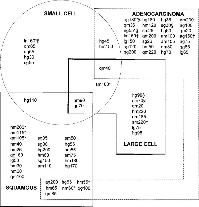
Fig. 5
Fig. 5
Diagrammatic summary of the results of four independent CART analyses identifying variables of importance in predicting the diagnosis of small cell carcinoma (circular grouping), squamous cell carcinoma (solid rectangular grouping), adenocarcinoma (dashed rectangular grouping), and large cell carcinoma (double-lined rectangular grouping). Variables identified by CART analysis as important in predicting each lung cancer cell type as a specific independent cancer and their overlapping relationships are represented in the diagram. For this analysis, it is assumed that these lung cancers are independent unrelated diseases. Some variables seem restricted to one cancer type, some are commonly identified in two cancer types, and some are identified in three cancer types. Annotations are included to further compare contributions of variables identified from separate analyses in Table 2 (using all of the lung cancer patients together as one disease) that potentially predict diagnosis (*), cancer cell type (§), and PFS (†). Antigen nomenclature is presented in Table 2.

References

    1. Abelev GI, Perova DS, Kheamkova NI, Posnikova ZA, Irlin IS. Production of embryonal α globulin by transplantable mouse hepatomas. Transplantation. 1963;1:174–180. - PubMed
    1. Day EA. The Immunochemistry of Cancer. Springfield, IL: C. C. Thomas; 1965.
    1. Gold P, Freedman SO. Specific carcinoembryonic antigens of the human digestive system. J. Exp. Med. 1965;122:467–481. - PMC - PubMed
    1. Knuth A, Danowski B, Oettgen HF, Old LJ. T-cell-mediated cytotoxicity against autologous malignant melanoma: analysis with interleukin 2-dependent T-cell cultures. Proc. Nat. Acad. Sci. USA. 1984;81:3511–3515. - PMC - PubMed
    1. van der Bruggen P, Traversari C, Chomez P, Lurquin C, De Plaen E, Van den Eynde B, Knuth A, Boon T. A gene encoding an antigen recognized by cytolytic T lymphocytes on a human melanoma. Science (Washington DC) 1991;254:1643–1647. - PubMed

Publication types

MeSH terms

LinkOut - more resources