. 1999 Jul;43(7):1638-43.
doi: 10.1128/AAC.43.7.1638.

Effective treatment of acute and chronic murine tuberculosis with liposome-encapsulated clofazimine

Affiliations

Effective treatment of acute and chronic murine tuberculosis with liposome-encapsulated clofazimine

L B Adams et al. Antimicrob Agents Chemother. 1999 Jul.

Abstract

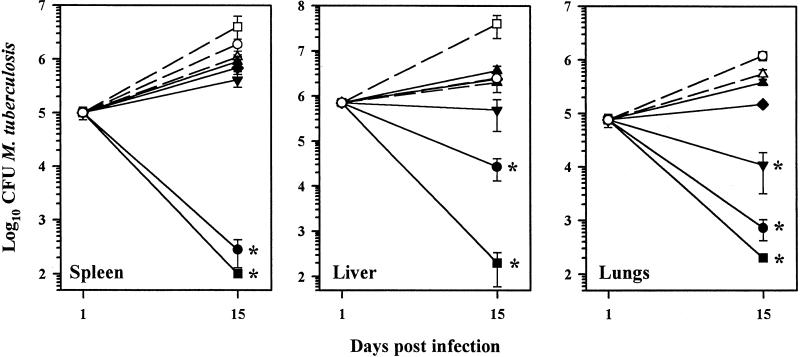
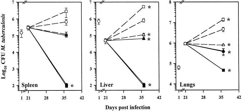
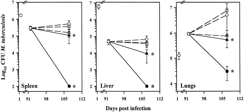
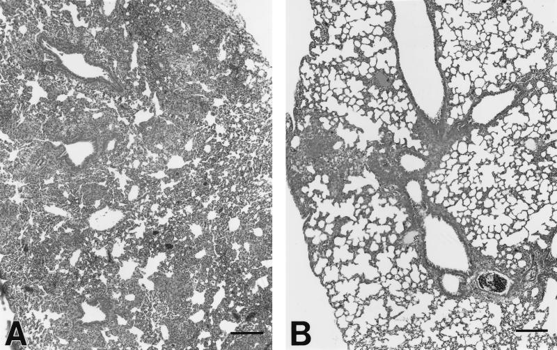
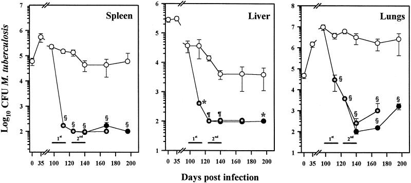
The therapeutic efficacy of liposomal clofazimine (L-CLF) was studied in mice infected with Mycobacterium tuberculosis Erdman. Groups of mice were treated with either free clofazimine (F-CLF), L-CLF, or empty liposomes twice a week for five treatments beginning on day 1 (acute), day 21 (established), or day 90 (chronic) postinfection. One day after the last treatment, the numbers of CFU of M. tuberculosis in the spleen, liver, and lungs were determined. F-CLF at the maximum tolerated dose of 5 mg/kg of body weight was ineffective; however, 10-fold-higher doses of L-CLF demonstrated a dose response with significant CFU reduction in all tissues without any toxic effects. In acutely infected mice, 50 mg of L-CLF/kg reduced CFU 2 to 3 log units in all three organs. In established or chronic infection, treated mice showed no detectable CFU in the spleen or liver and 1- to 2-log-unit reduction in the lungs. A second series of L-CLF treatments cleared M. tuberculosis in all three tissues. L-CLF appears to be bactericidal in the liver and spleen, which remained negative for M. tuberculosis growth for 2 months. Thus, L-CLF could be useful in the treatment of tuberculosis.


Figures

FIG. 1
FIG. 1
L-CLF treatment in acutely infected mice. BALB/c mice (n = 4 per group) were infected i.v. with 106 M. tuberculosis Erdman organisms and left untreated (○) or treated i.v. with L-CLF (■, 100 mg/kg; ●, 50 mg/kg; ▾, 25 mg/kg; ⧫, 10 mg/kg; and ▴, 5 mg/kg), F-CLF (▵, 5 mg/kg), or empty liposomes (□, lipid content equivalent to 100-mg/kg dose) on days 1, 5, 8, 11, and 14 postinfection. The mice were sacrificed 1 day after the last drug injection (day 15), and their spleens, livers, and lungs were homogenized and plated for CFU. The results are shown as means ± standard deviations. ✻, P < 0.01.
FIG. 2
FIG. 2
L-CLF treatment in mice with established infection. BALB/c mice (n = 4 per group) were infected i.v. with 106 M. tuberculosis Erdman organisms. Beginning on day 21 postinfection, the mice were left untreated (○) or treated i.v. every 3 to 4 days over a 2-week period (total, five injections) with L-CLF (■, 100 mg/kg; ●, 50 mg/kg; ▴, 5 mg/kg) F-CLF (▵, 5 mg/kg), or empty liposomes (□, equivalent to 100-mg/kg dose). The mice were sacrificed 1 day after the last drug injection (day 36), and the spleens, livers, and lungs were homogenized and plated for CFU. The results are shown as means ± standard deviations. ✻, P < 0.01.
FIG. 3
FIG. 3
L-CLF treatment in chronically infected mice. BALB/c mice (n = 4 per group) were infected i.v. with 106 M. tuberculosis Erdman organisms. Beginning on day 92 postinfection, the mice were left untreated (○) or treated i.v. every 3 to 4 days over a 2-week period (total, five injections) with L-CLF (●, 50 mg/kg; ▴, 5 mg/kg), F-CLF (▵, 5 mg/kg), or empty liposomes (□, lipid content equivalent to 50-mg/kg dose). The mice were sacrificed 1 day after the last drug injection (day 107), and the spleens, livers, and lungs were homogenized and plated for CFU. The results are shown as means ± standard deviations. ✻, P < 0.01.
FIG. 4
FIG. 4
Effect of L-CLF administration on early lung granuloma formation in M. tuberculosis-infected mice. (A) In control mice, there was an intense mononuclear cell infiltration into the lung parenchyma. An early granulomatous response with aggregates of epithelioid macrophages interspersed with lymphocytes was observed. (B) In contrast, mice treated with L-CLF exhibited perivascular and peribronchiolar cuffing and more localized granuloma formation without extensive involvement of lung parenchyma. The tissues were stained with hematoxylin-eosin. Bars = 150 μm.
FIG. 5
FIG. 5
Clearance and recovery of M. tuberculosis in tissues of mice after treatment with L-CLF. BALB/c mice (n = 4 per group) were infected i.v. with 106 M. tuberculosis Erdman organisms. Beginning on day 97 postinfection, the mice were left untreated (○) or were treated i.v. every 3 to 4 days over a 2-week period (1st) (total, five injections) with 50 mg of L-CLF/kg (⊙). Groups of mice were sacrificed 1 day after the last drug injection (day 112), 2 weeks posttreatment, 1 month posttreatment, and 2 months posttreatment. Another set of mice (●) were administered a second round (2nd) of L-CLF treatments (50 mg/kg) beginning 2 weeks after the completion of the first round (day 125). Groups of mice were sacrificed 1 day after the last drug injection (day 140), 1 month posttreatment, and 2 months posttreatment. The spleens, livers, and lungs were homogenized and plated for CFU. The results are shown as means ± standard deviations. ✻, P < 0.01; ¶, P < 0.001; §, P < 0.0001.

References

    1. Adams L B, Dinauer M C, Morgenstern D E, Krahenbuhl J L. Comparison of the roles of reactive oxygen and nitrogen intermediates in the host response to Mycobacterium tuberculosis using transgenic mice. Tuber Lung Dis. 1997;78:237–246. - PubMed
    1. Adams L B, Mason C M, Kolls J K, Scollard D, Krahenbuhl J L, Nelson S. Exacerbation of acute and chronic murine tuberculosis by administration of a TNF receptor-expressing adenovirus. J Infect Dis. 1995;171:400–405. - PubMed
    1. Agarwal A, Kandpal H, Gupta H P, Singh N B, Gupta C M. Tuftsin-bearing liposomes as rifampin vehicles in treatment of tuberculosis in mice. Antimicrob Agents Chemother. 1994;38:588–593. - PMC - PubMed
    1. Bermudez L E. Use of liposomal preparations to treat mycobacterial infections. Immunobiology. 1994;191:578–583. - PubMed
    1. Bermudez L E M, Wu M, Young L S. Intracellular killing of Mycobacterium avium complex by rifapentine and liposome-encapsulated amikacin. J Infect Dis. 1987;156:510–513. - PubMed

Publication types