. 1999 Jul;104(1):49-57.
doi: 10.1172/JCI6642.

Active participation of CCR5(+)CD8(+) T lymphocytes in the pathogenesis of liver injury in graft-versus-host disease

Affiliations

Active participation of CCR5(+)CD8(+) T lymphocytes in the pathogenesis of liver injury in graft-versus-host disease

M Murai et al. J Clin Invest. 1999 Jul.

Abstract

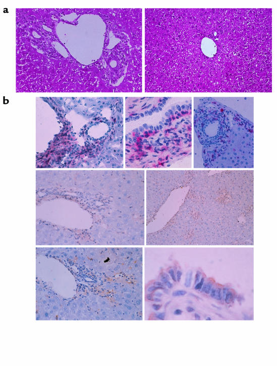
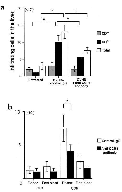
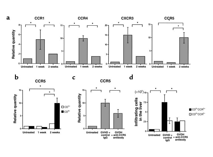
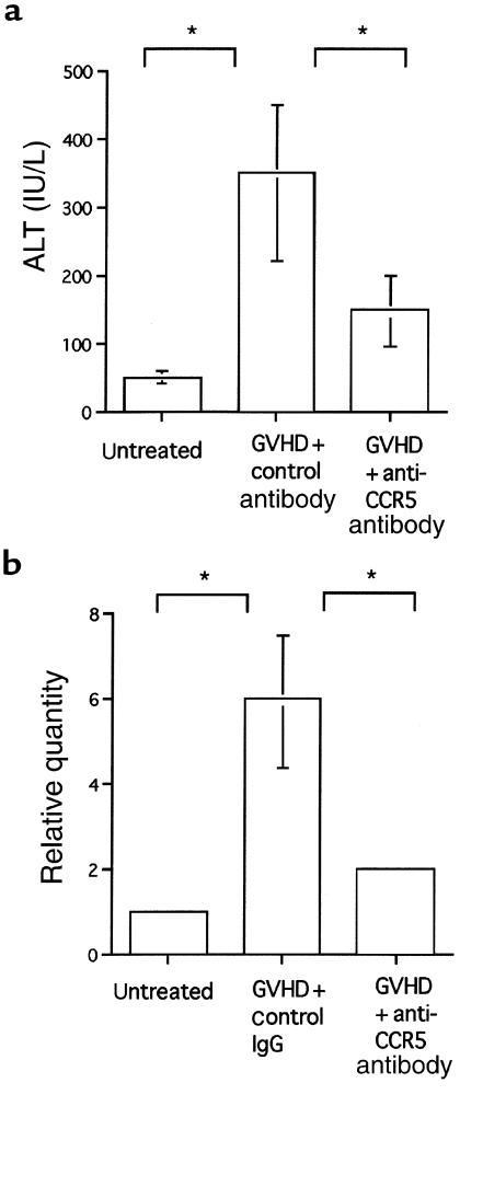
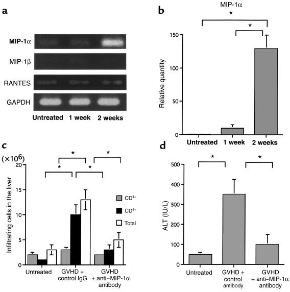
We examined the molecular pathogenesis of graft-versus-host disease-associated (GVHD-associated) liver injury in mice, focusing on the role of chemokines. At the second week after cell transfer in the parent-into-F1 model of GVHD, CD8(+) T cells -- especially donor-derived CD8(+) T cells -- infiltrated the liver, causing both portal hepatitis and nonsuppurative destructive cholangitis (NSDC). These migrating cells expressed CCR5. Moreover, macrophage inflammatory protein-1alpha (MIP-1alpha), one of the ligands for CCR5, was selectively expressed on intralobular bile duct epithelial cells, endothelial cells, and infiltrating macrophages and lymphocytes. Administration of anti-CCR5 antibody dramatically reduced the infiltration of CCR5(+)CD8(+) T lymphocytes into the liver, and consequently protected against liver damage in GVHD. The levels of Fas ligand (FasL) mRNA expression in the liver were also decreased by anti-CCR5 antibody treatment. Anti-MIP-1alpha antibody treatment also reduced liver injury. These results suggest that MIP-1alpha-induced migration of CCR5-expressing CD8(+) T cells into the portal areas of the liver plays a significant role in causing liver injury in GVHD; thus, CCR5 and its ligand may be the novel target molecules of therapeutic intervention of hepatic GVHD.


Figures

Figure 1
Figure 1
(a) Light-micrographic analysis of liver tissues. Histopathological examination was performed using hematoxylin and eosin–stained liver tissues obtained from GVHD-induced mice treated with control IgG (left) or anti-CCR5 antibody (right) at the second week after cell transfer. ×200. (b) Characterization of liver inflammatory foci during GVHD. GVHD-induced mouse livers were harvested and tissue sections prepared at the second week after induction. All micrographs show immunohistochemically stained frozen sections of areas encompassing a portal vein. Top row: left, CD8 staining, ×100; center, CD8 staining, ×400; right, CD4 staining, ×100. Center row: CCR5 staining, ×100. Bottom row: left, MIP-1a staining, ×100; right, MIP-1a staining, ×600. CD8 and CD4 are stained with red precipitates; CCR5 and MIP-1a are stained with brown precipitates.
Figure 2
Figure 2
Effect of anti-CCR5 antibody on intrahepatic infiltration of mononuclear cells. (a) The number of CD4+ (dotted bars) and CD8+ (filled bars) T cells was determined by multiplying the total leukocyte number (open bars) by the fractions representing the CD4+ and CD8+ populations. Liver-infiltrating leukocytes were prepared from untreated mice and GVHD-induced mice treated with either control antibody or anti-CCR5 antibody. (b) Effect of anti-CCR5 antibody on the chimerism at the second week after induction. The number of donor-derived cells and recipient-derived cells was determined by multiplying the total leukocyte number by the fractions representing the H-2Dd–negative and H-2Dd–positive populations, respectively. Moreover, by multiplying the donor cell number and the recipient cell number by the fraction of CD4+ and CD8+ cells, the chimerism was determined in the GVHD-induced mice. Open bars: control antibody; filled bars: anti-CCR5 antibody. The mean ± SD of 6 mice is shown here. Five independent experiments were performed. *P < 0.05.
Figure 3
Figure 3
(a) Kinetics of CCR1, CCR4, CXCR3, and CCR5 mRNA expression in liver CD8+ T cells. Sorted liver CD8+ T cells were prepared from untreated and GVHD-induced mice during the 2 weeks after parental cell transfer. The amounts of CCR1, CCR4, CXCR3, and CCR5 were normalized to the level of GAPDH at each time point. Each normalized chemokine receptor value from untreated liver-infiltrating leukocytes was designated as the calibrator, and final relative quantity of mRNA was expressed relative to the calibrator. PCR was performed in triplicate for each experiment. (b) Kinetics of CCR5 mRNA expression in liver CD4+ and CD8+ T cells. Sorted liver CD4+ (open bars) and CD8+ (filled bars) T cells were prepared from untreated and GVHD-induced mice during the 2 weeks after parental cell transfer. (c) Effect of anti-CCR5 antibody on CD8+ T cells in the liver. Relative quantity of CCR5 mRNA of sorted liver CD8+ T cells prepared from untreated mice and GVHD-induced mice treated with either anti-CCR5 antibody or control antibody, at the second week after induction. (d) The number of CCR5+CD8+ T cells (filled bars) and CCR5CD8+ T cells (open bars) was determined by multiplying the total leukocyte number by the fraction of CD3+, CD8+, and CCR5+ cells and totaling the results. Liver-infiltrating leukocytes were prepared from untreated mice and GVHD-induced mice treated with either anti-CCR5 antibody or control antibody, at the second week after induction. For each graph, data represent the mean ± SD of 6 mice. Each graph is representative of the results obtained from 3 independent experiments. *P < 0.05.
Figure 4
Figure 4
(a) Effect of anti-CCR5 antibody on serum ALT levels. The serum ALT levels of untreated mice and GVHD-induced mice treated with either anti-CCR5 antibody or control antibody, at the second week after induction. (b) Effect of anti-CCR5 antibody on FasL mRNA in the liver. Total RNA was isolated from liver tissue of untreated mice and GVHD-induced mice treated with either control antibody or anti-CCR5 antibody, at the second week after induction. The data represent the mean ± SD of 6 mice. Each graph is representative of the results obtained from 3 independent experiments. *P < 0.05.
Figure 5
Figure 5
(a) Kinetics of MIP-1α, MIP-1β, and RANTES mRNA expression in the liver. Total RNA was isolated from liver tissues of untreated and GVHD-induced mice during the second week after induction. RT-PCR cDNA products were generated and amplified using oligonucleotide primers specific to MIP-1α, MIP-1β, RANTES, or the housekeeping gene GAPDH. (b) The amount of MIP-1α was normalized to the level of GAPDH at each time point. Each normalized MIP-1α value of untreated liver was designated as the calibrator, and final relative quantity of mRNA was expressed relative to the calibrator. PCR was performed in triplicate for each experiment. (c) Effect of anti–MIP-1α antibody on intrahepatic infiltration of mononuclear cells. The number of CD4+ (dotted bars) and CD8+ (filled bars) cells was determined by multiplying the total leukocyte number (open bars) by the fraction of CD4+ and CD8+ cells. Liver-infiltrating leukocytes were prepared from untreated mice and GVHD-induced mice treated with either control antibody or anti–MIP-1α antibody. (d) Effect of anti–MIP-1α on serum ALT levels. Shown are serum ALT levels of untreated mice and GVHD-induced mice treated with either control antibody or anti–MIP-1α antibody, at the second week after induction. The data represent the mean ± SD of 6 mice. Graph is representative of results obtained from 3 independent experiments. *P < 0.05.

References

    1. Ferrara JLM, Deeg HJ. Graft-versus-host disease. N Engl J Med. 1991;324:667–674. - PubMed
    1. Gleichmann E, Pals ST, Rolink AG, Radaszkiewicz T, Gleichmann H. Graft-versus-host reactions: clues to the ethiopathology of a spectrum of immunological diseases. Immunol Today. 1984;5:324–332. - PubMed
    1. Rus V, Svetic A, Nguyen P, Gause WC, Via CS. Kinetics of Th1 and Th2 cytokine production during the early course of acute and chronic murine graft-versus-host disease. J Immunol. 1995;155:2396–2406. - PubMed
    1. Bobe P, et al. Fas-mediated liver damage in MRL hemopoietic chimeras undergoing lpr-mediated graft-versus-host disease. J Immunol. 1997;159:4197–4204. - PubMed
    1. Baker BMB, Altman N, Podack E, Levy RB. The role of cell-mediated cytotoxicity in acute GVHD after MHC-matched allogeneic bone marrow transplantation in mice. J Exp Med. 1996;183:2645–2656. - PMC - PubMed

MeSH terms