. 1999 Jul;104(1):73-81.
doi: 10.1172/JCI5535.

Circulating immune complexes in IgA nephropathy consist of IgA1 with galactose-deficient hinge region and antiglycan antibodies

Affiliations

Circulating immune complexes in IgA nephropathy consist of IgA1 with galactose-deficient hinge region and antiglycan antibodies

M Tomana et al. J Clin Invest. 1999 Jul.

Abstract

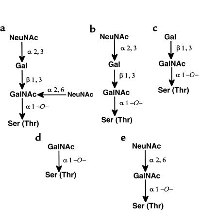
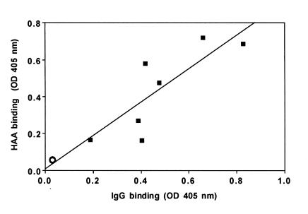
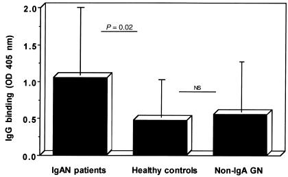
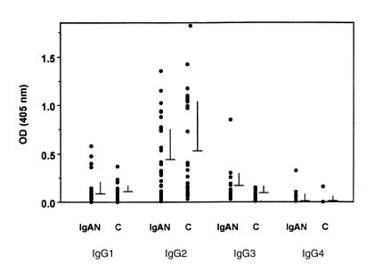
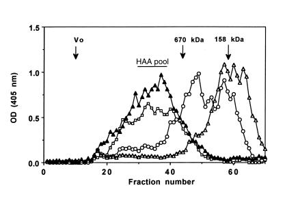
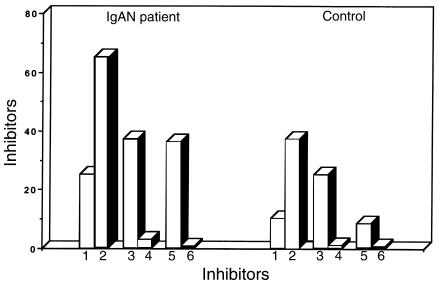
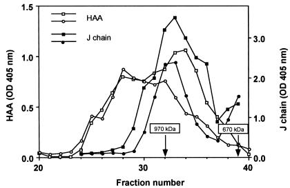
Circulating immune complexes (CICs) isolated from sera of patients with IgA nephropathy (IgAN) consist of undergalactosylated, mostly polymeric, and J chain-containing IgA1 and IgG antibodies specific for N-acetylgalactosamine (GalNAc) residues in O-linked glycans of the hinge region of IgA1 heavy chains. Antibodies with such specificity occur in sera of IgAN patients, and in smaller quantities in patients with non-IgA proliferative glomerulonephritis and in healthy controls; they are present mainly in the IgG (predominantly IgG2 subclass), and less frequently in the IgA1 isotype. Their specificity for GalNAc was determined by reactivity with IgA1 myeloma proteins with enzymatically removed N-acetylneuraminic acid (NeuNAc) and galactose (Gal); removal of the O-linked glycans of IgA1 resulted in significantly decreased reactivity. Furthermore, IgA2 proteins that lack the hinge region with O-linked glycans but are otherwise structurally similar to IgA1 did not react with IgG or IgA1 antibodies. The re-formation of isolated and acid-dissociated CICs was inhibited more effectively by IgA1 lacking NeuNAc and Gal than by intact IgA1. Immobilized GalNAc and asialo-ovine submaxillary mucin (rich in O-linked glycans) were also effective inhibitors. Our results suggest that the deficiency of Gal in the hinge region of IgA1 molecules results in the generation of antigenic determinants containing GalNAc residues that are recognized by naturally occurring IgG and IgA1 antibodies.


Figures

Figure 1
Figure 1
Possible structures of O-glycans in the hinge region of human IgA1.
Figure 2
Figure 2
Correlation of binding of HAA and normal serum IgG to IgA myeloma proteins. The binding of serum IgG to IgA1 (filled squares) and IgA2 (open circle) myeloma proteins. The binding of IgG to IgA1 significantly correlated with HAA binding (r = 0.875, P = 0.044), indicating requirement of terminal GalNAc residues for IgG binding.
Figure 3
Figure 3
The binding of serum IgG to Fab fragment of IgA1 (Ste) myeloma protein. Wells of microtiter plates were coated with Fab fragment of IgA1, incubated with diluted sera from 20 IgAN patients, 20 healthy controls, and 20 patients with non-IgA GN and subsequently with biotinylated mAb specific for IgG, avidin-alkaline phosphatase, and phosphatase substrate. Data shown are OD at 405 nm, mean and SD. Statistical significance is noted; NS, not significant.
Figure 4
Figure 4
Subclass specificity of serum IgG binding to a,a-IgA1. Wells of microtiter plates were coated with myeloma a,a-IgA1, incubated with sera (diluted 1:10) from 30 IgAN patients and 30 healthy controls (C) and subsequently with biotinylated mAb specific for IgG1, IgG2, IgG3, and IgG4; avidin-alkaline phosphatase; and phosphatase substrate. Data shown are OD at 405 nm, mean and SD.
Figure 5
Figure 5
Distribution of IgM, IgA, IgG, and reactivity with HAA in serum fractions from Superose 6 column. Serum from an IgAN patient was fractionated on a Superose 6 column (0.9 × 58.5 cm); 0.25-mL fractions were collected and analyzed by ELISA for the content of IgM (squares), IgA (circles), IgG (open triangles), and reactivity with HAA (filled triangles). Fractions containing aberrantly glycosylated IgA were pooled (HAA pool) and used in the inhibition experiment.
Figure 6
Figure 6
Inhibition of IgA1-IgG complex formation. IgA1-IgG CICs from an IgAN patient (left columns 1–6) and a healthy control (right columns 1–6) prepared by size-exclusion chromatography on Superose 6 were dissociated at pH 3.0. Aliquots of dissociated CICs were incubated with agarose- or Sepharose-immobilized inhibitors: 1, IgA1(Mce); 2, a,a-IgA1(Mce); 3, GalNAc; 4, GlcNAc; 5, a-OSM; and 6, HSA. The mixture was adjusted to neutral pH to allow re-formation of immune complexes, and the affinity of IgG toward the particular inhibitor was determined as described in Methods.
Figure 7
Figure 7
Distribution of HAA-reactive protein and J chain in serum fractions. Sera from an IgAN patient and a healthy control were fractionated on HPLC size-exclusion column TSK-5000. Aliquots of 0.25-mL fractions from the IgAN patient (open squares) and the control (open circles) were assayed for terminal GalNAc with biotinylated HAA. Aliquots of the same fractions from the IgAN patient (filled squares) and the control (filled circles) were assayed for J chain.

References

    1. D’Amico G. The commonest glomerulonephritis in the world: IgA nephropathy. Q J Med. 1987;64:709–727. - PubMed
    1. Julian BA, Waldo FB, Rifai A, Mestecky J. IgA nephropathy, the most common glomerulonephritis worldwide. A neglected disease in the United States? Am J Med. 1988;84:129–132. - PubMed
    1. Galla JH. IgA nephropathy. Kidney Int. 1995;47:377–387. - PubMed
    1. Russell MW, Mestecky J, Julian BA, Galla JH. IgA-associated renal diseases: antibodies to environmental antigens in sera and deposition of immunoglobulins and antigens in glomeruli. J Clin Immunol. 1986;6:74–86. - PubMed
    1. Czerkinsky C, et al. Circulating immune complexes and immunoglobulin A rheumatoid factor in patients with mesangial immunoglobulin A nephropathies. J Clin Invest. 1986;77:1931–1938. - PMC - PubMed

Publication types

MeSH terms