

Endostatin binds to blood vessels in situ independent of heparan sulfate and does not compete for fibroblast growth factor-2 binding
- PMID: 10393839
- PMCID: PMC1866664
- DOI: 10.1016/S0002-9440(10)65101-2
Endostatin binds to blood vessels in situ independent of heparan sulfate and does not compete for fibroblast growth factor-2 binding
Abstract
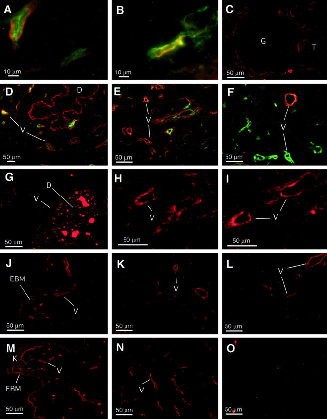
Endostatin is a carboxyl-terminal proteolytic fragment of collagen XVIII and a potent inhibitor of angiogenesis. The mechanism of action is unknown, but the crystal structure of endostatin predicts a prominent heparan sulfate binding site, suggesting that endostatin competitively inhibits heparin-binding angiogenic factors, such as basic fibroblast growth factor (FGF-2). The goal of the study was to map endostatin binding sites in intact human tissues and to determine whether this binding is heparan sulfate dependent. In situ binding was performed with recombinant epitope-tagged murine endostatin. Endostatin predominantly binds to blood vessels of different calibers in a saturable fashion. In addition, binding to some epithelial basement membranes is seen. The localization pattern is similar to that reported for collagen XVIII, endostatin's parent molecule. In breast carcinomas, endostatin co-localizes largely with FGF-2. In a surprising contrast to FGF-2, endostatin binding is resistant to treatment with heparitinase, demonstrating that binding is not mediated by heparan sulfate proteoglycans. Furthermore, FGF-2 and heparin do not compete for endostatin binding, providing additional evidence for the discreteness of endostatin and FGF-binding sites.
Figures
References
-
- Hanahan D, Folkman J: Patterns and emerging mechanisms of the angiogenic switch during tumorigenesis. Cell 1996, 86:353-364 - PubMed
-
- O’Reilly MS, Boehm T, Shing Y, Fukai N, Vasios G, Lane WS, Flynn E, Birkhead JR, Olsen BR, Folkman J: Endostatin: an endogenous inhibitor of angiogenesis and tumor growth. Cell 1997, 88:277-285 - PubMed
-
- Boehm T, Folkman J, Browder T, O’Reilly MS: Antiangiogenic therapy of experimental cancer does not induce acquired drug resistance. Nature 1997, 390:404-407 - PubMed
-
- Standker L, Schrader M, Kanse SM, Jurgens M, Forssmann WG, Preissner KT: Isolation and characterization of the circulating form of human endostatin. FEBS Lett 1997, 420:129-133 - PubMed
MeSH terms
Substances
LinkOut - more resources
Full Text Sources
Other Literature Sources
