. 1999 Jul;155(1):145-58.
doi: 10.1016/S0002-9440(10)65109-7.

Injection of pre-psoriatic skin with CD4+ T cells induces psoriasis

Affiliations

Injection of pre-psoriatic skin with CD4+ T cells induces psoriasis

B J Nickoloff et al. Am J Pathol. 1999 Jul.

Abstract

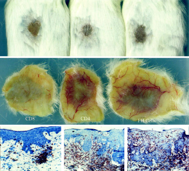
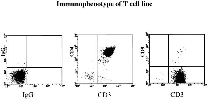
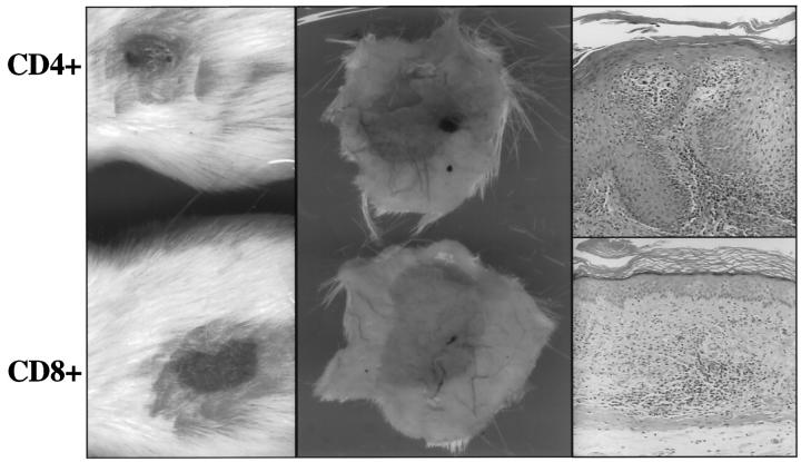
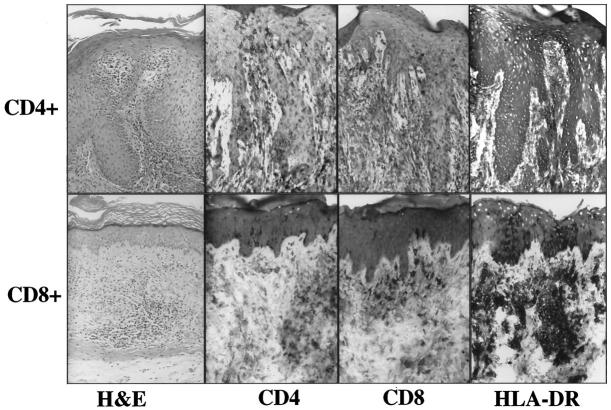
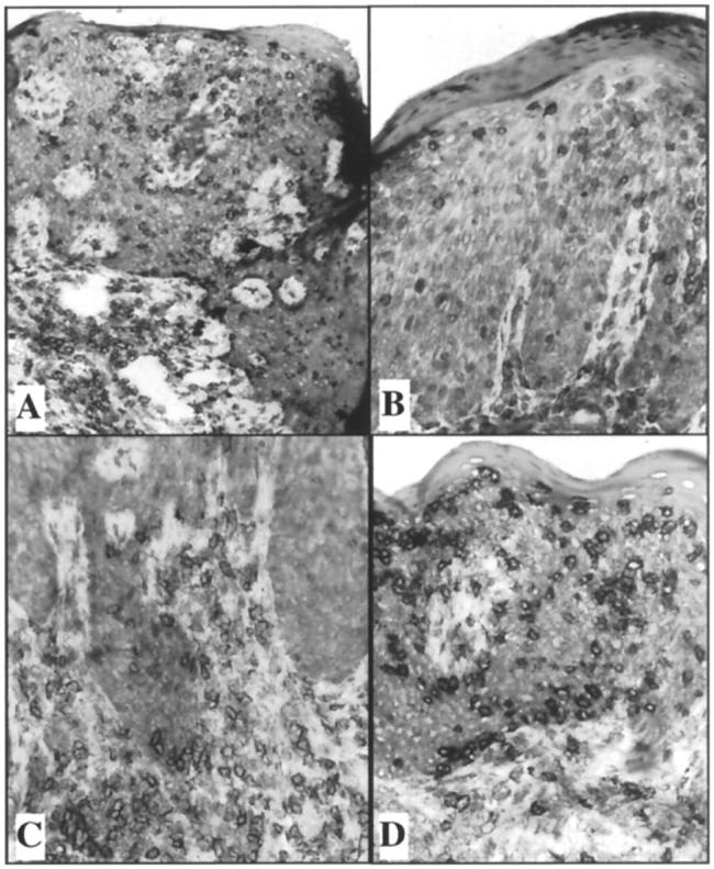
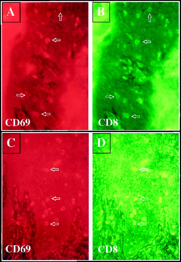
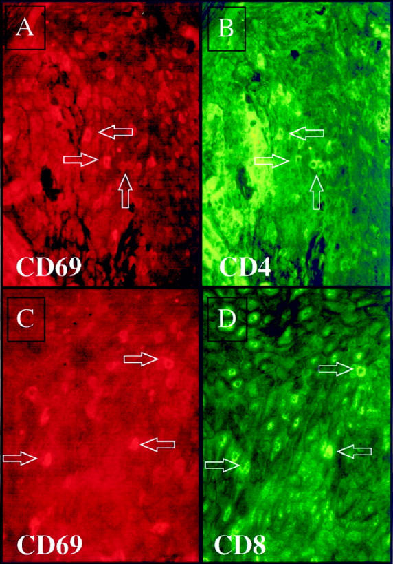
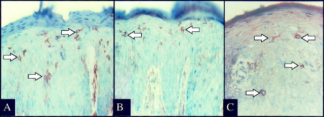
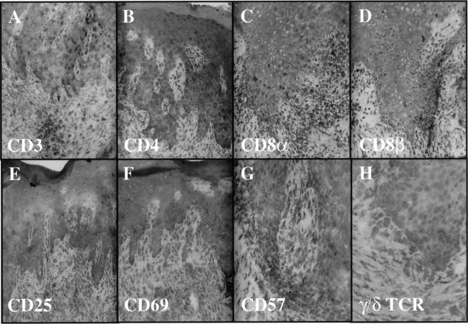
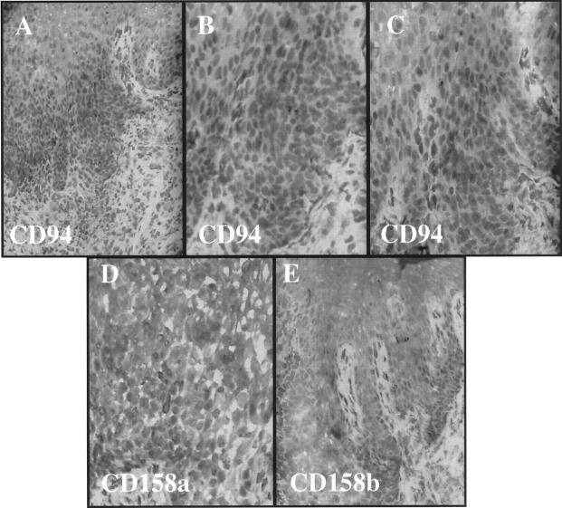
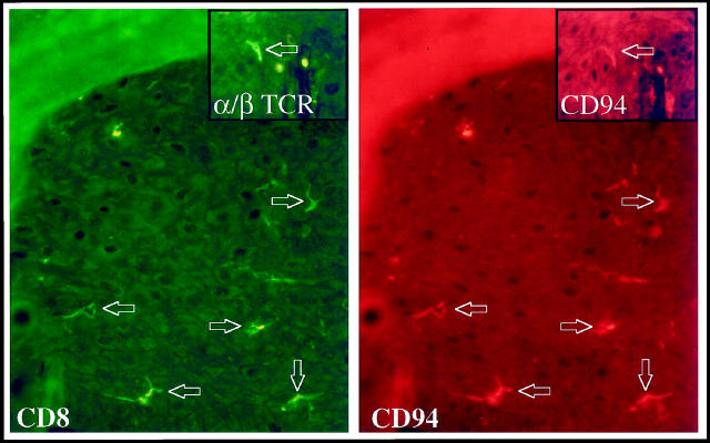
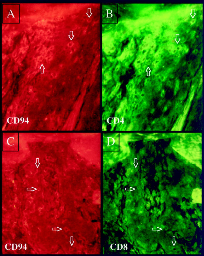
Psoriasis is an immunologically mediated skin disease linked to several different class I major histocompatibility complex alleles. However, the phenotype of the pathogenic lymphocyte and nature of the T cell activating event which triggers conversion of symptomless (PN) skin into psoriatic plaques (PP skin) is unknown. This study extends our previous observations in which autologous blood-derived immunocytes were injected into PN skin engrafted onto SCID mice to produce full-fledged PP lesions. The first question addressed is whether injected CD4+ T cells or CD8+ T cells were responsible for phenotypic conversion of PN to PP skin. In five different patients only CD4+ but not CD8+ T cell lines produced psoriatic lesions. Next, immunological events occurring within PN skin following injection of CD4+ T cells in grafts that had sufficient tissue available for detailed analysis was examined. In two patients, intraepidermal resident CD8+ T cells were induced to proliferate during lesion development, expressing acute activation markers CD25 and CD69. In another patient, injection of CD4+ T cells revealed CD69 expression by intraepidermal CD4+ as well as CD8+ T cells. To explore the molecular basis for local T cell activation and proliferation, we discovered that intraepidermal immunocytes, including both CD4 and CD8+ T cells, expressed surface receptors (ie, CD94, CD158a, CD158b) typically confined to natural killer cells (ie, natural killer receptors; NKRs) accumulated immediately before onset of acute lesions. The presence of NKR bearing immunocytes was also observed in 10 of 15 different biopsies of chronic plaques taken directly from patients, whereas PN skin (n = 8) or normal skin from healthy donors (n = 8), did not contain such NKR positive immunocytes. Of particular relevance to psoriasis is that these NKRs recognize various class I alleles including those typically inherited by psoriatic family members such as HLA-C and HLA-B allotypes. We conclude that injection of CD4+ T cells into PN skin triggers a series of local immunologically mediated stimulatory events that produce further T cell activation and appearance of both CD4 and CD8+ T cells that express NKRs.


Figures

Figure 1.
Figure 1.
Compared to unfractionated Ficoll-Hypaque derived immunocytes (right side panels), injected CD4+ T cells also induce PN-to-PP conversion (middle panels) , but CD8+ T cells do not cause psoriasis lesions (left side panels). Upper and middle horizontal panels portray clinical features in which surface scale production and underlying dermal angiogenesis is seen for PN skin converted to PP skin. Lower horizontal panels reveal immunostaining to detect CD3 (pan T cell marker).
Figure 2.
Figure 2.
Flow cytometry profile of CD4+ T cell line (day 23 in culture) established from peripheral blood using bacterial superantigens and IL-2, followed by negative selection to remove all other cell types and continued propagation using IL-2. Note that compared to isotype controls, the cell population is CD3+ CD4+ T cells (>95%), with only rare CD8+ T cells.
Figure 3.
Figure 3.
Using immobilized anti-CD3 mAb and IL-2, activated CD4+ T cells but not CD8+ T cells induce PN-to-PP conversion (thick, scaly skin with angiogenic tissue response, and epidermal hyperplasia). After CD4+ T cell injection, there is loss of granular cell layer, parakeratosis, elongation of rete pegs and influx of lymphocytes into epidermis (all histological hallmarks of psoriasis). In contrast, after CD8+ T cell injection, numerous lymphocytes are present in dermis, but with no change in the stratum corneum, granular cell layer, or epidermal thickness.
Figure 4.
Figure 4.
Following intradermal injection of CD4+ T cell, numerous CD4+ T cells are present in the hyperplastic epidermis with only rare CD8+ T cells in the epidermis and diffuse keratinocyte HLA-DR expression. Following intradermal injection of CD8+ T cells, only rare scattered CD4+ T cells in the dermis are seen as well as CD4+ Langerhans cells in the epidermis. Upper dermal collections of CD8+ T cells include HLA-DR-positive mononuclear cells and focal epidermal keratinocyte HLA-DR expression.
Figure 5.
Figure 5.
Appearance of CD8+ T cells in epidermis following dermal injection of highly purified CD4+ T cells. CD3 staining of injected CD4+ T cells (A). CD4+ staining demonstrating scattered lymphocytes in epidermis (B) and dermis (C). CD8+ staining demonstrating numerous intraepidemal CD8+ lymphocytes (D). Note even though only CD4+ T cells were injected into the dermis, CD8+ T cells outnumber CD4+ T cells in the epidermis.
Figure 6.
Figure 6.
Following injection of CD4+ T cells into PN skin, psoriatic lesions developed that contained CD69 positive cells (A, 3 days after injection; C, 2 weeks after injection). Note scattered rhodamine-stained CD69 positive cells (red) are present in the epidermis at both time points (arrows). Double staining to detect CD8 (FITC-green) revealed that most CD69-positive cells were also expressing CD8 (B, 3 days after injection; D, 2 weeks after injection).
Figure 7.
Figure 7.
Twenty-one days after injection of a CD4+ T cell line into PN skin, scattered intraepidermal immunocytes express rhodamine-stained CD69 (A and C). FITC-labeled antibody staining revealed that both CD4+ T cells (B), as well as CD8+ T cells (D) were CD69-positive. Arrows indicate double-positive cells.
Figure 8.
Figure 8.
Fourteen days after injection of CD4+ T cells into PN skin many intraepidermal immunocytes express CD94 (A), CD158a (B), and CD158b (C). Note the presence of numerous cytoplasmic spikes extending from the positively stained cells (arrows).
Figure 9.
Figure 9.
Twenty-one days after injection of CD4+ T cells into PN skin, induction of psoriasis is accompanied by influx of CD3-positive T cells into epidermis (A), including CD4 (B), CD8α (C), and rare CD8β (D) T cells. Acute activation markers CD25 (E) and CD69 (F) are present predominantly on intraepidermal lymphocytes with rare to no CD57 (G) or γ/δ TCR+ cells (H).
Figure 10.
Figure 10.
Serial sections from Figure 9 ▶ reveal conspicuous CD94+ cells in epidermis (A-C) with occasional CD158a- (D) and CD158b- (E) positive cells in epidermis.
Figure 11.
Figure 11.
Three days after injection of CD4+ T cells into PN skin many CD8+ immunocytes (FITC-green stained) also express CD94 (rhodamine-red stained) (arrows). The inset demonstrates that CD94+ cells express the α/β TCR, thereby confirming these as T lymphocytes.
Figure 12.
Figure 12.
Twenty-one days after injection of CD4+ T cells into PN skin many intraepidermal CD94 positive immunocytes are present (rhodamine-red stained) as shown in panels A and C; double staining reveals that both CD4+ T cells (FITC-green) (B) and CD8+ T cells (FITC-green) (D) are also CD94+ (arrows).
Figure 13.
Figure 13.
Chronic psoriatic plaque contains numerous CD3+ T cells in dermis and epidermis (A), with only a single CD57-positive NK cell identified in dermis (inset). Serial sections of identical plaque reveal scattered intraepidermal immunocytes (arrows) expressing CD94 (B), CD158a (C), and CD158b (D).

References

    1. Farber EM, Reush MK, Glinski W, Karasek MA, Recht B, Nickoloff BJ: Perspectives on psoriasis. Farber EM Noll L Morhenn V Jacobs PH eds. Psoriasis: Proceedings of the Fourth International Symposium. 1987, :pp 3-9 Elsevier Press, New York
    1. Elder JT, Nair RP, Guo S, Henseler T, Christophers E, Voorhees JJ: The genetics of psoriasis. Arch Dermatol 1996, 127:881-884 - PubMed
    1. Takada S, Engleman E: Evidence for an association between CD8 molecules and the T cell receptor complex on apoptotic T cells. J Immunol 1987, 139:3231-3235 - PubMed
    1. Bos JD, Hulsebosch HJ, Krieg SR, Baxter PM, Cormane RM: Immunocompetent cells in psoriasis. Arch Dermatol Res 1983, 275:181-189 - PubMed
    1. Hanney H, Gu SQ, Johannesson A, Sundkrist KG, Biberfeld P: Subpopulations of mononuclear cells in microscopic lesions of psoriatic patients. Selective accumulation of suppressor/cytotoxic T cells in epidermis during the evolution of the lesion. J Invest Dermatol 1989, 83:416-420 - PubMed