. 1999 Jul;155(1):275-84.
doi: 10.1016/s0002-9440(10)65121-8.

Nitric oxide inhibits HIV tat-induced NF-kappaB activation

Affiliations

Nitric oxide inhibits HIV tat-induced NF-kappaB activation

F Chen et al. Am J Pathol. 1999 Jul.

Abstract

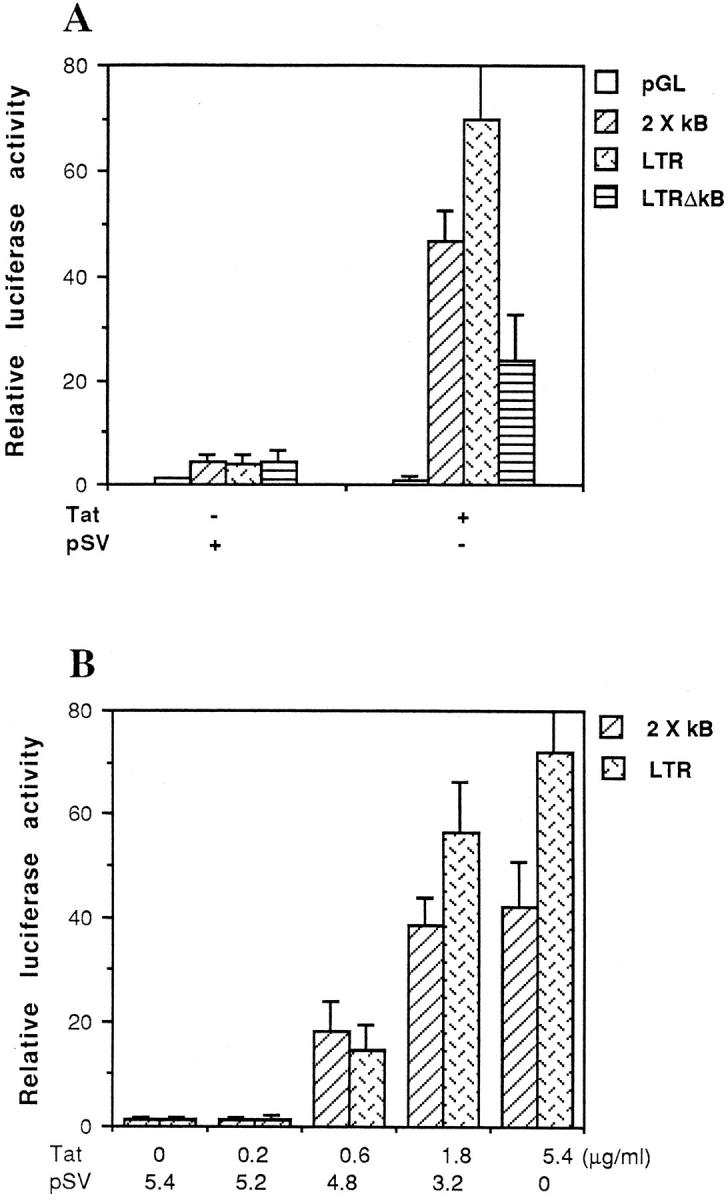
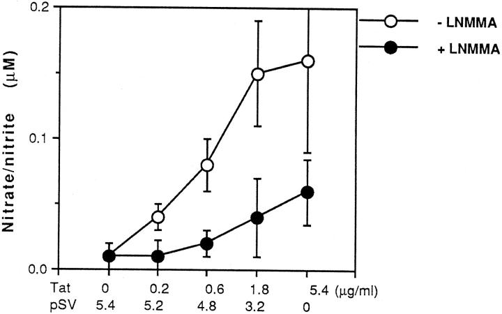
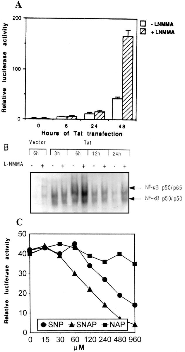
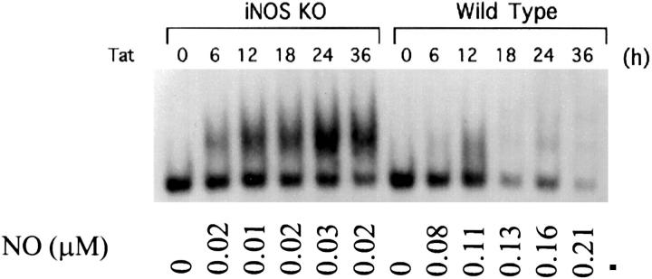
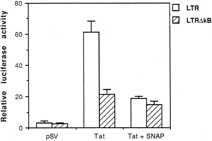
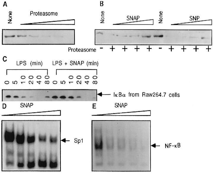
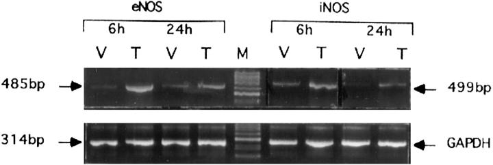
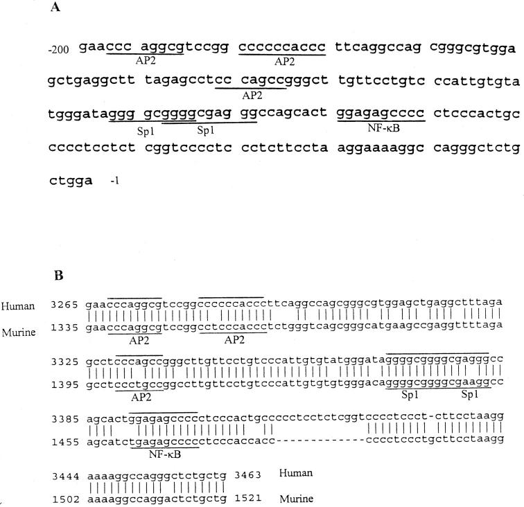
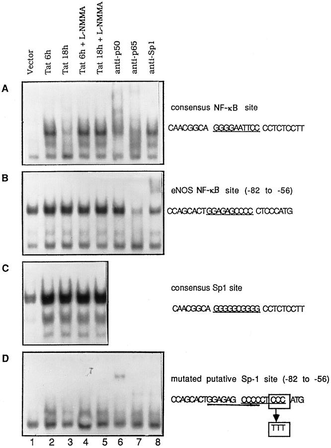
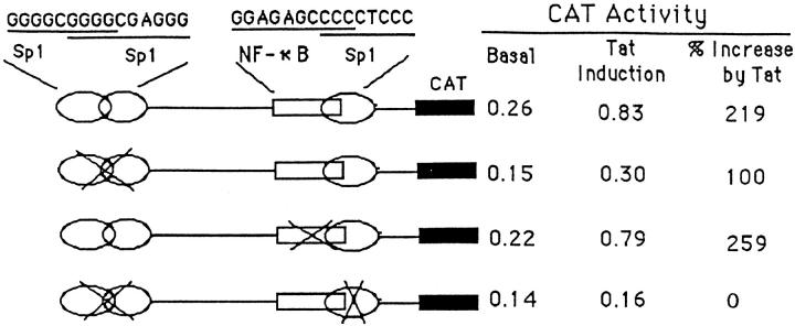
To evaluate the roles of nitric oxide (NO) on human immunodeficiency virus (HIV) Tat-induced transactivation of HIV long terminal repeat (HIV-LTR), we examined the effect of NO in the regulation of nuclear factor (NF)-kappaB, a key transcription factor involved in HIV gene expression and viral replication. In the present study, we demonstrate that HIV Tat activates NF-kappaB and that this activation can be attenuated by endogenous or exogenous NO. Inhibition of endogenous NO production with the NO synthase (NOS) inhibitor L-NMMA causes a significant increase in Tat-induced NF-kappaB activity. In addition, NO attenuates signal-initiated degradation of IkappaBalpha, an intracellular inhibitor of NF-kappaB, and blocks the DNA binding activity of the NF-kappaB p50/p50 homodimer and p50/p65 heterodimer. To determine how NO is induced by HIV Tat, reverse transcription polymerase chain reaction was used to demonstrate the induction of NOS-2 and NOS-3 mRNA by Tat. Although a putative NF-kappaB binding site was identified in the -74 GGAGAGCCCCC -64 region of the NOS-3 gene promoter, gel mobility shift assays and site-directed mutation analyses suggest that the putative NF-kappaB site is not of primary importance. Rather, several Sp-1 sites adjoining the putative NF-kappaB binding site in the promoter region of NOS-3 gene are required for the induction of NOS-3 gene expression by Tat.


Figures

Figure 1.
Figure 1.
HIV Tat activates NF-κB-dependent gene expression. A: Luciferase reporter assay of RAW264.7 cells co-transfected with reporter constructs and empty expression vector (pSV) or expression vector for Tat as indicated. B: Dose-dependent transactivation of the 2 × κB and wild-type HIV-LTR reporter constructs by Tat. RAW264.7 cells were co-transfected with 2 μg of 2 × κB or wild-type HIV-LTR reporter construct, and 0, 0.2, 0.6, 1.8, or 5.4 μg of expression vector for Tat. Total DNA transfected was made up to 5.4 μg with empty expression vector (pSV). The results of luciferase were normalized to β-galactosidase expression. Data shown are from representative experiments where transfection was performed in triplicate.
Figure 2.
Figure 2.
Tat induces NO production that can be blocked by the NOS inhibitor l-NMMA. RAW264.7 cells were transfected with increasing dose of expression vector for Tat as indicated in Figure 1B ▶ in the absence or presence of 200 μmol/L l-NMMA. At 48 h, cell culture supernatant was harvested and analyzed for NO production by the Griess reaction. Total DNA transfected was made up to 5.4 μg with empty expression vector (pSV). Data shown are from representative experiments where transfection was performed in triplicate.
Figure 3.
Figure 3.
Inhibition of endogenous NO production by l-NMMA amplifies Tat-induced NF-κB activity. A: Luciferase reporter assay of RAW264.7 cells co-transfected with a 2 × κB luciferase reporter and expression vector for Tat in the absence or presence of 200 μmol/L NOS inhibitor l-NMMA for 6, 24, or 48 hours. B: EMSA with nuclear extracts from RAW264.7 cells and an oligonucleotide probe containing a NF-κB binding site. RAW264.7 cells were transfected with expression vector for Tat in the absence (−) or presence (+) of 200 μmol/L l-NMMA for 3, 6, 12, and 24 hours. C: Luciferase reporter assay of RAW264.7 cells co-transfected with a 2 × κB luciferase reporter and expression vector for Tat in the presence of various concentrations of SNAP, SNP, or NAP for 48 hours.
Figure 4.
Figure 4.
Tat induces sustained NF-κB activation in NOS-2 gene knockout macrophages. EMSA was performed with nuclear extracts from mouse peritoneal macrophages and an oligonucleotide containing a NF-κB binding site. Peritoneal macrophages were harvested form wild-type or NOS-2 gene knockout B6 mice (iNOS-KO). Macrophages were immortalized by repeating passage and transfected with expression vector for Tat for different time periods as indicated. The lowest bands in the gel shift panel are nonspecific bands. Bottom numbers indicate relative NO production (μmol/L) from the cells transfected with Tat.
Figure 5.
Figure 5.
κB element-dependent inhibition of Tat-induced HIV-LTR transactivation by NO, as shown by luciferase reporter assay of RAW264.7 cells co-transfected with wild-type HIV-LTR or κB-mutated HIV-LTR (ΔκB) and an empty vector (pSV) or expression vector for Tat in the absence or presence of 200 μmol/L SNAP. Data shown are from representative experiments where transfections were performed in triplicate.
Figure 6.
Figure 6.
Exogenous NO inhibits IκBα degradation and DNA binding of NF-κB. A: Recombinant IκBα (0.25 μg) was incubated with reaction buffer alone (None) or in recombinant 20s proteasome (0.0625, 0.125, 0.25, 0.5, and 1 μg) in 20 μl of reaction buffer at 68°C for 30 minutes. The reactions were terminated by adding 8 μl of SDS reducing buffer and boiling for 5 minutes. The protein bands were visualized by SDS-PAGE in 12% gel and Western blot analysis using the ECL system. B: Recombinant IκBα (0.25 μg) was incubated with reaction buffer alone (None) or 0.5 μg of recombinant 20s proteasome in the presence of various concentrations of SNAP (25, 50, 100, and 200 μmol/L) or SNP (100, 200, 400, and 800 μmol/L). The reaction condition was as described in A. C: RAW264.7 cells were treated with LPS (5 μg/ml) in the absence (lanes 1 to 6) or presence (lanes 7 to 12) of SNAP (200 μmol/L). Total cellular proteins were extracted at various time points as indicated and subjected to Western blot for the detection of IκBα protein. D and E: Nuclear proteins extracted from LPS-stimulated RAW264.7 cells were incubated with SNAP (0, 25, 50, 100, and 200 μmol/L) for 20 minutes followed by addition of 32P-labeled Sp-1 binding DNA probe (D) or NF-κB binding probe (E) and incubated for another 15 minutes and subjected to EMSA.
Figure 7.
Figure 7.
Tat induces NOS-3 (eNOS) and NOS-2 (iNOS) mRNA expression. Total RNA (1 μg) isolated from vector (V) or Tat (T) transfected RAW264.7 cells was used for RT-PCR analysis using specific primers for NOS-3 or NOS-2 (upper panel) and GAPDH (lower panel) as indicated in Materials and Methods.
Figure 8.
Figure 8.
Proximal promoter region of NOS-3 gene. A: The 200-bp promoter region of human NOS-3 gene. The transcription factor binding sites for Sp-1, NF-κB, and AP-2 are underlined. B: Alignment of the 200-bp promoter sequence of human NOS-3 (upper line, GenBank ID AF032908) with the same region of murine NOS-3 (lower line, GenBank ID AF045940)
Figure 9.
Figure 9.
Characterization of functional activity of putative κB motif in NOS-3 gene promoter regions. A: EMSA using the probe containing a consensus NF-κB binding site (underlined). B: EMSA using the probe from the −82 to −56 region of NOS-3 (eNOS) gene promoter, which contained a putative κB motif (underlined). C: EMSA using a Sp-1 probe that encompasses a consensus Sp-1 binding site (underlined). D: 3′ CCC of an unidentified Sp-1 binding site (−68 to −60) that overlapped with the putative κB motif in the −74 to −64 region of NOS-3 promoter was mutated to TTT and used as probe for EMSA. In all panels, the nuclear proteins were extracted from RAW264.7 cells transfected with vector or Tat in the absence or presence of 50 μmol/L l-NMMA for 6 or 18 hours. Lanes 6 to 8, nuclear protein extracted from Tat transfected cells was incubated with antibodies against NF-κB p50 (lane 6.), NF-κB p65 (lane 7), and Sp-1 (lane 8), respectively, for 30 minutes followed by EMSA. The bottom bands in each panel are nonspecific bands.
Figure 10.
Figure 10.
CAT activity assay of RAW264.7 cells transfected with a plasmid containing wild-type or mutated NOS-3 proximal promoter region (116 bp) in the presence of an empty vector (basal) or expression vector for Tat. Sp-1 sites or the NF-κB site are mutated using a QuikChange site-directed mutagenesis kit (Stratagene) and confirmed by DNA sequencing. CAT activity was determined 48 hours after transfection using 50 μg of whole cellular extract and a CAT ELISA kit. The values of basal and Tat induction were represented by the absorbance at 405 nm measured with a microplate reader and calibrated by transfection efficiency with β-galactosidase staining.

References

    1. Nathan C: Inducible nitric oxide synthase: what difference does it make? J Clin Invest 1997, 100:2417-2423 - PMC - PubMed
    1. MacMicking J, Xie QW, Nathan C: Nitric oxide and macrophage function. Annu Rev Immunol 1997, 15:323-350 - PubMed
    1. Guidotti LG, Chisari FV: To kill or to cure: options in host defense against viral infection. Curr Opin Immunol 1996, 8:478-483 - PubMed
    1. Tucker PC, Griffin DE, Choi S, Bui N, Wesselingh S: Inhibition of nitric oxide synthesis increases mortality in Sindbis virus encephalitis. J Virol 1996, 70:3972-3977 - PMC - PubMed
    1. Peterhans E: Reactive oxygen species and nitric oxide in viral diseases. Biol Trace Element Res 1997, 56:107-116 - PubMed

Publication types

MeSH terms