. 1999 Jul;155(1):315-24.
doi: 10.1016/S0002-9440(10)65125-5.

Development of keratoacanthomas and squamous cell carcinomas in transgenic rabbits with targeted expression of EJras oncogene in epidermis

Affiliations

Development of keratoacanthomas and squamous cell carcinomas in transgenic rabbits with targeted expression of EJras oncogene in epidermis

X Peng et al. Am J Pathol. 1999 Jul.

Abstract

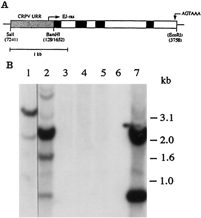
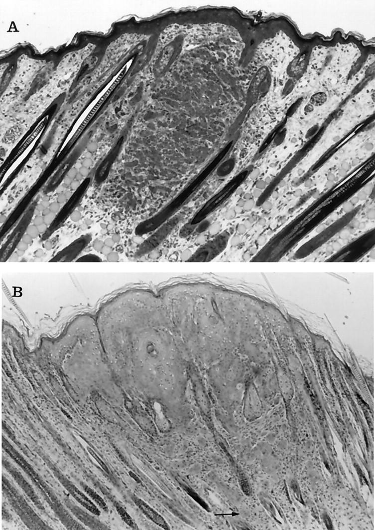
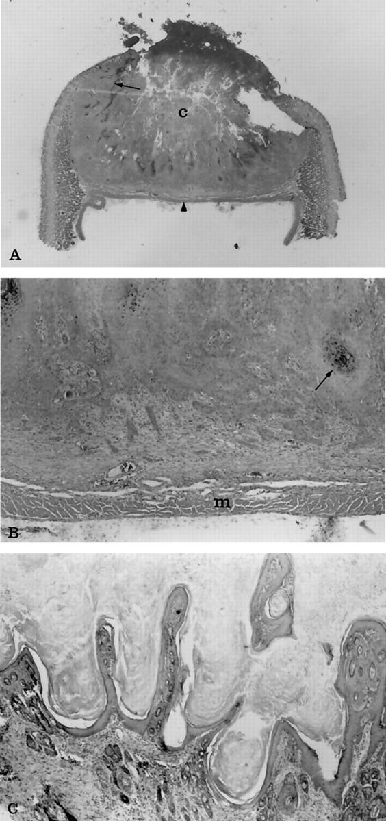
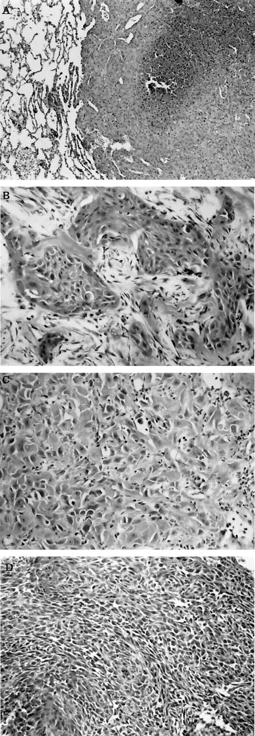
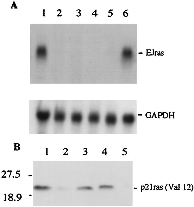
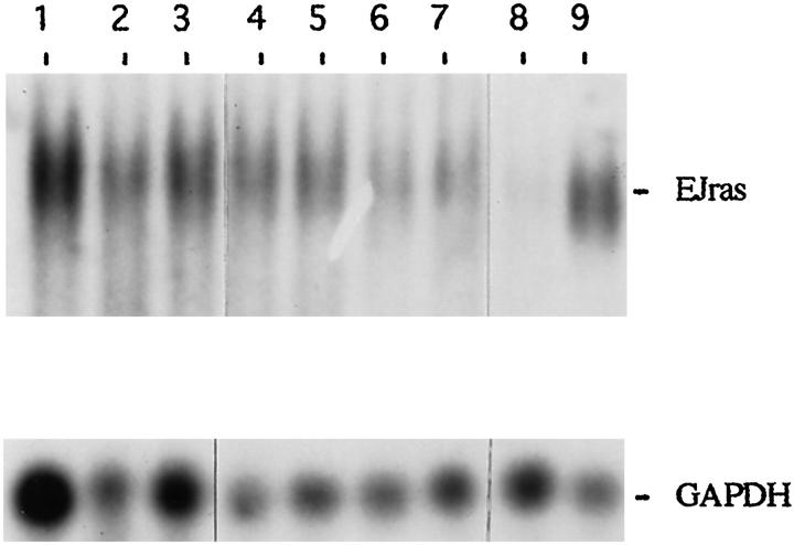
Activated ras genes have been frequently identified in both benign and malignant human tumors, including keratoacanthoma and squamous cell carcinoma. In this study, we developed two lines of transgenic rabbits in which the expression of EJras has been specifically targeted to the rabbit epidermal keratinocytes, using the upstream regulatory region of cottontail rabbit papillomavirus. All of the F1 transgenic progenies developed multiple keratoacanthomas at about 3 days after birth. The rabbits developed an average of 20 tumors, which usually reached the size of approximately 1 cm in diameter and then spontaneously regressed in about 2 months, similar to keratoacanthoma regression in humans. In addition, up to 18% of the rabbits then developed squamous cell carcinoma at about 5 months of age. The expression of EJras was detectable in all of the keratoacanthomas and squamous cell carcinomas. These results strongly support the involvement of the ras oncogene in both the initiation and regression of keratoacanthoma, and in the development of squamous cell carcinomas. These novel transgenic rabbits, with their consistent tumorigenic phenotype at an early age, high similarity to the human lesions, and easy accessibility for examination, manipulation, biopsy, and treatment, should provide a unique model system for studying ras activation-related tumor initiation, regression, and progression, and for evaluating antitumor therapies.


Figures

Figure 1.
Figure 1.
A: diagram showing the structure of the URR-EJras transgene. The URR-EJras transgene contains a 0.76-kb regulatory fragment (URR) from CRPV and a 2.1-kb coding sequence of EJras. The fragment of CRPV URR (diagonal box) was ligated to the coding sequence of EJras, which contains all of the exons (black box), introns (open box), and a 3′ flanking sequence carrying the polyadenylation signal as indicated. B: Southern blot analysis of EJras in the transgenic founders. Each lane contains 10 μg DNA, which was isolated from the biopsies of the rabbit ear skin and digested by BamHI and EcoRI. DNAs were isolated from transgenic founders TR-ras1 (lane 1) and TR-ras2 (lane 2) and four nontransgenic rabbits (lanes 3 to 6). DNA in lane 7 was the SalI- and EcoRI-released URR-EJras DNA reconstructed with normal rabbit skin DNA as a control. The two fragments produced by BamHI (EcoRI does not cut unintegrated URR-EJras) digestion in lane 7 indicate the appropriate separation of URR (0.76 kb) and the EJras coding sequence (2.1 kb). The fragment patterns shown in lanes 1 and 2 indicate that the TR-ras 1 rabbit may contain a single copy of integrated URR-EJras, and the TR-ras 2 rabbit contains multiple copies of the integrated URR-EJras, with head-to-tail tandem repeats.
Figure 2.
Figure 2.
Photomicrographs of keratoacanthomas from transgenic rabbits at 1 and 3 days of age. A: At 18 hours after birth the small mass of epithelial cells appeared to be associated with hair follicles, although the overlying epidermis was also slightly thickened. The cells within the tumor were arranged in small clusters that were separated by blood vessels and connective tissue. Some of the cells contained cytoplasmic vacuoles, suggesting sebaceous differentiation. At higher power (not shown) intercellular bridges were visible between cells. B: At 3 days the tumor cells had spread laterally to replace and compress hair follicles and were contiguous with the overlying epidermis. The neoplastic cells shown squamous differentiation, including clusters of “glassy cells” and keratin horn formation. Notice the clusters of neoplastic cells that have invaded the deeper areas of the dermis separate from the main tumor mass (arrow). Toluidine blue and H&E stains, respectively. Original magnification, ×50.
Figure 3.
Figure 3.
Photomicrographs of histological changes of regressing and almost regressed keratoacanthomas. A: The entire tumor from a 20-day-old transgenic rabbit at ×1.5 original magnification. Notice the characteristic necrotic central core (c) and the wall of epithelial cells that extend laterally under the epidermis (arrow). Typically the tumors terminated at the panniculus muscle (arrowhead), which on section gave the tumors a flattened side that was evident grossly as well as histologically. B: The same tumor at ×30 original magnification. Notice the multiple cords and clusters of neoplastic epithelial cells that have invaded deeper areas of dermis. The neoplastic cells shown squamous differentiation, including keratin production (arrow), dyskeratosis, and intercellular bridges, which are not seen at this magnification. Small clusters of cells at the leading edge of the tumor were associated with a fibrous connective tissue response. The neoplastic cells were not found to invade past the layer of panniculus muscle (m) in any of the specimens examined. C: An almost totally regressed tumor from a 60-day-old transgenic rabbit at ×50 original magnification. Notice the papillary projections of epidermal cells and hyperkeratosis that resembles papilloma. H&E stain.
Figure 4.
Figure 4.
Photomicrographs of metastatic lesion and SCC with different grades. A: Metastatic lesion within lung from a transgenic rabbit that developed SCC. Notice the sheets of poorly differentiated neoplastic epithelial cells that have replaced lung, and the focal tumor necrosis. B: Cutaneous SCC from a transgenic rabbit that resembles grade II squamous cell carcinoma. Notice the clusters and cords of neoplastic epithelial cells with squamous differentiation separated by desmoplastic connective tissue. C: Cutaneous SCC from a transgenic rabbit that resembles grade III squamous cell carcinoma. Notice the sheets of poorly differentiated neoplastic epithelial cells with occasional intracellular keratin production. D: Cutaneous SCC from a transgenic rabbit that resembles grade IV squamous cell carcinoma. Notice the sheets of poorly differentiated neoplastic epithelial cells that show slight whorl patterns. H&E stain. Original magnification, ×50.
Figure 5.
Figure 5.
Detection, of EJras expression in transgenic rabbits. A: EJras mRNA was analyzed by Northern blot hybridization to 32P-labeled EJras DNA probe. Total cellular RNAs (15 μg/lane) were isolated from keratoacanthoma (lane 1), tongue (lane 2), kidney (lane 3), spleen (lane 4), and brain (lane 5 ) of a F1 progeny in the TR-ras transgenic line. Lane 6 is RNA isolated from the URR-EJras-transfected rabbit epidermal keratinocytes. The EJras transcript (1.2 kb) was detected only in keratoacanthoma (lane 1) and positive control (lane 6), but not in any of the normal organ tissues (lanes 2 to 5). The transcription of GAPDH is shown as loading control of RNA in each lane. The GAPDH hybridization was carried out after the previous probe on the same membrane was stripped off. The mRNA size was determined in comparison to the relative mobility of mRNA standards. B: The p21ras (Val12) protein was detected by Western blot with monoclonal antibody pan-ras (Val12). This antibody specifically recognizes the activated Ha-ras p21 with valine at the position of the codon 12, but not the normal Ha-ras with glycine at the same position. Lanes 1 to 4 are proteins isolated from keratoacanthomas of F1 progeny in the TR-ras1 transgenic line, and lane 5 is protein from normal rabbit skin. The Val p21 Ha-ras protein is shown in all of the tested keratoacanthomas, which is consistent with the results shown by the Northern bolt analysis. Prestained molecular mass standards (kd) are shown on the left.
Figure 6.
Figure 6.
Northern blot analysis of EJras transcription in tumors of TR-ras1 transgenic rabbits. Each lane contains 15 μg total cellular RNA. Lanes 1 to 3 are RNAs isolated from SCCs of 3 transgenic rabbits, and lanes 4 to 7 are RNAs isolated from keratoacan-thomas of four transgenic rabbits, respectively. RNA in lane 8 was isolated from the normal skin of a rabbit, and RNA in lane 9 was isolated from URR-EJras-transfected rabbit epidermal keratinocytes. The probe and hybridization were the same as described in Figure 5 ▶ . The transcription of GAPDH is shown as loading control of RNA in each lane.

References

    1. Barbacid M: ras genes. Annu Rev Biochem 1987, 56:779-827 - PubMed
    1. Trahey M, McComick F: A cytoplasmic protein stimulates normal N-ras p21 GTPase, but does not affect oncogenic mutants. Science 1987, 238:542-545 - PubMed
    1. Polakis P, McCormick F: Structural requirements for the interaction of p21ras with GAP exchange factors, and its biological effector target. J Biol Chem 1993, 268:9157-9160 - PubMed
    1. Bos JL: ras oncogenes in human cancer: a review. Cancer Res 1989, 49:4682-4689 - PubMed
    1. Corominas M, Kamino H, Leon J, Pellicer A: Oncogene activation in human benign tumors of the skin (keratoacanthoma): is Hras involved in differentiation as well as proliferation? Proc Natl Acad Sci USA 1989, 86:6372-6376 - PMC - PubMed

Publication types

Substances