Review
. 1999 Jul 6;96(14):7705-9.
doi: 10.1073/pnas.96.14.7705.

Pain perception: is there a role for primary somatosensory cortex?

Affiliations
Review

Pain perception: is there a role for primary somatosensory cortex?

M C Bushnell et al. Proc Natl Acad Sci U S A. .

Abstract

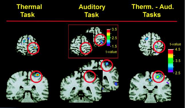
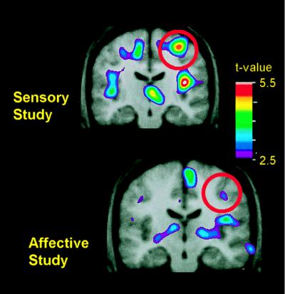
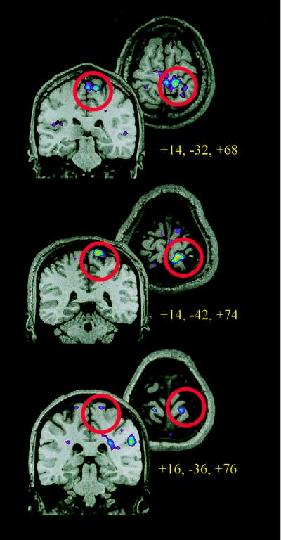
Anatomical, physiological, and lesion data implicate multiple cortical regions in the complex experience of pain. These regions include primary and secondary somatosensory cortices, anterior cingulate cortex, insular cortex, and regions of the frontal cortex. Nevertheless, the role of different cortical areas in pain processing is controversial, particularly that of primary somatosensory cortex (S1). Human brain-imaging studies do not consistently reveal pain-related activation of S1, and older studies of cortical lesions and cortical stimulation in humans did not uncover a clear role of S1 in the pain experience. Whereas studies from a number of laboratories show that S1 is activated during the presentation of noxious stimuli as well as in association with some pathological pain states, others do not report such activation. Several factors may contribute to the different results among studies. First, we have evidence demonstrating that S1 activation is highly modulated by cognitive factors that alter pain perception, including attention and previous experience. Second, the precise somatotopic organization of S1 may lead to small focal activations, which are degraded by sulcal anatomical variability when averaging data across subjects. Third, the probable mixed excitatory and inhibitory effects of nociceptive input to S1 could be disparately represented in different experimental paradigms. Finally, statistical considerations are important in interpreting negative findings in S1. We conclude that, when these factors are taken into account, the bulk of the evidence now strongly supports a prominent and highly modulated role for S1 cortex in the sensory aspects of pain, including localization and discrimination of pain intensity.


Figures

Figure 1
Figure 1
Pain-related activity when attention is directed to the painful heat stimulus (Left) or to an auditory stimulus (Center) is revealed by subtracting PET data recorded when a warm stimulus (32–38°C) was presented from those recorded when a painfully hot stimulus (46.5–48.5°C) was presented during each attentional state. Differences in pain-related activity during the two attentional conditions are revealed (Right) by subtracting PET data recorded during the auditory task from that recorded during the heat-discrimination task (using only painful stimulus trials—46.5–48.5°C). PET data, averaged across nine subjects, are illustrated against an MRI from one subject. Horizontal and coronal slices through S1 are centered at the activation peaks. Red circles surround the region of S1. Whereas there was a significant activation of S1 when subjects attended to the painful stimulus (Left), there was no significant activation when subjects attended to the auditory stimulus (Center). However, there was a subsignificant activation in S1 during the auditory task, as shown in the Inset. The direct comparison of pain in the two attentional conditions (Right) shows a significant difference in pain-related S1 activity during the two attentional states.
Figure 2
Figure 2
Changes in pain-related activity associated with previous hypnotic training by using suggestions for modulating pain sensation (Upper) or pain unpleasantness (Lower). Both images represent data from control scans, in which no hypnotic suggestions were given. Each image represents the subtraction of PET data recorded when the hand was submerged in thermally neutral water (35°C) from data recorded when the hand was submerged in painfully hot water (47°C). PET data were averaged across 10 experimental sessions in the sensory study (Upper) and, in a different group of subjects, 11 experimental sessions in the affective study (Lower). The PET data are illustrated against the average MRI for that subject group. Coronal slices through S1 are centered at the activation peaks, and red circles surround the region of S1.
Figure 3
Figure 3
Functional MRI data from three subjects, using a 1.5-T scanner and standard head coil. Each horizontal and coronal image represents the anatomical and functional data from a single subject during one session, which included a high-resolution anatomical scan and five to eight runs of 120 whole-brain functional MRI scans (–13) 7-mm slices acquired at 3-sec intervals. Thermal stimuli were applied to the left calf on separate runs. Thermal runs consisted of 9 s alternating cycles of rest, painful (45–46°C), rest, and neutral (35–36°C) stimulation by using a 9 cm2 thermode. Activation maps were generated by using Spearman’s rank order correlation, comparing painful to neutral heat. The coronal and horizontal slices through S1 are centered at the activation peaks, and red circles surround the region of S1.

References

    1. Head H, Holmes G. Brain. 1911;34:102–254.
    1. Penfield W, Boldrey E. Brain. 1937;60:389–443.
    1. Kenshalo D R, Jr, Isensee O. J Neurophysiol. 1983;50:1479–1496. - PubMed
    1. Kenshalo D R, Jr, Chudler E H, Anton F, Dubner R. Brain Res. 1988;454:378–382. - PubMed
    1. White J C, Sweet W H. Pain and the Neurosurgeon: A Forty-Year Experience. Springfield, IL: Thomas; 1969.

Publication types

MeSH terms