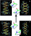
Validation of the single-stranded channel conformation of gramicidin A by solid-state NMR
- PMID: 10393921
- PMCID: PMC22161
- DOI: 10.1073/pnas.96.14.7910
Validation of the single-stranded channel conformation of gramicidin A by solid-state NMR
Abstract
The monovalent cation selective channel formed by a dimer of the polypeptide gramicidin A has a single-stranded, right-handed helical motif with 6.5 residues per turn forming a 4-A diameter pore. The structure has been refined to high resolution against 120 orientational constraints obtained from samples in a liquid-crystalline phase lipid bilayer. These structural constraints from solid-state NMR reflect the orientation of spin interaction tensors with respect to a unique molecular axis. Because these tensors are fixed in the molecular frame and because the samples are uniformly aligned with respect to the magnetic field of the NMR spectrometer, each constraint restricts the orientation of internuclear vectors with respect to the laboratory frame of reference. The structural motif of this channel has been validated, and the high-resolution structure has led to precise models for cation binding, cation selectivity, and cation conductance efficiency. The structure is consistent with the electrophysiological data and numerous biophysical studies. Contrary to a recent claim [Burkhart, B. M., Li, N., Langs, D. A., Pangborn, W. A. & Duax, W. L. (1998) Proc. Natl. Acad. Sci. USA 95, 12950-12955], the solid-state NMR constraints for gramicidin A in a lipid bilayer are not consistent with an x-ray crystallographic structure for gramicidin having a double-stranded, right-handed helix with 7.2 residues per turn.
Figures


Similar articles
-
High-resolution conformation of gramicidin A in a lipid bilayer by solid-state NMR.Science. 1993 Sep 10;261(5127):1457-60. doi: 10.1126/science.7690158. Science. 1993. PMID: 7690158
-
Tryptophan dynamics and structural refinement in a lipid bilayer environment: solid state NMR of the gramicidin channel.Biochemistry. 1995 Oct 31;34(43):14138-46. doi: 10.1021/bi00043a019. Biochemistry. 1995. PMID: 7578011
-
High-resolution polypeptide structure in a lamellar phase lipid environment from solid state NMR derived orientational constraints.Structure. 1997 Dec 15;5(12):1655-69. doi: 10.1016/s0969-2126(97)00312-2. Structure. 1997. PMID: 9438865
-
Gramicidin channels.IEEE Trans Nanobioscience. 2005 Mar;4(1):10-20. doi: 10.1109/tnb.2004.842470. IEEE Trans Nanobioscience. 2005. PMID: 15816168 Review.
-
Correlations of structure, dynamics and function in the gramicidin channel by solid-state NMR spectroscopy.Novartis Found Symp. 1999;225:4-16; discussion 16-22. Novartis Found Symp. 1999. PMID: 10472044 Review.
Cited by
-
Design, synthesis, and biological evaluation of stable β6.3-Helices: Discovery of non-hemolytic antibacterial peptides.Eur J Med Chem. 2018 Apr 10;149:193-210. doi: 10.1016/j.ejmech.2018.02.057. Epub 2018 Feb 20. Eur J Med Chem. 2018. PMID: 29501941 Free PMC article.
-
Orientation determination of interfacial beta-sheet structures in situ.J Phys Chem B. 2010 Jul 1;114(25):8291-300. doi: 10.1021/jp102343h. J Phys Chem B. 2010. PMID: 20504035 Free PMC article.
-
Modulation of concentration fluctuations in phase-separated lipid membranes by polypeptide insertion.Biophys J. 2002 Jul;83(1):334-44. doi: 10.1016/S0006-3495(02)75173-4. Biophys J. 2002. PMID: 12080124 Free PMC article.
-
Proton mobilities in water and in different stereoisomers of covalently linked gramicidin A channels.Biophys J. 2000 Apr;78(4):1825-34. doi: 10.1016/S0006-3495(00)76732-4. Biophys J. 2000. PMID: 10733963 Free PMC article.
-
A synthetic S6 segment derived from KvAP channel self-assembles, permeabilizes lipid vesicles, and exhibits ion channel activity in bilayer lipid membrane.J Biol Chem. 2011 Jul 15;286(28):24828-41. doi: 10.1074/jbc.M110.209676. Epub 2011 May 18. J Biol Chem. 2011. PMID: 21592970 Free PMC article.
References
Publication types
MeSH terms
Substances
Grants and funding
LinkOut - more resources
Full Text Sources

