. 1999 Jul 6;96(14):7962-7.
doi: 10.1073/pnas.96.14.7962.

Nephrin is specifically located at the slit diaphragm of glomerular podocytes

Affiliations

Nephrin is specifically located at the slit diaphragm of glomerular podocytes

V Ruotsalainen et al. Proc Natl Acad Sci U S A. .

Abstract

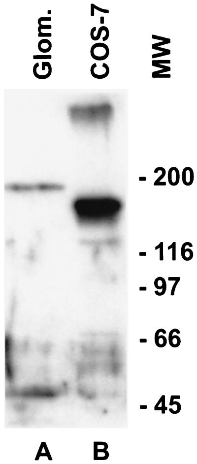
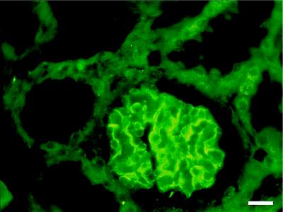
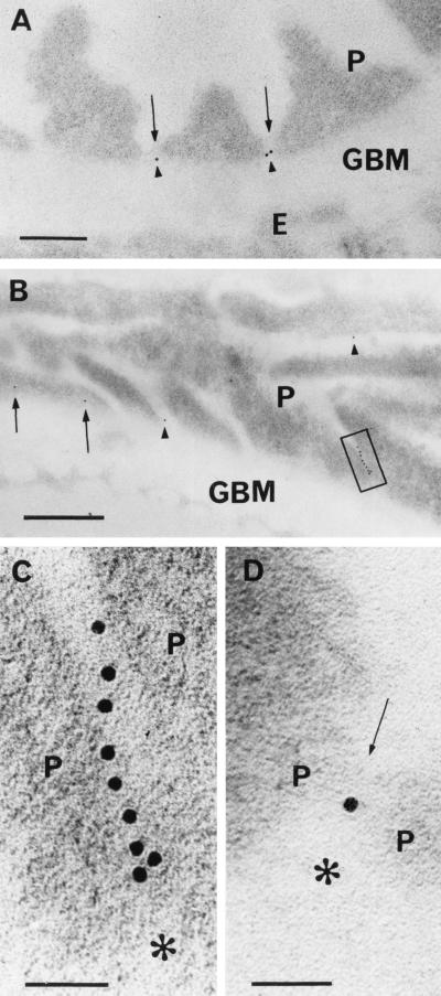
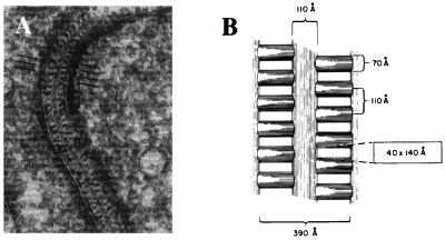
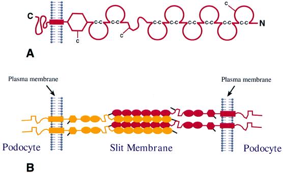
We describe here the size and location of nephrin, the first protein to be identified at the glomerular podocyte slit diaphragm. In Western blots, nephrin antibodies generated against the two terminal extracellular Ig domains of recombinant human nephrin recognized a 180-kDa protein in lysates of human glomeruli and a 150-kDa protein in transfected COS-7 cell lysates. In immunofluorescence, antibodies to this transmembrane protein revealed reactivity in the glomerular basement membrane region, whereas the podocyte cell bodies remained negative. In immunogold-stained thin sections, nephrin label was found at the slit between podocyte foot processes. The congenital nephrotic syndrome of the Finnish type (NPHS1), a disease in which the nephrin gene is mutated, is characterized by massive proteinuria already in utero and lack of slit diaphragm and foot processes. These features, together with the now demonstrated localization of nephrin to the slit diaphragm area, suggests an essential role for this protein in the normal glomerular filtration barrier. A zipper-like model for nephrin assembly in the slit diaphragm is discussed, based on the present and previous data.


Figures

Figure 1
Figure 1
Western blot analysis of glomerular and recombinant nephrin. A polyclonal anti-nephrin antibody reacts with a 180-kDa protein in a Triton X-100 extract of isolated normal human glomeruli (A) and with a 150-kDa protein from the lysate of COS-7 cells transiently transfected with full length nephrin cDNA (B). An additional band of over 300 kDa in the front of the gel can also be seen in the COS-7 cell extract. The nonreduced sample from human glomerular extracts did not reveal any immunoreactive bands, indicating extensive disulfide crosslinking of nephrin, and only the 300-kDa band from COS-7 cell extract was present (not shown). The preimmune serum did not reveal any staining (not shown).
Figure 2
Figure 2
Immunohistochemical localization of nephrin in human kidney. Immunofluorescence staining was carried out on a 2-mo-old human kidney with antibodies against recombinant human nephrin. Immunoreactivity is seen in the glomerulus, presumably at the podocyte-GBM junction. No staining is present in mesangial or endothelial cells. Bar = 20 nm.
Figure 3
Figure 3
Immunoelectron microscopic localization of nephrin in human renal glomeruli. Indirect postembedding staining for nephrin by using affinity purified IgG against extracellular region of recombinant human nephrin and 10 nm gold-coupled secondary antibody. (A) Paraformaldehyde (PF)-fixed kidney embedded in Lowicryl. Note gold label (arrowheads) between foot processes of podocytes (P). The label is located in the central area of the slit, between the GBM and the faintly visible slit diaphragm (arrows). Endothelium (E) is unlabeled. Bar = 200 nm. (B) Several gold particles in a row (box) can be seen between tangentially sectioned podocyte (P) foot processes. Gold particles in two other podocyte–podocyte interspaces are shown with arrowheads. Apparent intracellular staining of a podocyte (arrows) above GBM could be caused by grazing section of slit. Sample treated as in A. Bar = 500 nm. (C) Blow-up of B. The row of nine gold particles lies in tangentially cut, ca. 40-nm-wide slit between two podocytes (P). GBM is marked with asterisk. Bar = 50 nm. (D) Gold particle between slit diaphragm (arrow) joining two podocytes (P) and GBM (asterisk) in cross section. Sample fixed in 3.5% PF with 0.01% glutaraldehyde and embedded in LR White resin. Bar = 50 nm.
Figure 4
Figure 4
Morphological structure of the glomerular podocyte slit diaphragm. Reproduced and modified from R. Rodewald and M. J. Karnovsky (31) [reproduced from The Journal of Cell Biology (1974) 60, 423–433, by copyright permission of The Rockefeller University Press]. (A) Electron microscopy of tannic acid-stained and glutaraldehyde-fixed rat glomerulus reveals the presence of a central filament and cross bridges. Density of the cytoplasm opposite the points of attachment of the slit diaphragm can be observed. Discontinuities in the diaphragm represent regions where the diaphragm has left the plane of section. (B) Schematic model of the slit diaphragm. The average cross section dimensions of the pores between cross bridges are indicated within the rectangle.
Figure 5
Figure 5
Hypothetical model of nephrin assembly to form the isoporous filter of the podocyte slit diaphragm. (A) Schematic domain structure of nephrin. The Ig repeats are shown by incomplete circles connected by disulfide bridges (C-C). The locations of free cysteine residues are indicated by a —C. (B) Possible mode of interdigitating association of four nephrin molecules in the slit between two foot processes. For the sake of clarity, nephrin molecules from opposite foot processes are illustrated in different colors. In this model, it is assumed that Ig repeats 1–6 of a nephrin molecule of one foot process associate in an interdigitating fashion with Ig repeats 1–6 in a neighboring molecule from the opposite foot process. Cysteine residues are depicted by black lines and two potential disulfide bridges crosslinking four nephrin molecules in the center of the slit are illustrated. The remaining single free cysteine present in the fibronectin domain may react with another nephrin molecule, or some other as yet unknown molecule, which may connect with the plasma membrane or cytoskeleton.

References

    1. Tisher C C, Madsen K M. In: Anatomy of the Kidney. Brenner B M, Rector F C, editors. Vol. 1. Philadelphia: Saunders; 1996. pp. 3–71.
    1. Bohrer M P, Bayliss C, Humes H D, Glassock R J, Robertson C R, Brenner B M. J Clin Invest. 1978;61:72–78. - PMC - PubMed
    1. Brenner B M, Hostetter T H, Humes H D. N Engl J Med. 1978;298:826–832. - PubMed
    1. Batsford S R, Rohrbach R, Vogt A. Kidney Int. 1987;31:710–717. - PubMed
    1. Kanwar Y S, Liu Z Z, Wallner E I. Semin Nephrol. 1991;11:390–413. - PubMed

Publication types

Associated data

LinkOut - more resources