. 1999 Jul 6;96(14):8034-9.
doi: 10.1073/pnas.96.14.8034.

The nop-1 gene of Neurospora crassa encodes a seven transmembrane helix retinal-binding protein homologous to archaeal rhodopsins

Affiliations

The nop-1 gene of Neurospora crassa encodes a seven transmembrane helix retinal-binding protein homologous to archaeal rhodopsins

J A Bieszke et al. Proc Natl Acad Sci U S A. .

Abstract

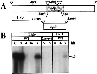
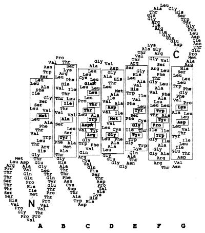
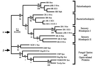
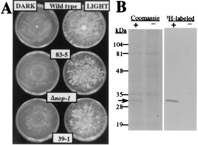
Opsins are a class of retinal-binding, seven transmembrane helix proteins that function as light-responsive ion pumps or sensory receptors. Previously, genes encoding opsins had been identified in animals and the Archaea but not in fungi or other eukaryotic microorganisms. Here, we report the identification and mutational analysis of an opsin gene, nop-1, from the eukaryotic filamentous fungus Neurospora crassa. The nop-1 amino acid sequence predicts a protein that shares up to 81.8% amino acid identity with archaeal opsins in the 22 retinal binding pocket residues, including the conserved lysine residue that forms a Schiff base linkage with retinal. Evolutionary analysis revealed relatedness not only between NOP-1 and archaeal opsins but also between NOP-1 and several fungal opsin-related proteins that lack the Schiff base lysine residue. The results provide evidence for a eukaryotic opsin family homologous to the archaeal opsins, providing a plausible link between archaeal and visual opsins. Extensive analysis of Deltanop-1 strains did not reveal obvious defects in light-regulated processes under normal laboratory conditions. However, results from Northern analysis support light and conidiation-based regulation of nop-1 gene expression, and NOP-1 protein heterologously expressed in Pichia pastoris is labeled by using all-trans [3H]retinal, suggesting that NOP-1 functions as a rhodopsin in N. crassa photobiology.


Figures

Figure 1
Figure 1
nop-1 gene structure and expression. (A) Gene structure. The bar represents the 5′ → 3′ genomic region of nop-1. The shaded area corresponds to nop-1 ORF. Triangles indicate the two intron positions. The hph gene replacement between the EcoRI and BglII sites of nop-1 is shown by dotted lines. (B) Northern analysis. Samples contained 25 μg of total RNA isolated from light or dark-grown cultures as indicated. Δnop-1 strains are 39-1 (left) and 83-5 (right). WT, wild type; C, conidia; S, SCM plates; 4, 4-hr germlings; 16, 16-hr germlings; V, VM plates.
Figure 2
Figure 2
Predicted topology of the NOP-1 protein. The seven transmembrane α-helices are designated A through G, and helix boundaries are based on those of BR (18). Arg-128 in helix C has been included in the pocket in accordance with the crystal structure of BR (30). Retinal-binding pocket residues conserved among the archaeal transport and sensory rhodopsins (31, 32) are boxed. ∗ mark positions homologous to Asp-85 and Asp-96 in BR. There are two in-frame methionines upstream of helix A; it is not known whether one or both are used for translation.
Figure 3
Figure 3
Phylogenetic relationship between NOP-1 and related proteins from fungi and archaea. Phylogeny of the opsins was estimated by ML quartet puzzling, NJ of γ distances with α = 2, and MP. Analyses were conducted by using the complete alignment. Estimates of branch lengths were obtained by ML, assuming the PAM model of evolution with empirical amino acid frequencies, and the scale bar indicates 0.1 estimated amino acid substitutions per site (EAASS) by using this model. Support for the monophyly of subgroups of archaeal rhodopsins and all groupings within the fungal proteins is shown, with the quartet puzzling values above branches and the NJ and MP bootstrap proportions below branches. Cases in which alternative groupings were supported by NJ or parsimony are indicated by ∗ (within the fungal proteins, NJ and parsimony bootstrap consensus trees reverse the branching order of C. versicolor Hsp30 and the S. pombe conserved hypothetical protein). Two possible tree roots, α and β, are indicated by arrows. Cve, Coriolus versicolor; Eni, Emericella nidulans (the sexual form of Aspergillus nidulans); Hsa, Halobacterium salinarum; Hva, Haloarcula vallismortis; Ncr, Neurospora crassa; Nph, Natronomonas pharaonis; Sce, Saccharomyces cerevisiae; Spo, Schizosaccharomyces pombe. International Collaboration (IC) accession numbers: HR Hsa, P16102; cHR-3 Hva, P94853; HR SG1, P25964; HR Nph, P15647; CR-2 Hva, Q53496; BR Hsa, P02945; SR-1 SG1, P19585; AR-2 AUS-2, P29563; SR-I Hsa, P25964; SR-I SG1, P33743; cSR-3 Hva, Q48334; SR-II Hsa, P71411; SR-II Nph, P42196; SR-II Hva, P42197; NOP-1 Ncr, AF135863; ORP Eni, AA787158, AA785169, AA786492; Hsp30 Cve, AB003518; CHP Spo, AL031824; Hsp30p Sce, S31838; YDR033W Sce, S61586; Yro2p Sce, P38079.
Figure 4
Figure 4
Oligomycin-dependent phenotype of Δnop-1 N. crassa strains and retinal binding of NOP-1 heterologously expressed in P. pastoris. (A) Sensitivity to oligomycin. Wild-type and Δnop-1 N. crassa strains were cultured on 100 ng/ml oligomycin-containing medium in constant light or darkness for 4 days at room temperature and then photographed. (B) Binding of all-trans retinal to NOP-1. Crude membrane fractions from P. pastoris strains containing nop-1 overexpression (+) or control (−) plasmid were incubated with all-trans [3H]retinal, the linkage reduced by sodium cyanoborohydride, and samples analyzed by using SDS/PAGE followed by fluorography. The position of the NOP-1 protein is indicated by the arrow.

References

    1. Rando R R. Chem Biol. 1996;3:255–262. - PubMed
    1. Spudich J L. Mol Microbiol. 1998;28:1051–1058. - PubMed
    1. Fryxell K J, Meyerowitz E M. J Mol Evol. 1991;33:367–378. - PubMed
    1. Skulachev V P. Q Rev Biophys. 1993;26:177–199. - PubMed
    1. Khorana H G. Proc Natl Acad Sci USA. 1993;90:1166–1171. - PMC - PubMed

Publication types

MeSH terms

Associated data

LinkOut - more resources