. 1999 Jul 6;96(14):8161-6.
doi: 10.1073/pnas.96.14.8161.

Targeting tumor vasculature endothelial cells and tumor cells for immunotherapy of human melanoma in a mouse xenograft model

Affiliations

Targeting tumor vasculature endothelial cells and tumor cells for immunotherapy of human melanoma in a mouse xenograft model

Z Hu et al. Proc Natl Acad Sci U S A. .

Abstract

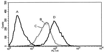
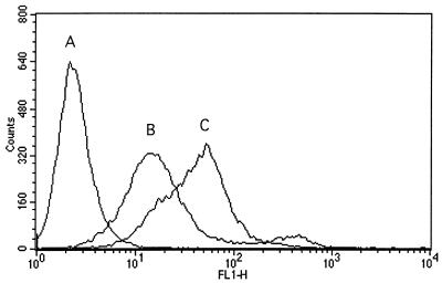
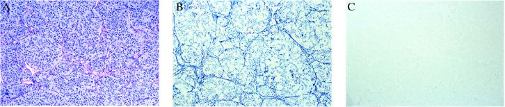
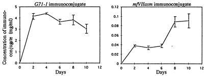
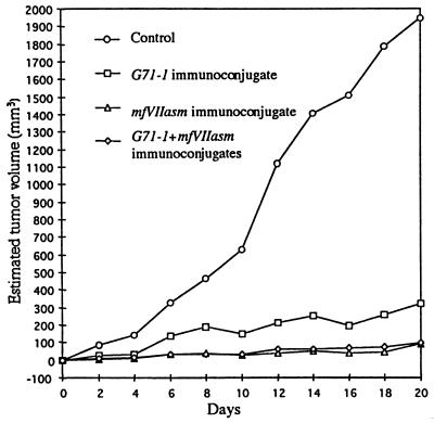
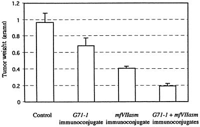
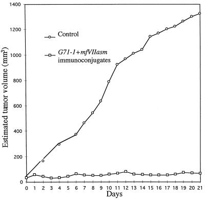
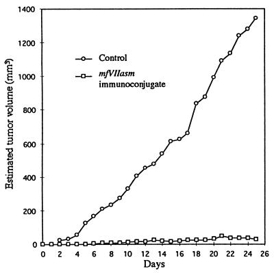
An immunotherapy treatment for cancer that targets both the tumor vasculature and tumor cells has shown promising results in a severe combined immunodeficient mouse xenograft model of human melanoma. The treatment involves systemic delivery of an immunoconjugate molecule composed of a tumor-targeting domain conjugated to the Fc effector domain of human IgG1. The effector domain induces a cytolytic immune response against the targeted cells by natural killer cells and complement. Two types of targeting domains were used. One targeting domain is a human single-chain Fv molecule that binds to a chondroitin sulfate proteoglycan expressed on the surface of most human melanoma cells. Another targeting domain is factor VII (fVII), a zymogen that binds with high specificity and affinity to the transmembrane receptor tissue factor (TF) to initiate the blood coagulation cascade. TF is expressed by endothelial cells lining the tumor vasculature but not the normal vasculature, and also by many types of tumor cells including melanoma. Because the binding of a fVII immunoconjugate to TF might cause disseminated intravascular coagulation, the active site of fVII was mutated to inhibit coagulation without affecting the affinity for TF. The immunoconjugates were encoded as secreted molecules in a replication-defective adenovirus vector, which was injected into the tail vein of severe combined immunodeficient mice. The results demonstrate that a mutated fVII immunoconjugate, administered separately or together with a single-chain Fv immunoconjugate that binds to the tumor cells, can inhibit the growth or cause regression of an established human tumor xenograft. This procedure could be effective in treating a broad spectrum of human solid tumors that express TF on vascular endothelial cells and tumor cells.


Figures

Figure 1
Figure 1
Competition between human fVIIa and the mfVIIasm immunoconjugate for binding to TF2 cells. The assays were done by fluorescence-activated cell sorting. Curve A: Control without fVIIa or mfVIIasm immunoconjugate. Curve B: Equimolar mixture of fVIIa and mfVIIasm immunoconjugate (25 nM each). Curve C: 10× molar excess of fVIIa to mfVIIasm immunoconjugate (250 nM/25 nM). Curve D: mfVIIasm immunoconjugate only (25 nM).
Figure 2
Figure 2
Fluorescence-activated cell sorting assays for binding of the mfVIIasm immunoconjugate to LXSN and TF2 cells. Curve A: TF2 cells without mfvIIasm; curve B: LXSN cells with mfvIIasm; curve C: TF2 cells with mfvIIasm.
Figure 3
Figure 3
Immunohistochemical assay for binding of the mfVIIasm immunoconjugate to tumor cells and tumor vascular endothelial cells in an LXSN/VEGF xenograft grown in SCID mice. The second antibody was anti-human γ-chain labeled with alkaline phosphatase, and the substrate was 5-bromo-4-chloro-3-indolyl phosphate/nitroblue tetrazolium, which produces a blue color; the counterstain was methyl green. (A) Control stained with hematoxylin + eosin showing extensive vascularization of the xenograft. (B) Immunohistochemistry with the mfVIIasm immunoconjugate showing intense staining of both the vasculature and tumor cells. (C) Immunohistochemical control without the mfVIIasm immunoconjugate. Magnification: ×85.
Figure 4
Figure 4
Concentrations of the G71–1 and mfVIIasm immunoconjugates in the blood of SCID mice after i.v. injections of the adenovirus encoding each immunoconjugate. The mice were injected on days 0 and 7 with 2 × 10¹¹ adenovirus encoding the G71–1 immunoconjugate or with 4 × 10¹¹ adenovirus encoding the mfVIIasm immunoconjugate. The concentration of the encoded immunoconjugate in the blood was determined by ELISA. Each point is the average of the concentration for the five mice in each group.
Figure 5
Figure 5
Inhibitory effect of the G71–1 and mfVIIasm immunoconjugates on the growth of a TF2 xenograft. For each curve five SCID mice were injected s.c. with 5 × 10⁵ TF2 cells. When the xenografts had grown to a palpable size, the mice received tail vein injections on days 0, 7, and 14 of the adenoviruses indicated. The amount of adenovirus injected was 4 × 10¹¹ for the control, 2 × 10¹¹ for the adenovirus encoding the G71–1 immunoconjugate, and 4 × 10¹¹ for the adenovirus encoding the mfVIIasm immunoconjugate. The estimated tumor volumes are the averages for the five mice in each group.
Figure 6
Figure 6
Tumor weights of the xenografts from the experiment reported in Fig. 5. The xenografts were dissected from the mice on day 20, which was 6 days after the last injection of adenovirus. The bar heights are the average weights for the five mice in each group.
Figure 7
Figure 7
Inhibitory effect of the G71–1 and mfVIIasm immunoconjugates on the growth of a larger TF2 xenograft. Each mouse was injected s.c. with 5 × 10⁵ TF2 cells, and the xenografts were allowed to grow to an estimated tumor volume of 50 mm3 on the skin surface (day 1). A mixture of 2 × 10¹¹ adenoviruses encoding the G71–1 immunoconjugate and 7 × 10¹¹ adenoviruses encoding the mfVIIasm immunoconjugate was injected into the tail vein of five mice on days 1, 6, 12, and 19. As a control five mice were injected with 4 × 10¹¹ adenoviruses that did not encode an immunoconjugate. The estimated tumor volumes are the averages for the five mice in each group. One of the mice injected with the adenoviruses encoding the immunoconjugates was found dead on day 17; the estimated tumor volumes on subsequent days are the averages for the remaining four mice.
Figure 8
Figure 8
Inhibitory effect of the mfVIIasm immunoconjugate on the growth of an LXSN xenograft. The mice were injected s.c. with 5 × 10⁵ LXSN cells, and when the xenograft had grown to a palpable size (day 0) five mice were injected with 9 × 10¹¹ adenoviruses encoding the mfVIIasm immunoconjugate, and five mice were injected with 4 × 10¹¹ control adenoviruses. Additional injections were done on days 7, 13, 21, and 24, and on day 25 the mice were dissected for morphological and histochemical examination. The estimated tumor volumes are the averages for the five mice in each group.
Figure 9
Figure 9
Histochemistry of the LXSN xenografts from the experiment reported in Fig. 8. The xenografts were dissected on day 25 and embedded in paraffin, and sections were stained with hemotoxylin + eosin. (A) Xenograft from a control mouse injected with the adenovirus that does not encode an immunoconjugate. (B) Xenograft from a mouse injected with the adenovirus encoding the mfVIIasm immunoconjugate. Magnification: ×245.

References

    1. Wang B, Chen Y, Ayalon O, Bender J, Garen A. Proc Natl Acad Sci USA. 1999;96:1627–1632. - PMC - PubMed
    1. Abdel-Wahab Z, Weltz C, Hester D, Pickett N, Vervaert C, Barber J R, Jolly D, Seigler H F. Cancer. 1997;80:401–412. - PubMed
    1. Cai X, Garen A. Proc Natl Acad Sci USA. 1997;94:9261–9266. - PMC - PubMed
    1. Pluschke G, Vanek M, Evans A, Dittmar T, Schmid P, Itin P, Filardo E J, Reisfeld R. Proc Natl Acad Sci USA. 1998;93:9710–9713. - PMC - PubMed
    1. Nemerson Y. Blood. 1988;71:1–8. - PubMed

Publication types

MeSH terms