. 1999 Jul 6;96(14):8178-83.
doi: 10.1073/pnas.96.14.8178.

Structural basis of chaperone self-capping in P pilus biogenesis

Affiliations

Structural basis of chaperone self-capping in P pilus biogenesis

D L Hung et al. Proc Natl Acad Sci U S A. .

Abstract

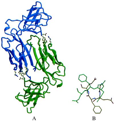
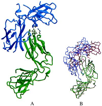
PapD is an immunoglobulin-like chaperone that mediates the assembly of P pili in uropathogenic strains of Escherichia coli. It binds and caps interactive surfaces on pilus subunits to prevent their premature associations in the periplasm. We elucidated the structural basis of a mechanism whereby PapD also interacts with itself, capping its own subunit binding surface. Crystal structures of dimeric forms of PapD revealed that this self-capping mechanism involves a rearrangement and ordering of the C2-D2 and F1-G1 loops upon dimerization which might ensure that a stable dimer is not formed in solution in spite of a relatively large dimer interface. An analysis of site directed mutations revealed that chaperone dimerization requires the same surface that is otherwise used to bind subunits.


Figures

Figure 1
Figure 1
Structure of the R8A PapD dimer. (A) molscript (33) ribbon drawing of the R8A PapD dimer. The view is looking down the dimer twofold axis. One chaperone subunit is shown in blue and the second subunit in green. In the dimer, contacts between the two N-terminal domains are mediated mostly by the two G1 edge strands across the dimer twofold axis. The interactions between two residues in the C2–D2 loop, Glu-167 and Phe-168, of one subunit with residues Pro-30, Leu-32, Ile-93, Pro-95, and Arg-58 at the lip of the second subunit are shown as ball-and-stick models. (B) Comparison of the C2–D2 loop conformation in monomeric PapD (green) and in the R8A PapD dimer (yellow). The figure was generated after superpositioning of C-terminal domains in monomeric WT PapD and dimeric R8A PapD with an rms deviation of 0.565 Å for 88 Cα atoms.
Figure 2
Figure 2
Specificity of chaperone–chaperone interaction. (A) Coomassie blue-stained SDS/12.5% PAGE showing purified Q108C PapD and WT PapD proteins run in nonreducing conditions (lanes 1 and 3, respectively) and in reducing conditions (lanes 2 and 4, respectively). (B) Silver-stained acidic native PAGE gels of purified WT PapD protein (lane 1), purified Q108C PapD protein (lane 2), and periplasmic extracts expressing the following: no chaperone (lane 3), WT PapD (lane 4), Q108C PapD (lane 5), N89C PapD (lane 6), and K110C PapD (lane 7). D1 indicates where WT PapD (monomer) migrates and D2 indicates where dimeric PapD migrates. Arrow indicates the direction of migration. (C) Autoradiogram showing radiolabeled periplasmic extracts of cells expressing WT PapD (Left) and Q108C PapD (Right) after various times of chase analyzed by acidic native PAGE (18) on Gradient 10–15 (Pharmacia) gels and silver stained on a Pharmacia Phast. The first lane of each gel (labeled neg.) is radiolabeled periplasmic extract from cells expressing no chaperone at 30 min into the chase. D1, D2, and the arrow are as in B. (D) (Inset) Autoradiogram showing radiolabeled Q108C PapD immunoprecipitated after various times of chase from periplasmic extracts of cells expressing only Q108C PapD (pQ108C) (upper gel) and in cells expressing Q108C PapD (pQ108C) in trans with a papD pap operon, pDH1 (lower gel). The samples were analyzed on nonreducing SDS/12.5% PAGE gels. For quantification, triplicate gels of each experiment were exposed to the Bio-Rad Molecular Imaging Screen-CS of the Bio-Rad Molecular Imager System, model GS-363, and the amount of PapD dimer was quantified by using Molecular Analyst/Macintosh Software, Version 2.0. The averages are graphed. The y axis equals the density of the PapD dimer band.
Figure 3
Figure 3
Structure of the Q108C PapD dimer and comparison of R8A PapD and Q108C PapD dimers. (A) molscript ribbon drawing of the Q108C PapD dimer with one chaperone subunit shown in blue and the second subunit in green. The orientation of the green subunit is the same as for the green R8A PapD subunit in Fig. 1A. (B) Comparison of R8A PapD (green and blue) and Q108C PapD (light green and red) PapD dimers. N-terminal domains in one subunit from each dimer were superimposed by using the lsq options in o (29). The pivotal point for the rotation is at the tip of the C2–D2 loop (indicated by a red arrow), which remains in essentially the same position in both dimers relative to the superimposed N-terminal domains.
Figure 4
Figure 4
Effect of mutants on chaperone–subunit interactions and chaperone dimerization. (A) Effect of mutations on PapD dimerization. WT PapD, F168R PapD, and G1 β strand mutants I105A PapD, I105E PapD, L107A PapD, and L107E PapD were induced for expression 5 min prior to pulse-labeling and then chased for 20 min. Periplasm was isolated from the cells and then subjected to glutaraldehyde crosslinking, and PapD was immunoprecipitated with anti-PapDK antiserum. The immunoprecipitates were subjected to electrophoresis on reducing SDS/12.5% polyacrylamide gels. Triplicate gels of each experiment were quantified as in Fig. 2D. (B) Curve showing the binding of purified WT PapD (○), F168R PapD (▴), native (□), or alkylated Q108C PapD (IAA-Q108C) (■) to immobilized MBP/G175–314 protein quantified by ELISA.

References

    1. Normark S, Baga M, Goransson M, Lindberg F P, Lund B, Norgren M, Uhlin B E. In: Microbial Lectins and Agglutinins: Properties and Biological Activity. Mirelman D, editor. New York: Wiley Interscience; 1986. pp. 113–143.
    1. Hultgren S J, Lindberg F, Magnusson G, Kihlberg J, Tennent J M, Normark S. Proc Natl Acad Sci USA. 1989;86:4357–4361. - PMC - PubMed
    1. Hultgren S J, Abraham S N, Caparon M G, Falk P, St. Geme J W, III, Normark S. Cell. 1993;73:887–901. - PubMed
    1. Lindberg F, Tennent J M, Hultgren S J, Lund B, Normark S. J Bacteriol. 1989;171:6052–6058. - PMC - PubMed
    1. Kuehn M J, Heuser J, Normark S, Hultgren S J. Nature (London) 1992;356:252–255. - PubMed

Publication types

MeSH terms

LinkOut - more resources