. 1999 Jul 6;96(14):8184-9.
doi: 10.1073/pnas.96.14.8184.

A single amino acid substitution in the cyclin D binding domain of the infected cell protein no. 0 abrogates the neuroinvasiveness of herpes simplex virus without affecting its ability to replicate

Affiliations

A single amino acid substitution in the cyclin D binding domain of the infected cell protein no. 0 abrogates the neuroinvasiveness of herpes simplex virus without affecting its ability to replicate

C Van Sant et al. Proc Natl Acad Sci U S A. .

Abstract

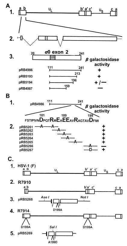
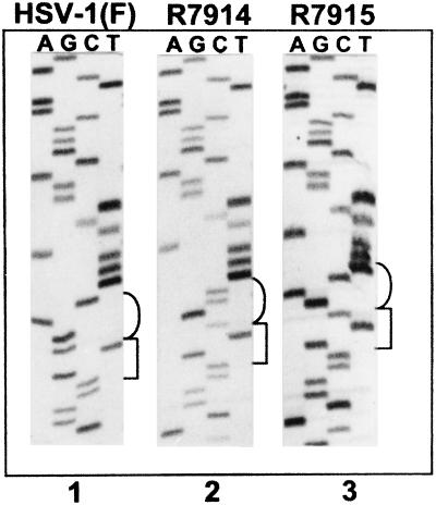
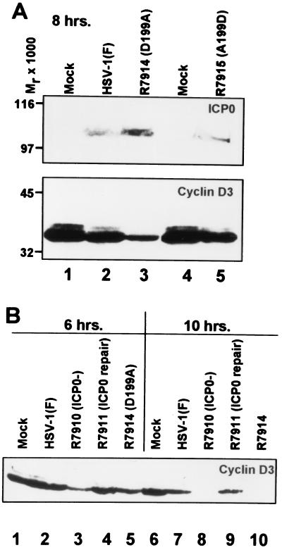
The infected cell protein no. 0 (ICP0) of herpes simplex virus 1 is a promiscuous transactivator shown to enhance the expression of genes introduced into cells by infection or transfection. The protein interacts with several viral and cellular proteins. Earlier studies have shown that ICP0 binds and stabilizes cyclin D3 but does interfere with the phosphorylation of retinoblastoma protein, its major function. Cyclin D3 plays a key role in the transition from G1 to S phase. To define the role of cyclin D3 in productive infection, the ICP0 binding site for cyclin D3 was mapped and mutagenized by substitution of aspartic acid codon 199 with the alanine codon. We report that the substitution precluded the interaction of this protein with cyclin D3 in the yeast two-hybrid system and the stabilization of cyclin D3 in infected cells. A recombinant virus carrying this mutation could not be differentiated from wild-type parent with respect to replication in dividing cells but yielded 10-fold less progeny from infected resting cells and serum-deprived or contact-inhibited human fibroblasts. In mice, the mutant was only slightly less pathogenic than the wild-type parent by intracerebral route but was significantly less neuroinvasive after peripheral inoculation. Replacement of the mutated amino acid with aspartic acid restored wild-type phenotype. Stabilization of cyclin D3 therefore is linked to higher virus yields in nondividing cells and potentially higher virulence in experimental and natural hosts. One function of ICP0 is to scavenge the cell for proteins that could bolster viral replication.


Figures

Figure 1
Figure 1
(A) Schematic diagram of the sequence arrangements of the HSV-1 genome and the location of the α0 gene. Line 1, a linear representation of the HSV-1 genome showing the unique long (UL) and short (US) sequences. The terminal repeats flanking UL and US, ab and ca, respectively, are represented as rectangles. Line 2, expansion of a portion of the repeat sequence ab showing the location of one of the copies of the α0 gene. Line 3, an expanded section of exon II (codons 20–241). The shaded area represents the ICP0 zinc ring finger. Plamsid pRB4986 (3) carries the HSV-1 strain F [HSV-1(F)] α0 coding sequence that specifically interacts with cyclin D3. Plasmids pRB5193, pRB5194, and pRB4987 carrying truncated portions of α0 binding domain from pRB4986 were analyzed in the yeast two-hybrid system as described for the ability of the expressed peptides to bind cyclin D3 (3). (B) Line 1, a representation of exon II domain cloned in pRB4986. Line 2, the amino acid sequence of the minimal binding domain mapped by truncation of the pRB4986 sequence in A. The large letters represent codons substituted with alanine codon. Also shown are the results of the yeast assays to determine whether the mutagenized fragments yields a product that interacts with cyclin D3. (C) Lines 1 and 2, schematic representation of the wild-type HSV-1(F) and R7910 (ICP0) genomes, respectively. In R7910, both copies of the α0 gene had been deleted. The rescue of R7910 virus was effected by transfection of rabbit skin cells with the DNA cloned in pRB5268 (line 3), followed by superinfection with R7910 (line 2), which yielded the virus R7914 carrying the D199A substitution within the ICP0 reading frame. Rescue of the R7914 by cotransfection of its DNA with that of pRB5269 yielded the recombinant R7915 (line 5) in which the A199D substitution rendered the α0 gene wild type.
Figure 2
Figure 2
Autoradiographic image of ICP0 exon II sequence from wild-type HSV-1(F) and recombinant viruses R7914 and R7915. Cytoplasmic viral DNA was obtained from infected Vero cells, and a 561-bp fragment was PCR-amplified and then sequenced. The sequences of HSV-1(F) (lane 1) and recombinant viruses R7914 and R7915 (lanes 2 and 3, respectively) were identical with the exception of the desired mutations shown in brackets. The square bracket to the right of the images represents V198 (codon usage GTN), and the round brackets represent codon 199 (codon usage for aspartic acid is GAC or GAT, and codon usage for alanine is GCN).
Figure 3
Figure 3
(A) Immunoblots of lysates of cells mock-infected (lanes 1 and 4) or exposed to 0.5 PFU of HSV-1(F) (lane 2), R7914 (lane 3), or R7915 (lane 5) per cell. The cells were harvested after infection, solubilized in buffer containing SDS, electrophoretically separated in a denaturing polyacrylamide gel, and probed with mAb to ICP0 (H1083, ref. 18) or the mAb to cyclin D3 (Pharmingen, #G107–565). Molecular weights are shown on the left. (B) The cells were mock-infected (lanes 1 and 6) or infected with HSV-1(F) (lanes 2 and 7), the ICP0 null recombinant R7910 (lanes 3 and 8), recombinant R7911 (repair of R7910) (lanes 4 and 9), or recombinant R7914 (lanes 5 and 10) and harvested 6 hr after infection (lanes 1–5) or 10 hr. after infection (lanes 6–10), and processed as above. The electrophoretically separated proteins were reacted with the mAb to cyclin D3.

References

    1. Roizman B, Sears A E. In: Fields’ Virology. 3rd Ed. Fields B N, Knipe D M, Howley P, Chanock R M, Hirsch M S, Melnick J L, Monath T P, Roizman B, editors. New York: Raven; 1996. pp. 2231–2295.
    1. Sacks W, Schaffer P A. J Virol. 1987;61:829–839. - PMC - PubMed
    1. Kawaguchi Y, Van Sant C, Roizman B. J Virol. 1997;71:7328–7336. - PMC - PubMed
    1. Cai W, Schaffer P A. J Virol. 1993;67:7501–7512. - PMC - PubMed
    1. Everett R, Preston C, Stow N. In: Herpes Virus Transcription: Functional and Genetic Analysis of the Role of Vmw110 in Herpes Simplex Virus Replication. Wagner E K, editor. Boca Raton FL: CRC; 1991. pp. 49–76.

Publication types

MeSH terms

LinkOut - more resources