Case Reports
. 1999 Feb;83(2):185-9.
doi: 10.1136/bjo.83.2.185.

Confocal microscopy in cornea guttata and Fuchs' endothelial dystrophy

Affiliations
Case Reports

Confocal microscopy in cornea guttata and Fuchs' endothelial dystrophy

A G Chiou et al. Br J Ophthalmol. 1999 Feb.

Abstract

Aims: To report the appearances of cornea guttata and Fuchs' endothelial dystrophy from white light confocal microscopy.

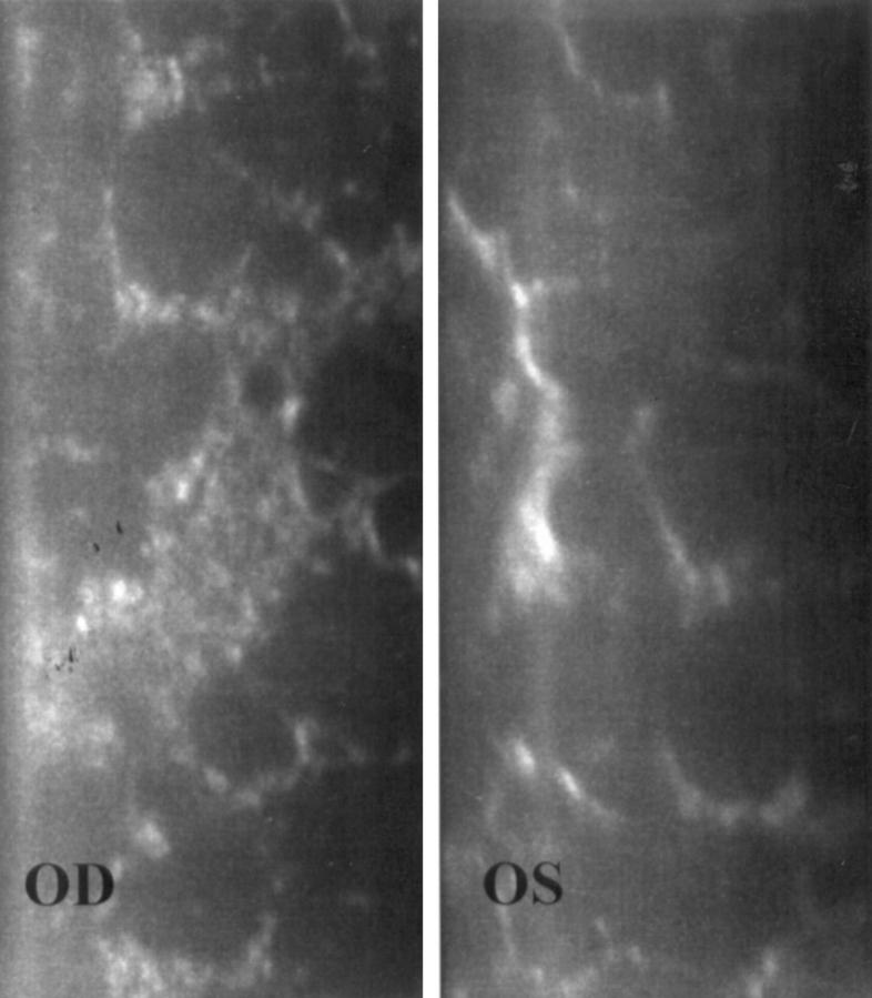
Methods: Seven eyes of four consecutive patients with cornea guttata were prospectively examined. Of the seven eyes, three also had corneal oedema (Fuchs' dystrophy). In vivo white light tandem scanning confocal microscopy was performed in all eyes. Results were compared with non-contact specular microscopy.

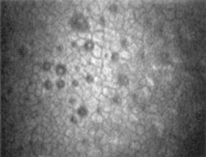
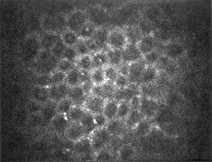
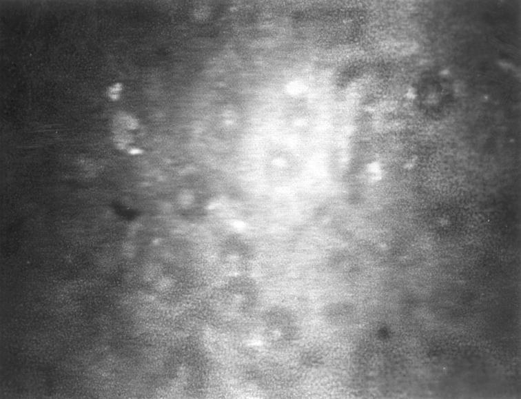
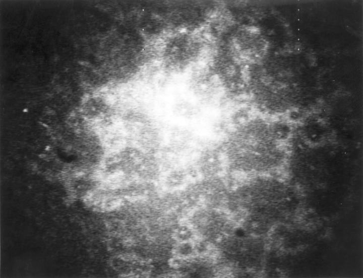
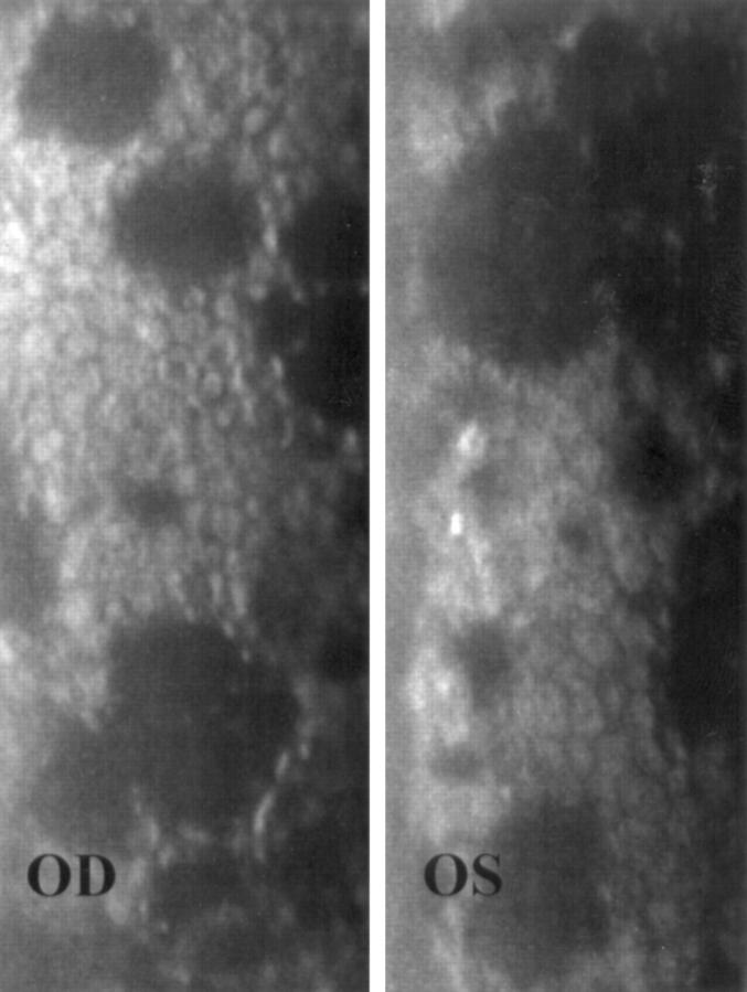
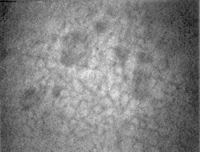
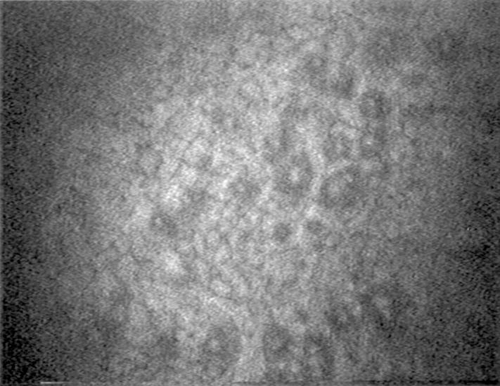
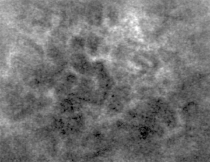
Results: Specular microscopy was precluded by corneal oedema in one eye. In the remaining six eyes, it demonstrated typical changes including pleomorphism, polymegathism, and the presence of guttae appearing as dark bodies, some with a central bright reflex. In all seven eyes, confocal microscopy revealed the presence of round hyporeflective images with an occasional central highlight at the level of the endothelium. Changes in cell morphology and size were readily appreciated.

Conclusion: By comparison with specular microscopy, the hyporeflective images with an occasional central highlight seen on confocal microscopy are consistent with the presence of guttae. Confocal microscopy may confirm the diagnosis of cornea guttata and Fuchs' endothelial dystrophy by demonstrating the presence of guttae. This technique is especially valuable in cases of corneal oedema, where specular microscopy may fail to visualise the endothelium. However, specular microscopy should remain the method of choice to evaluate the endothelium, principally because it is easier to use.


Figures

Figure 1
Figure 1
Patient 1. Specular microscopy in a case of cornea guttata with asymmetric presentation. Dark round bodies are more predominant in the right eye (OD). OS indicates left eye.
Figure 2
Figure 2
Patient 1. Confocal microscopy (magnification ×210) of the left eye. A few hyporeflective images with an occasional central highlight were seen at the level of the endothelium.
Figure 3
Figure 3
Patient 1. Confocal microscopy (magnification ×210) of the right eye. Compared with the left eye, significantly more hyporeflective images were seen. Hyperreflective endothelial cells were found between the lesions.
Figure 4
Figure 4
Patient 2. Specular microscopy in the right (OD) and left (OS) eyes in a case of bilateral corneal guttae and oedema (Fuchs' dystrophy).
Figure 5
Figure 5
Patient 2. Confocal microscopy (magnification ×210) of the right eye. Hyporeflective images with a central highlight were seen in the endothelium.
Figure 6
Figure 6
Patient 2. Confocal microscopy (magnification ×210) of the left eye. Confluent hyporeflective images with a sporadic central highlight were demonstrated.
Figure 7
Figure 7
Patient 3. Specular microscopy in a case of bilateral cornea guttata. OD indicates right eye and OS indicates left eye.
Figure 8
Figure 8
Patient 3. Confocal microscopy (magnification ×210) of the right eye. Hyporeflective images were seen in the endothelium.
Figure 9
Figure 9
Patient 3. Confocal microscopy (magnification ×210) of the left eye. Hyporeflective images with a central highlight were demonstrated in the endothelium.
Figure 10
Figure 10
Patient 4. Confocal microscopy (magnification ×210) revealed the presence of confluent hyporeflective images in the endothelium. Hyperreflective endothelial cells were found between the lesions. The hyperreflective image on the upper part of the photograph is compatible with fibrous changes. Specular microscopy in this case of Fuchs' endothelial dystrophy was prevented by corneal oedema.

References

    1. Am J Ophthalmol. 1976 Jun;81(6):773-85 - PubMed
    1. Metab Pediatr Syst Ophthalmol (1985). 1995;18(1-4):1-4 - PubMed
    1. Arch Ophthalmol. 1978 Apr;96(4):653-5 - PubMed
    1. Br J Ophthalmol. 1997 Feb;81(2):168-9 - PubMed
    1. Arch Ophthalmol. 1997 Nov;115(11):1461-3 - PubMed

Publication types