

Histochemical localisation of mitochondrial enzyme activity in human optic nerve and retina
- PMID: 10396204
- PMCID: PMC1722931
- DOI: 10.1136/bjo.83.2.231
Histochemical localisation of mitochondrial enzyme activity in human optic nerve and retina
Abstract
Aims: To demonstrate the quantitative distribution of mitochondrial enzymes within the human optic nerve and retina in relation to the pathogenesis of ophthalmic disease.
Methods: Enucleations were performed at the time of multiple organ donation and the optic nerve and peripapillary retina immediately excised en bloc and frozen. Reactivities of the mitochondrial enzymes cytochrome c oxidase and succinate dehydrogenase were demonstrated in serial cryostat sections using specific histochemical assays.
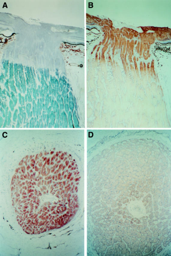
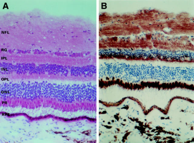
Results: In the optic nerve the unmyelinated prelaminar and laminar regions were rich in both cytochrome c oxidase and succinate dehydrogenase. Myelination of fibres as they exited the lamina cribrosa was associated with an abrupt reduction in enzyme activity. Within the retina, high levels of enzyme activity were found localised within the retinal ganglion cells and nerve fibre layer, the outer plexiform layer, inner segments of photoreceptors, and the retinal pigment epithelium.
Conclusions: Mitochondrial enzyme activity is preserved in human optic nerve and retina retrieved at the time of multiple organ donation. The distribution of enzyme activity within the eye has implications for the understanding of the pattern of ophthalmic involvement seen in mitochondrial diseases and the site of ganglion cell dysfunction in those patients with optic nerve involvement.
Figures
References
Publication types
MeSH terms
Substances
LinkOut - more resources
Full Text Sources
Medical
Research Materials