. 1999 Jul;10(7):2251-64.
doi: 10.1091/mbc.10.7.2251.

The plant vesicle-associated SNARE AtVTI1a likely mediates vesicle transport from the trans-Golgi network to the prevacuolar compartment

Affiliations
Free PMC article

The plant vesicle-associated SNARE AtVTI1a likely mediates vesicle transport from the trans-Golgi network to the prevacuolar compartment

H Zheng et al. Mol Biol Cell. 1999 Jul.
Free PMC article

Abstract

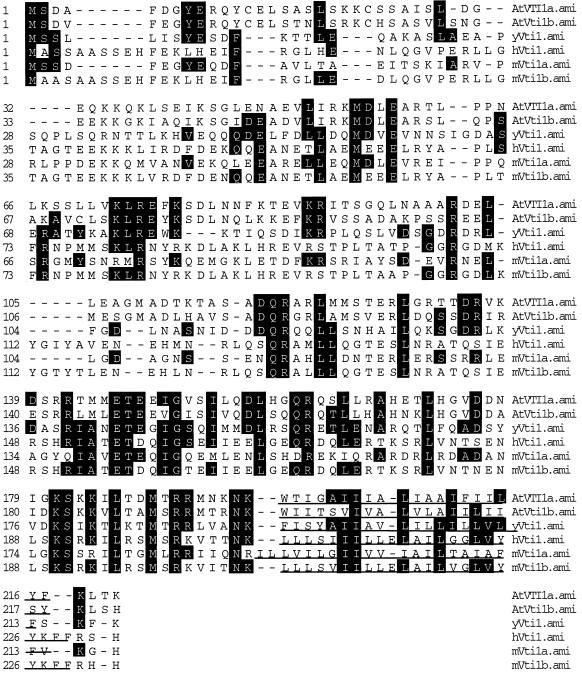
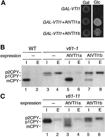
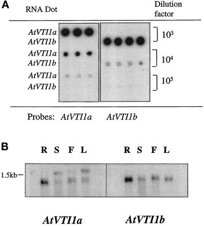
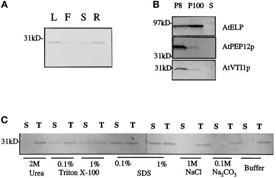
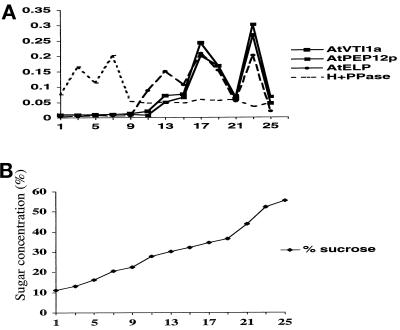
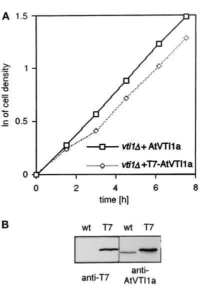
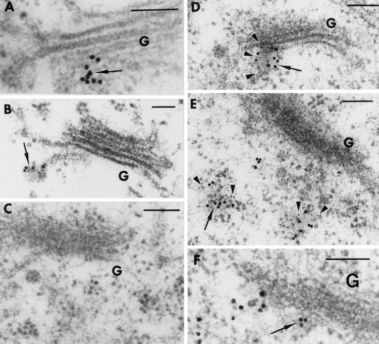
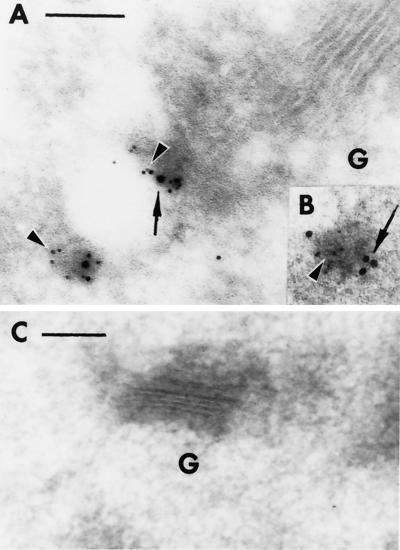
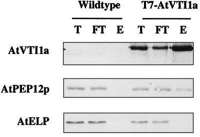
Membrane traffic in eukaryotic cells relies on recognition between v-SNAREs on transport vesicles and t-SNAREs on target membranes. Here we report the identification of AtVTI1a and AtVTI1b, two Arabidopsis homologues of the yeast v-SNARE Vti1p, which is required for multiple transport steps in yeast. AtVTI1a and AtVTI1b share 60% amino acid identity with one another and are 32 and 30% identical to the yeast protein, respectively. By suppressing defects found in specific strains of yeast vti1 temperature-sensitive mutants, we show that AtVTI1a can substitute for Vti1p in Golgi-to-prevacuolar compartment (PVC) transport, whereas AtVTI1b substitutes in two alternative pathways: the vacuolar import of alkaline phosphatase and the so-called cytosol-to-vacuole pathway used by aminopeptidase I. Both AtVTI1a and AtVTI1b are expressed in all major organs of Arabidopsis. Using subcellular fractionation and immunoelectron microscopy, we show that AtVTI1a colocalizes with the putative vacuolar cargo receptor AtELP on the trans-Golgi network and the PVC. AtVTI1a also colocalizes with the t-SNARE AtPEP12p to the PVC. In addition, AtVTI1a and AtPEP12p can be coimmunoprecipitated from plant cell extracts. We propose that AtVTI1a functions as a v-SNARE responsible for targeting AtELP-containing vesicles from the trans-Golgi network to the PVC, and that AtVTI1b is involved in a different membrane transport process.


Figures

Figure 1
Figure 1
Sequence comparison of AtVTI1a and AtVTI1b with other members of the family including yVti1p (Saccharomyces cerevisiae, accession number 2497184), hVti1p (Homo sapiens, accession number 268740), mVti1a (Mus musculus, accession number 3213227), and mVti1b (Mus musculus, accession number 3213229). The sequence comparison was generated using the J. Hein method in MegAlign (DNAStar program). Amino acids identical to yVti1p are shaded in black. The C-terminal hydrophobic domain is underlined.
Figure 2
Figure 2
Expression of either AtVTI1a or AtVTI1b allows yeast cells to grow in the absence of Vti1p, but only AtVTI1a functions in TGN-to-PVC traffic. (A) Growth of the vti1Δ GAL1-VTI1 strain (FvMY6/pFvM16) alone or expressing AtVTI1a or AtVTI1b on plates containing galactose (Gal) or glucose (Glc). The vti1Δ GAL1-VTI1 strain could not grow on glucose after the expression of VTI1 was shut off, but expression of either AtVTI1a or AtVTI1b supported growth. (B and C) CPY traffic in wild-type, vti1-1 (FvMY7) (B) and vti1-11 (C) cells (FvMY21) alone (−) or expressing AtVTI1a or AtVTI1b. Cells were grown at 24°C, preincubated at 36°C for 15 min, pulse labeled for 10 min, and chased for 30 min at 36°C. CPY was immunoprecipitated from cellular extracts (I) and extracellular fractions (E) and analyzed by SDS-PAGE and autoradiography. The TGN-to-PVC traffic block in vti1-1 cells (FvMY7) was suppressed by AtVTI1a expression, as indicated by the presence of vacuolar mCPY, but not by AtVTI1b expression. (C) vti1-11 cells (FvMY21) accumulated p1CPY because of a block in retrograde traffic to the cis-Golgi. Expression of either AtVTI1a or AtVTI1b reduced the amount of p1CPY that accumulated.
Figure 3
Figure 3
AtVTI1b but not AtVTI1a could replace yeast Vti1p in ALP and API traffic to the vacuole, which are transported to the vacuole via two different biosynthetic pathways. (A) Wild-type and vti1-2 cells (FvMY24) alone (−) or expressing either AtVTI1a or AtVTI1b were grown at 24°C, preincubated at 36°C for 15 min, pulse labeled for 7 min, and chased for 0 or 30 min at 36°C. ALP was immunoprecipitated from cellular extracts and separated by SDS-PAGE. The accumulation of pALP in vti1-2 cells was suppressed by expression of AtVTI1b but not by AtVTI1a. (B) Wild-type and vti1-11 cells (FvMY21) alone (−) or expressing AtVTI1a or AtVTI1b were grown at 24°C, preincubated at 36°C for 15 min, labeled for 10 min, and chased for 0 or 120 min at 36°C. API was immunoprecipitated from cellular extracts and analyzed by SDS-PAGE. Vacuolar mAPI was found only in vti1-11 cells expressing AtVTI1b, not in vti1-11 cells expressing AtVTI1a.
Figure 4
Figure 4
Northern blot analyses of AtVTI1a and AtVTI1b. (A) Dot blot for testing the specificity of the AtVTI1a and AtVTI1b probes. One microliter of in vitro–transcribed mRNAs of AtVTI1a and AtVTI1b in serial 10× dilutions was applied to the Hybond-N membrane and hybridized with in vitro–transcribed [α-32P]UTP-labeled RNA probes at high stringency (65°C for 16 h). The blot was washed under highly stringent conditions (2× SSC and 1% SDS for 30 min at 65°C, 0.2× SSC and 1% SDS 10 min for three times at room temperature). The signal was then detected by autoradiography. (B) RNA gel blot analysis of AtVTI1 expression in several Arabidopsis organs. Twenty micrograms of total RNA extracted from roots (R), stems (S), flowers (F), and leaves (L) were separated on a denaturing gel and transferred to Hybond-N. The membrane was then hybridized with probes described in A following the same procedure.
Figure 5
Figure 5
AtVTI1a is an integral membrane protein. (A) Distribution of AtVTI1a in Arabidopsis organs. Equal amounts of total protein from leaves (L), flowers (F), stems (S), and roots (R) were separated by SDS-PAGE and immunoblotted with guinea pig antiserum against AtVTI1a. Molecular mass is indicated on the left. (B) AtVTI1a fractionates with heavy membranes during differential centrifugation. A postnuclear supernatant from 0.5 g of cultured roots was centrifuged at 8000 × g for 20 min. The pellet (P8) was solubilized in 200 μl of 2× Laemmli loading buffer. The supernatant (S8) was further ultracentrifuged at 100,000 × g for 2 h. The pellet (P100) was solubilized in 200 μl of 2× Laemmli buffer. Equal amounts of the supernatant (S100), P100, and P8 were separated by SDS-PAGE and immunoblotted with anti-AtVTI1a, anti-AtELP, or anti-AtPEP12p antiserum. Molecular mass is indicated on the left. (C) AtVTI1a is an integral membrane protein. Equal amounts of total membranes from Arabidopsis cultured cells were treated with 2 M urea, 0.1 or 1% Triton X-100, 0.1 or 1% SDS, 1 M NaCl, 0.1 M Na2CO3, pH 11, or extraction buffer alone. All treatments were performed at room temperature for 30 min. An aliquot of each treatment was saved as total. Membranes were pelleted by centrifugation at 100,000 × g for 1 h after the treatments. Equal volumes of supernatant or total were separated by SDS-PAGE and immunoblotted with anti-AtVTI1a antiserum. S, supernatant, T, total.
Figure 6
Figure 6
Subcellular fractionation of AtVTI1a by step sucrose gradient. Postnuclear membranes of Arabidopsis roots were loaded on a step sucrose gradient. After equilibrium by ultracentrifugation at 100,000 × g for 3 h, 0.5-ml fractions were collected from top (1) to bottom of the gradient (25). Equal volumes of odd-numbered fractions were loaded on an SDS-PAGE gel and immunoblotted with anti-AtVTI1a, anti-AtELP, anti-AtPEP12p, and anti-H+PPase antibodies. Blots were analyzed by densitometry, and the percentage of the total marker protein detected in each fraction for AtVTI1a, AtPEP12p, AtELP, and H+PPase was plotted in A. The sucrose concentration of each fraction was determined by refractometry and plotted in B.
Figure 7
Figure 7
T7 tag does not affect AtVTI1a function and is expressed in transgenic plants. (A) Growth curves of vti1Δ cells expressing AtVTI1a or T7-AtVTI1a. vti1Δ cells (FvMY6) expressing either AtVTI1a or epitope-tagged T7-AtVTI1a grew at similar rates, indicating that T7-AtVTI1a was functional. Cells were grown in a rich medium at 30°C at logarithmic phase. The cell density was determined by measuring the optical density at 600 nm. (B) T7 antibodies specifically recognized T7-AtVTI1a in transgenic plants. Equal amounts of postnuclear supernatant of Arabidopsis (T7-AtVTI1a transgenic plants or wild-type) were separated on SDS-PAGE gel and immunoblotted with monoclonal T7 antibody or polyclonal antiserum against AtVTI1a raised in guinea pig.
Figure 8
Figure 8
In situ localization of T7-AtVTI1a and AtELP on ultrathin sections of Arabidopsis roots from T7-AtVTI1a transgenic plants. T7-AtVTI1a and AtELP are localized on the TGN and on dense vesicles. (A and B) Ultrathin sections were incubated with T7 mAb followed by rabbit anti-mouse IgG and biotinylated goat anti-rabbit secondary antibody and were visualized with streptavidin conjugated to 10-nm colloidal gold. (C) Control. The ultrathin sections were treated with the same procedure as described in A and B, except T7 mAb was substituted with 2% BSA in PBS. T7-AtVTI1a and AtELP are colocalized on the TGN (D) and on dense vesicles (E). (D and E) Ultrathin sections were incubated with T7 mAb followed by rabbit anti-mouse IgG and biotinylated goat anti-rabbit secondary antibody and were visualized with streptavidin conjugated to 10-nm colloidal gold. After the second fixation step (see MATERIALS AND METHODS), the same sections were incubated with antiserum to AtELP, followed by biotinylated goat anti-rabbit secondary antibody, and then visualized with streptavidin conjugated to 5-nm colloidal gold. (F) Control section. The same procedures were used as in D and E, except preimmune serum was used in the place of AtELP antiserum. G, Golgi; arrow, AtVTI1a; arrowhead, AtELP; bar, 0.1 μm.
Figure 9
Figure 9
T7-AtVTI1a and AtPEP12p colocalize on the PVC in cryosections of Arabidopsis roots from T7-AtVTI1a transgenic plants. (A and B) Ultrathin sections were incubated with T7 mAb followed by rabbit anti-mouse IgG and biotinylated goat anti-rabbit secondary antibody and were visualized with streptavidin conjugated to 10-nm colloidal gold. After the second fixation step (see MATERIALS AND METHODS), the same sections were incubated with AtPEP12p antiserum, followed by biotinylated goat anti-rabbit IgG, and then detected by streptavidin conjugated to 5-nm gold particles. (C) Control section. The same procedures were used as in A and B, except first antibodies were substituted with 2% BSA in PBS for T7 mAb and AtPEP12p preimmune serum for AtPEP12p antiserum. G, Golgi; arrow, AtVTI1a; arrowhead, AtPEP12p; bar, 0.1 μm.
Figure 10
Figure 10
AtVTI1a associates with AtPEP12p. Postnuclear supernatant from three grams of T7-AtVTI1a transgenic or wild-type Arabidopsis plants (21 d old) were treated with 1% Triton X-100 to solubilize membrane proteins. An aliquot was saved as total protein. The Triton X-100–solubilized protein extract was then incubated with 100 μl of T7 antibody agarose (Novagen) for 5 h. Beads were pelleted, and the flow-through was collected. After being washed, proteins associated with the T7 antibody agarose were eluted. Equal volumes of total (T), flow-through (FT), and eluate (E) were separated by SDS-PAGE, followed by immunoblotting with antibodies against AtVTI1a, AtPEP12p, or AtELP.

Similar articles

Cited by

References

    1. Advani RJ, Bae HR, Bock JB, Chao DS, Doung YC, Prekeris R, Yoo JS, Scheller RH. Seven novel mammalian SNARE proteins localize to distinct membrane compartments. J Biol Chem. 1998;273:10317–10324. - PubMed
    1. Ahmed SU, Bar-Peled M, Raikhel NV. Cloning and subcellular location of an Arabidopsis receptor-like protein that shares common features with protein-sorting receptors of eukaryotic cells. Plant Physiol. 1997;114:325–336. - PMC - PubMed
    1. Altschul SF, Gish W, Miller W, Myers EW, Lipman DJ. Basic local alignment search tool. J Mol Biol. 1990;215:403–410. - PubMed
    1. Bar-Peled M, Raikhel NV. Characterization of AtSEC12 and AtSAR1. Proteins likely involved in endoplasmic reticulum and Golgi transport. Plant Physiol. 1997;114:315–324. - PMC - PubMed
    1. Bassham DC, Gal S, Conceição AS, Raikhel NV. An Arabidopsis syntaxin homologue isolated by functional complementation of a yeast pep12 mutant. Proc Natl Acad Sci USA. 1995;92:7262–7266. - PMC - PubMed

Publication types

MeSH terms

Associated data