Comparative Study
. 1999 Jul;10(7):2309-27.
doi: 10.1091/mbc.10.7.2309.

Polymerizing microtubules activate site-directed F-actin assembly in nerve growth cones

Affiliations
Free PMC article
Comparative Study

Polymerizing microtubules activate site-directed F-actin assembly in nerve growth cones

M W Rochlin et al. Mol Biol Cell. 1999 Jul.
Free PMC article

Abstract

We identify an actin-based protrusive structure in growth cones termed "intrapodium." Unlike filopodia, intrapodia are initiated exclusively within lamellipodia and elongate in a continuous (nonsaltatory) manner parallel to the plane of the dorsal plasma membrane causing a ridge-like protrusion. Intrapodia resemble the actin-rich structures induced by intracellular pathogens (e.g., Listeria) or by extracellular beads. Cytochalasin B inhibits intrapodial elongation and removal of cytochalasin B produced a burst of intrapodial activity. Electron microscopic studies revealed that lamellipodial intrapodia contain both short and long actin filaments oriented with their barbed ends toward the membrane surface or advancing end. Our data suggest an interaction between microtubule endings and intrapodia formation. Disruption of microtubules by acute nocodazole treatment decreased intrapodia frequency, and washout of nocodazole or addition of the microtubule-stabilizing drug Taxol caused a burst of intrapodia formation. Furthermore, individual microtubule ends were found near intrapodia initiation sites. Thus, microtubule ends or associated structures may regulate these actin-dependent structures. We propose that intrapodia are the consequence of an early step in a cascade of events that leads to the development of F-actin-associated plasma membrane specializations.


Figures
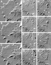
Figure 1
Figure 1
Two time-lapse sequences revealing intrapodia formation and dynamics. (A–C) Intrapodium initiated in the marginal zone. (A) At the onset of intrapodia formation a protuberance is initiated at the margin between the central, organelle-rich domain and the flattened, F-actin-rich domain (arrowhead). In this case, elongation is fairly straight, perhaps because the direction of elongation is orthogonal to the nearest leading edge and therefore is not being “side-swiped” by the retrograde flow. A second intrapodium is developing near the top. Time interval between images is 5 s. (D–K) Intrapodia that developed in the peripheral domain of a growth cone from a particle released from the leading edge and carried rearward by the retrograde flow. (D) Just after release of a particle from the leading edge (arrow). During the rearward movement, but before E, the particle increases in length but then shortens to assume its original size (our unpublished results). (E) The particle (arrow) continues to move rearward, carried back by the retrograde flow. (F) Two intrapodia extend away from the particle (arrowheads, see inset), growing in opposite directions. (G) Intrapodial growth continues, but the direction of growth begins to curve to the left. The intrapodium that initially grew toward the center of the growth cone (proximal intrapodium) elongates more rapidly than the intrapodium that initially grew toward the leading edge (distal intrapodium) against the retrograde flow. (H) The distal intrapodium appears to have discontinued net extension in the plane of the growth cone, whereas the proximal intrapodium continues to extend, now in a direction parallel to the leading edge and perpendicular to its original direction. (I) The proximal intrapodium (arrowhead) continues to grow in the same direction. The distal intrapodium grows much less and appears to be meandering. (J) The proximal intrapodium continues to extend toward linear elements emanating from the central region of the growth cone. These linear elements presumably contain microtubules and endoplasmic reticulum and vesicular structures known to extend along microtubules in peripheral regions of growth cones (Dailey and Bridgman, 1991). (K) A large portion of the tail is dissolved, leaving behind a few remnants. The tip of the proximal intrapodium continues to advance, but upon contacting the linear elements, it turned abruptly, coursing centripetally along the linear structure. Bar, 5 μm.
Figure 2
Figure 2
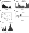
Histogram of intrapodia frequency in growth cones elongating exclusively on the laminin-coated coverglass (solid bars) or along a neurite (stippled bars). The number of intrapodia observed in 1-min intervals is plotted. In both situations, intrapodia frequency is episodic. The level of intrapodia activity is higher in the growth cone elongating along another neurite.
Figure 3
Figure 3
Comparison of elongation of individual intrapodia (IP) and filopodia (Filo). Intrapodia elongate more rapidly than filopodia. This is due in part to the saltatory nature of filopodial growth and also to a slower elongation rate during periods of growth.
Figure 4
Figure 4
Intrapodia are composed of F-actin and F-actin-associated proteins. Growth cones were fixed during live observation and stained for actin, β1-integrin, or capping protein. (A–K) A growth cone stained for actin and β1-integrin. (A–D) DIC images of growth cone before and after fixation (time is in seconds). (A) The intrapodium (arrowhead) is initiated in the marginal zone. (B) The tip of the intrapodium advances toward the leading edge, against the direction of the retrograde flow. (C) Moments before the arrival of the fixative, but during perfusion, the intrapodium continues to elongate toward the leading edge. In addition, two smaller intrapodia have begun to form nearby, one extending toward the central, thickened region of the growth cone and the other extending tangential to the marginal zone. Because of the perfusion, the focus drifts slightly. (D) After completion of fixation, the growth cone is thinner, but there is sufficient contrast to resolve the two intrapodia that were closest to the leading edge. (E) β1-Integrin staining is elevated along intrapodia (arrowheads) and also along the leading edge. (F) Overlay of β1-integrin staining on the DIC image of the fixed growth cone in D, showing the coincidence of the elevated integrin staining and the intrapodia (arrowheads). (G) 4× zoom of F, featuring the growth cone region containing the intrapodium. Note that the β1-integrin staining is punctate along the intrapodia. (H) Before immunostaining, the growth cone was labeled with rhodamine-phalloidin. Bar, 9 μm. (I–K) Growth cone stained for capping protein (Schafer et al., 1998). Capping protein staining was distributed along the length of the intrapodium (arrowhead) and also stained rib-like structures that presumably correspond to F-actin bundles that are perpendicular to the leading edge (arrow). Inset, colocalized capping protein staining (2× zoom). Bar, 5 μm.
Figure 5
Figure 5
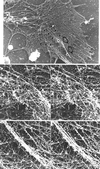
Electron micrograph of a growth cone–containing intrapodia reveals deformation of dorsal plasma membrane, a high concentration of actin filaments, and microtubule endings. (A) Low-magnification EM image of a rotary-shadowed growth cone cytoskeleton. An intrapodium (large arrows) that was identified and tracked by video-enhanced DIC recording (our unpublished data) before fixation and extraction of the membrane elongated from a region in close proximity to the ends of microtubules (arrowheads). (B) Higher-magnification stereo pairs of the two areas indicated by the asterisks in A. The first area contains the origin of the intrapodium. A relatively broad, dense mass of actin filaments (between the arrows) forms a ridge on the dorsal surface of the cone. A microtubule (arrowheads) that appears decorated with globular material ends on one side of the ridge (the microtubule end is adjacent to the arrowhead on the right). The second area contains the leading end of the intrapodium. A bundle of actin filaments forms a narrow ridge (between arrows). At the tip of the intrapodium, numerous filament ends can be seen (arrowheads). Bars, 2.6 μm (A); 360 nm (B).
Figure 6
Figure 6
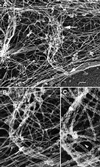
Polarity of actin filaments observed using myosin S1 decoration. (A) An intrapodium extends from its base (upper arrow) toward the cell margin (lower arrow). (B) Decorated filaments in the intrapodium (arrow) are superimposed upon those in the underlying lamellipodia. Stereo images of this intrapodium (our unpublished data) allowed identification of intrapodial-associated filaments. (C) A higher magnification allows the decorated filament orientation (indicated by the arrowhead) to be seen in a small portion of intrapodium-associated filaments. Bars, 360 nm (A); 180 nm (B); 90 nm (C).
Figure 7
Figure 7
Cytochalasin B washout causes a burst of intrapodial activity. In this case the SCG explant had been plated and maintained in a low dose of nocodazole. (A) Just after perfusion with cytochalasin. (B) One minute 33 s later. Note the more flattened appearance of the lamellipodium, the thickened leading edge, and the islands of cytoplasm that accumulate in the lamellipodium. (C and D). After perfusion of cytochalasin-free media. In this instance, intrapodia (arrowheads) emanated from one cytoplasmic island, producing a starburst appearance. Bar, 5 μm. *, Reverse shadow-cast vacuole.
Figure 8
Figure 8
Influence of nocodazole and Taxol on intrapodia frequency. The number of intrapodia initiated during the indicated period is plotted. (A) After overnight growth in a low dose of nocodazole (0.33 μM), perfusion of a high dose of nocodazole (3.3 μM) causes a reversible decrease in intrapodial frequency. (B) Effect of nocodazole treatment and washout on intrapodia frequency. Both the first and second washouts cause a spike in intrapodial activity followed by a slow decline, whereas readministration of a low dose of nocodazole slowly decreases intrapodial activity. (C) Because growth cones grown overnight in 0.33 μM nocodazole were larger than untreated growth cones, we assessed the contribution of the increased area to intrapodial frequency under the two conditions. In nocodazole, the rate of intrapodia formation is greater per unit area than in the untreated growth cones. The SD for the intrapodia rate in control growth cones averaged 1.2 and for the nocodazole treated cones averaged 2.5. (D) Changes in growth cone area at the leading edge (front 50%) during washout of 0.33 μM nocodazole. Positive values for area change indicate net protrusion, whereas negative values indicate net retraction. During the spike of intrapodia activity (shown in B), the growth cone leading edge retracts transiently. Protrusion then resumes and increases after several minutes delay. (E) Taxol treatment also affects intrapodia frequency. Washout of Taxol did not have an effect on intrapodia frequency, but readdition of Taxol (12 nM) caused a burst of intrapodial activity.
Figure 9
Figure 9
Nocodazole washout causes a burst of intrapodial frequency followed by leading edge protrusion. (A) Before washout of 0.33 μM nocodazole. (B) Just after washout (1 min, 18 s). Note the intrapodia (arrows) that formed at the edge of the marginal zone and elongated toward the leading edge. Protrusion at the leading edge is minimal compared with A. (C) Protrusion at the leading edge (arrowheads) is now observed (8 min, 8 s after washout). Bar, 7 μm.
Figure 10
Figure 10
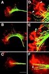
Microtubule endings are associated with intrapodial tails. Red corresponds to actin staining, green to microtubule staining. Growth cones in the presence or absence of microtubule-perturbing treatments were monitored during time-lapse DIC recordings and fixed during intrapodia formation. Arrowheads in D–F represent the initiation sites of intrapodia as determined before fixation during DIC observations. At high magnification, the microtubule staining sometimes appears discontinuous. Because similarly fixed samples preparedfor EM show no evidence of microtubule fragmentation, we suspect that the discontinuities in staining are due to the antibody failing to bind a subset of antigen sites. If we used higher antibody concentrations, we obtained more continuous staining but also higher background. (A) Untreated growth cone fixed during intrapodia formation. (D) Digital zoom (4×) of a portion of the same cone shown in A. Some intrapodia are associated with microtubules (arrowheads). (B) After washout of nocodazole (0.33 μM), most of the intrapodia appear to have been initiated in the vicinity of microtubule endings. (E) Digital zoom of the intrapodia-rich area showing the close relationship between intrapodia (arrowheads) and microtubules. (C) After perfusion with medium containing 12 nM Taxol, intrapodia are initiated at sites rich in microtubule endings. (F) Digital zoom of the intrapodia-rich area (arrowheads) showing the relationship with microtubule ends. Bar, 18 μm.

Similar articles

Cited by

References

    1. Beckerle MC. Spatial control of actin filament assembly: lessons from Listeria. Cell. 1998;95:741–748. - PubMed
    1. Berg HC, Block SM. A miniature flow cell designed for rapid exchange of media under high-power microscope objectives. J Gen Microbiol. 1984;130:2915–2920. - PubMed
    1. Bershadsky A, Chausovsky A, Becker E, Lyubimova A, Geiger B. Involvement of microtubules in the control of adhesion-dependent signal transduction. Curr Biol. 1996;6:1279–1289. - PubMed
    1. Bershadsky AD, Vaisberg EA, Vasiliev JM. Pseudopodial activity at the active edge of migrating fibroblast is decreased after drug-induced microtubule depolymerization. Cell Motil Cytoskeleton. 1991;19:152–158. - PubMed
    1. Best A, Ahmed S, Kozma R, Lim L. The Ras-related GTPase Rac1 binds tubulin. J Biol Chem. 1996;271:3756–3762. - PubMed

Publication types

MeSH terms

LinkOut - more resources