. 1999 Jul;10(7):2343-60.
doi: 10.1091/mbc.10.7.2343.

UNC-11, a Caenorhabditis elegans AP180 homologue, regulates the size and protein composition of synaptic vesicles

Affiliations
Free PMC article

UNC-11, a Caenorhabditis elegans AP180 homologue, regulates the size and protein composition of synaptic vesicles

M L Nonet et al. Mol Biol Cell. 1999 Jul.
Free PMC article

Abstract

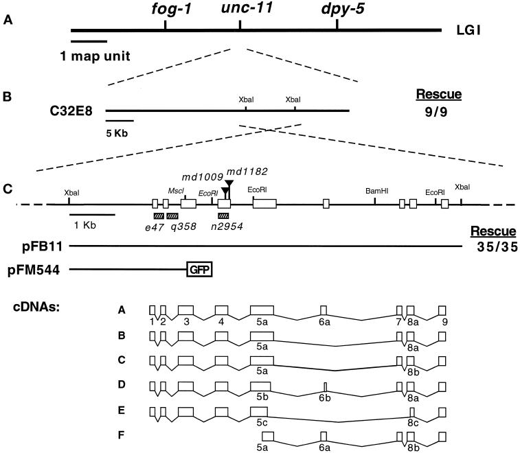
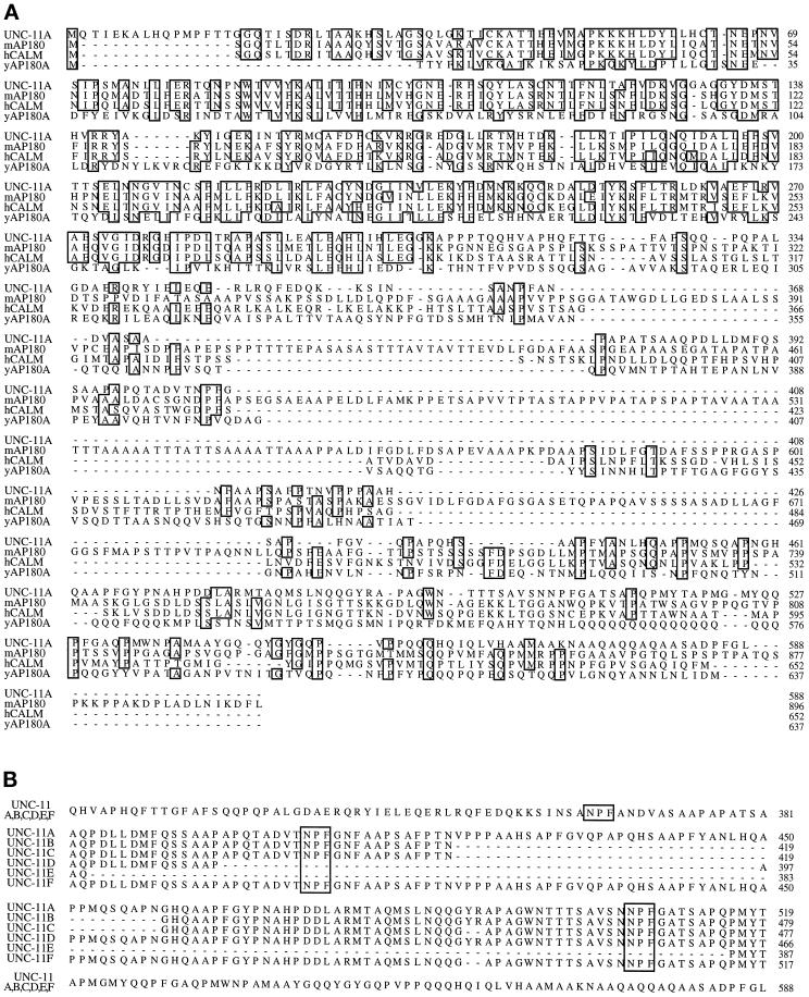
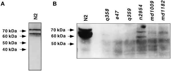
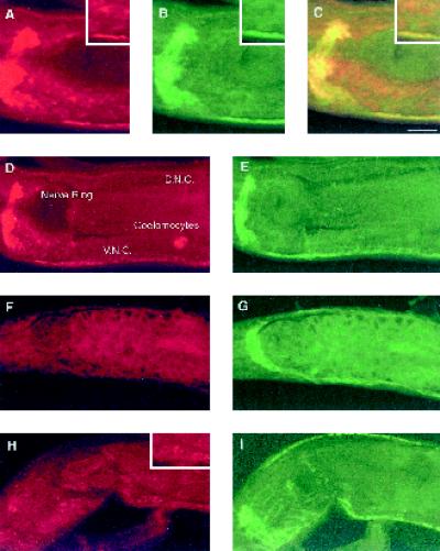
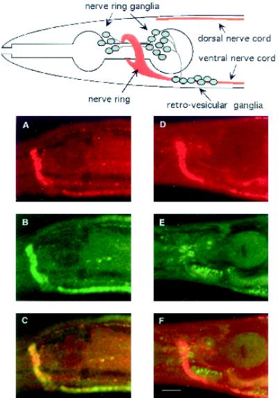
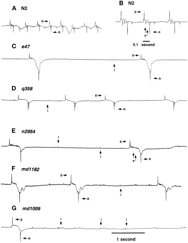
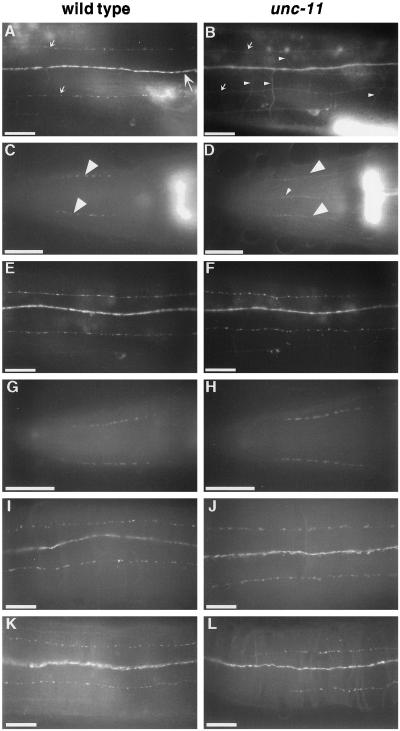
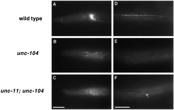
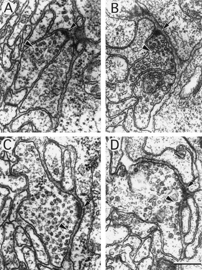
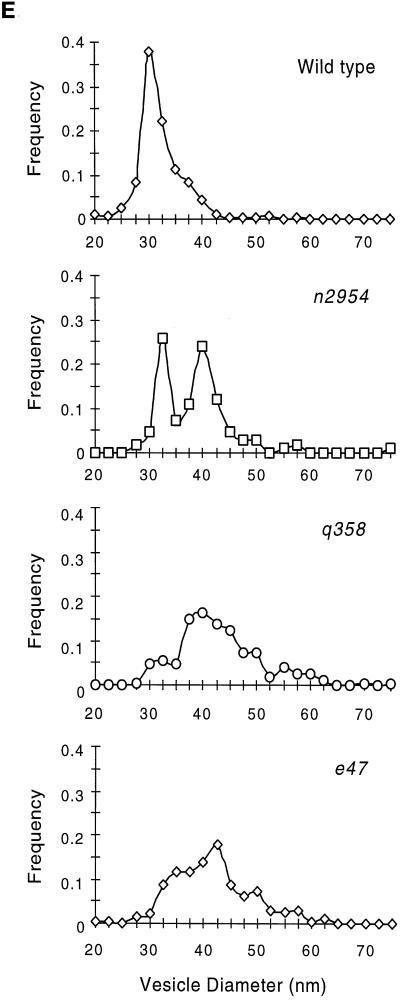
The unc-11 gene of Caenorhabditis elegans encodes multiple isoforms of a protein homologous to the mammalian brain-specific clathrin-adaptor protein AP180. The UNC-11 protein is expressed at high levels in the nervous system and at lower levels in other tissues. In neurons, UNC-11 is enriched at presynaptic terminals but is also present in cell bodies. unc-11 mutants are defective in two aspects of synaptic vesicle biogenesis. First, the SNARE protein synaptobrevin is mislocalized, no longer being exclusively localized to synaptic vesicles. The reduction of synaptobrevin at synaptic vesicles is the probable cause of the reduced neurotransmitter release observed in these mutants. Second, unc-11 mutants accumulate large vesicles at synapses. We propose that the UNC-11 protein mediates two functions during synaptic vesicle biogenesis: it recruits synaptobrevin to synaptic vesicle membranes and it regulates the size of the budded vesicle during clathrin coat assembly.


Figures

Figure 1
Figure 1
Molecular cloning of the unc-11 locus. (A) Genetic map of chromosome I of C. elegans. (B) Transgenic rescue of unc-11 mutants with DNA clones. Rescue data are provided as a fraction of stable lines that gave complete rescue of the unc-11 phenotype. (C) Organization of the unc-11 locus. A restriction map of the genomic unc-11 locus. Selected restriction sites are indicated on the linear map. Triangles delineate the positions of Tc1 transposons inserted in the unc-11 alleles listed in italics. Sequences deleted in unc-11 alleles are shown as hatched rectangles. Exons are represented as boxes. The structure of constructs used in this study are illustrated below the restriction map. A schematic representation of the six classes of unc-11 cDNAs is also shown. Exons are numbered 1–9. The a, b, or c suffixes represent alternative versions of these exons. GenBank accession numbers for the six cDNA forms are AF144257–AF144262.
Figure 2
Figure 2
Sequence comparison of UNC-11 and its vertebrate homologues. (A) An alignment of the UNC-11A isoform with mouse AP180 (Zhou et al., 1993), human CALM (Dreyling et al., 1996), and yeast AP180A protein sequences (Wendland and Emr, 1998). The alignment was obtained using the program ClustalW 1.5. Sequences identical in three or four of the proteins are boxed. (B) An alignment of the six UNC-11 isoforms illustrating the diversity in the C-terminal domains of products from this locus. NPF tripeptide motifs are boxed.
Figure 3
Figure 3
C. elegans expresses at least three UNC-11 isoforms. Western blots of protein extracts isolated from the wild type and unc-11 mutants. UNC-11 immunoreactivity was detected as specified in MATERIALS AND METHODS. The position of molecular weight markers is indicated on the left of the panels. (A) Total protein extracts (63 μg) from a mixed-staged culture of the wild type showing three UNC-11 isoforms. (B) Total protein extracts (150 μg) from mixed-stage cultures of the indicated strains. All UNC-11 iso-forms are severely reduced or absent in unc-11 mutants e47, q358, q359, md1009, and md1182. Two products of altered mobility are visible in n2954.
Figure 4
Figure 4
UNC-11 is enriched in synaptic regions of C. elegans. Confocal images of adult nematodes prepared for immunohistochemistry using both affinity-purified rabbit α-UNC-11 antisera and affinity-purified mouse α-synaptotagmin antisera. Animals were double stained for UNC-11 with rhodamine-conjugated secondary antibody and for synaptotagmin with FITC-conjugated secondary antibody: wild-type (A–E), unc-11(e47) (F and G), and unc-11 (n2954) (H and I). Anterior is left in all images. Panel C is the merge of panels A and B. A representative nerve ring, ventral nerve cord (V. N. C.), dorsal nerve cord (D. N. C.), and coelomocyte are labeled in panel D. Insets in panels A–C and H depict localization of UNC-11 staining in neuronal cell bodies. Bar, 10 μm.
Figure 5
Figure 5
Subcellular localization of UNC-11 in wild-type and unc-104 mutant nematodes. Confocal images of freeze-cracked nematodes double stained with affinity-purified rabbit α-UNC-11 antisera (first row); affinity purified mouse α-synaptotagmin antisera (second row), and merge (third row). (A–C) Wild-type. (D–F) unc-104(rh43). A diagram depicting the organization of nervous tissue in the C. elegans head including the position of neuronal cell bodies, the nerve ring neuropil, and the ventral and dorsal nerve cords is positioned above the confocal images. Synaptic-rich regions are depicted in red. Commissures, dendrites, minor process bundles, and many neuronal cell bodies of the ganglia were omitted for clarity. Bar, 10 μm.
Figure 6
Figure 6
Extracellular recording from wild-type and unc-11 animals. Representative traces of electropharygeogram recordings of the wild-type strain N2 (A and B), unc-11(e47) (C), unc-11(q358) (D), unc-11(n2954) (E), unc-11(md1182) (F), and unc-11(md1009) (G). Representative interpump transients (I), excitatory phase transients (E), relaxation phase transients (R), and plateau phase transients (P) are labeled. The wild-type and unc-11 mutants had the following mean pump lengths: wild-type (N2), 124 ± 10 ms; unc-11(e47), 310 ± 23 ms; unc-11(q358), 240 ± 11 ms; unc-11(n2954), 284 ± 11 ms; unc-11(md1182), 202 ± 5 ms; unc-11(md1009), 233 ± 7 ms; unc-11(e47), 310 ± 23 ms. All traces are millivolts versus time. All traces except for panel B share the same time scale indicated at the bottom of the trace in panel G.
Figure 7
Figure 7
Synaptobrevin, but not other synaptic vesicle proteins, is mislocalized in unc-11 mutants. The localization of vesicle proteins was examined in the wild type (first column) and in unc-11 animals (second column) using both GFP-tagged proteins and antibodies. (A and B) Images of the dorsal nerve cord of live immobilized adult animals expressing GFP-tagged synaptobrevin. The dorsal cord (large arrow), dorsal sublateral processes (small arrows), and commissural processes of motor neurons (small arrowheads) are labeled. The out-of-focus fluorescence in the lower right of each panel derives from the spermatheca that expresses synaptobrevin. (A) A wild-type animal. (B) unc-11(e47) animal. (C and D) Images of heads of adult worms fixed and stained with α-synaptobrevin antibodies and visualized with FITC-conjugated secondary antibodies. Synaptic varicosities of the SAB motor neuron processes are labeled with large arrowheads in a wild-type animal (C) and an unc-11(e47) animal (D). Note the appearance of synaptobrevin immunoreactivity in ventral processes (probably AVM and VA1) in panel D. (E and F) Images of the dorsal nerve cord of live immobilized adult animals expressing GFP-tagged synaptogyrin in a wild-type animal (E) and a unc-11(e47) animal (F). (G–J) Images of adult worms fixed andstained with α-RAB-3 antibodies and visualized with FITC-conjugated secondary antibodies in the head of a wild-type animal (G), and an unc-11(e47) animal (H); and a portion of the dorsal cord of a wild-type animal (I) and an unc-11(e47) animal (J). (K and L) Images of a portion of the dorsal cord of adult hermaphrodite worms fixed and stained with α-synaptotagmin antibodies and visualized with FITC-conjugated secondary antibodies in a wild-type animal (K) and an unc-11(n2954) animal (L). Anterior is left in all images. Bar, 10 μm.
Figure 8
Figure 8
UNC-11 functions in the soma of neurons. Images of L1 larvae expressing synaptobrevin-GFP. A lateral view of the head of a wild-type animal (A), an unc-104(e1265) animal (B), and an unc-11(e47); unc-104(e1265) animal (C). A dorsal view of the body of (D) a wild-type animal (D), an unc-104(e1265) animal (E), and and unc-11(e47); unc-104(e1265) animal (F). Note that GFP fluorescence is visible in the dorsal cord (arrow) of the double mutant. Anterior is to the left in all photographs. Bar, 10 μm.
Figure 9
Figure 9
Ultrastructure of neuromuscular junctions from wild-type and unc-11 mutants. Electron micrographs of the ventral nerve cord of young adult animals. The neuronal cell type was deduced from examination of serial sections. A thick electron-dense presynaptic specialization is visible at the apposition of nerve and muscle in each image (arrow), and a single synaptic vesicle is indicated (arrowhead) in each micrograph. (A) Cholinergic neuromuscular junction in a wild-type animal. (B) Cholinergic neuromuscular junction in an unc-11(q358) mutant. (C) GABAergic neuromuscular junctions in a wild-type animal. (D) GABAergic neuromuscular junction in an unc-11(e47) mutant. Note the increase in vesicles in contact with the membrane adjacent to the postsynaptic muscle. (E) Quantitation of vesicle diameter from wild-type and unc-11 neuromuscular junctions. Vesicle diameters were measured in sections containing active zones and binned into 2.5-nm intervals. The fraction of vesicles in each size class is plotted at the midpoint of each binning interval (wild-type, n = 305; unc-11(e47), n = 214; unc-11(q358), n = 197; unc-11(n2954), n = 109). Bar, 200 nm.
Figure 9
Figure 9
Ultrastructure of neuromuscular junctions from wild-type and unc-11 mutants. Electron micrographs of the ventral nerve cord of young adult animals. The neuronal cell type was deduced from examination of serial sections. A thick electron-dense presynaptic specialization is visible at the apposition of nerve and muscle in each image (arrow), and a single synaptic vesicle is indicated (arrowhead) in each micrograph. (A) Cholinergic neuromuscular junction in a wild-type animal. (B) Cholinergic neuromuscular junction in an unc-11(q358) mutant. (C) GABAergic neuromuscular junctions in a wild-type animal. (D) GABAergic neuromuscular junction in an unc-11(e47) mutant. Note the increase in vesicles in contact with the membrane adjacent to the postsynaptic muscle. (E) Quantitation of vesicle diameter from wild-type and unc-11 neuromuscular junctions. Vesicle diameters were measured in sections containing active zones and binned into 2.5-nm intervals. The fraction of vesicles in each size class is plotted at the midpoint of each binning interval (wild-type, n = 305; unc-11(e47), n = 214; unc-11(q358), n = 197; unc-11(n2954), n = 109). Bar, 200 nm.

Similar articles

Cited by

References

    1. Ahle S, Ungewickell E. Purification and properties of a new clathrin assembly protein. EMBO J. 1986;5:3143–3149. - PMC - PubMed
    1. Albertson DG. Formation of the first cleavage spindle in nematode embryos. Dev Biol. 1984;101:61–72. - PubMed
    1. Alfonso A, Grundahl K, McManus JR, Rand JB. Cloning and characterization of the choline acetyltransferase structural gene (cha-1) from C. elegans. J Neurosci. 1994;14:2290–2300. - PMC - PubMed
    1. Artalejo CR, Henley JR, McNiven MA, Palfrey HC. Rapid endocytosis coupled to exocytosis in adrenal chromaffin cells involves Ca2+, GTP and dynamin, but not clathrin. Proc Natl Acad Sci USA. 1995;92:8328–8332. - PMC - PubMed
    1. Avery L. Motor neuron M3 controls pharyngeal muscle relaxation timing in Caenorhabditis elegans. J Exp Biol. 1993a;175:283–297. - PubMed

Publication types

MeSH terms

LinkOut - more resources