. 1999 Jul;10(7):2441-59.
doi: 10.1091/mbc.10.7.2441.

Rho-dependent regulation of cell spreading by the tetraspan membrane protein Gas3/PMP22

Affiliations
Free PMC article

Rho-dependent regulation of cell spreading by the tetraspan membrane protein Gas3/PMP22

C Brancolini et al. Mol Biol Cell. 1999 Jul.
Free PMC article

Abstract

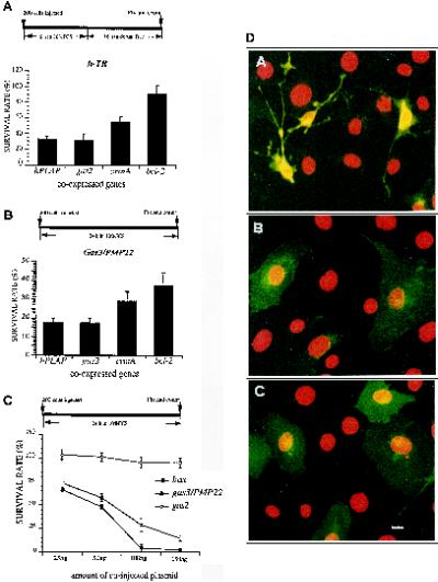
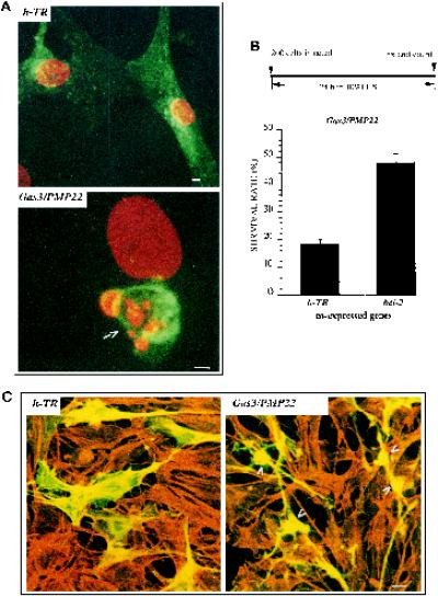
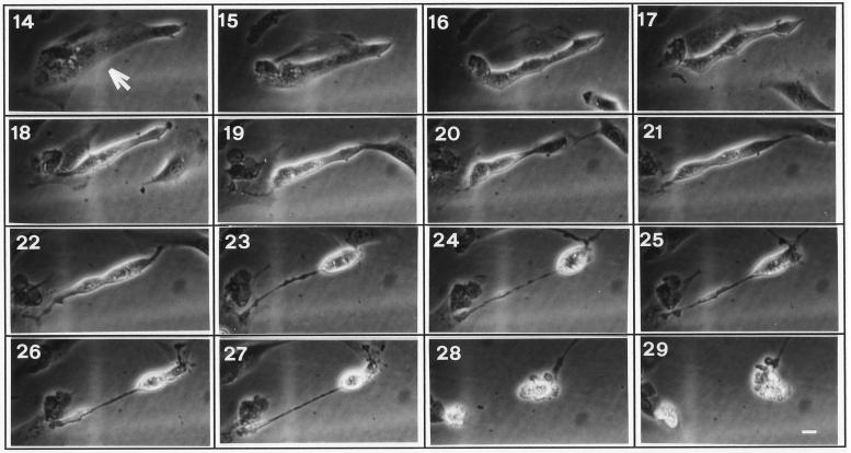
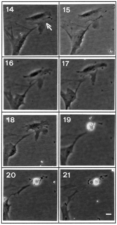
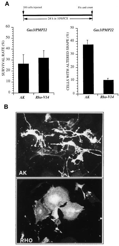
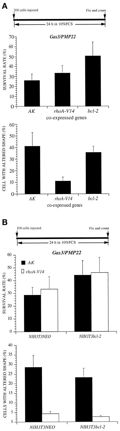
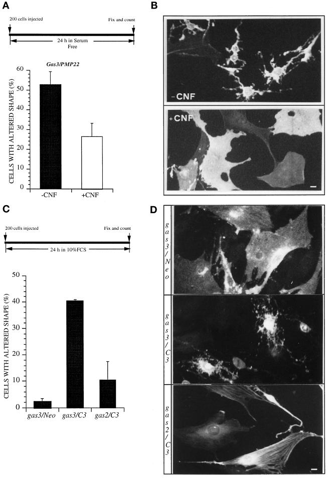
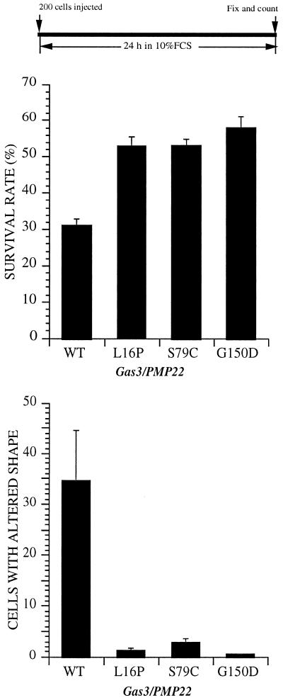
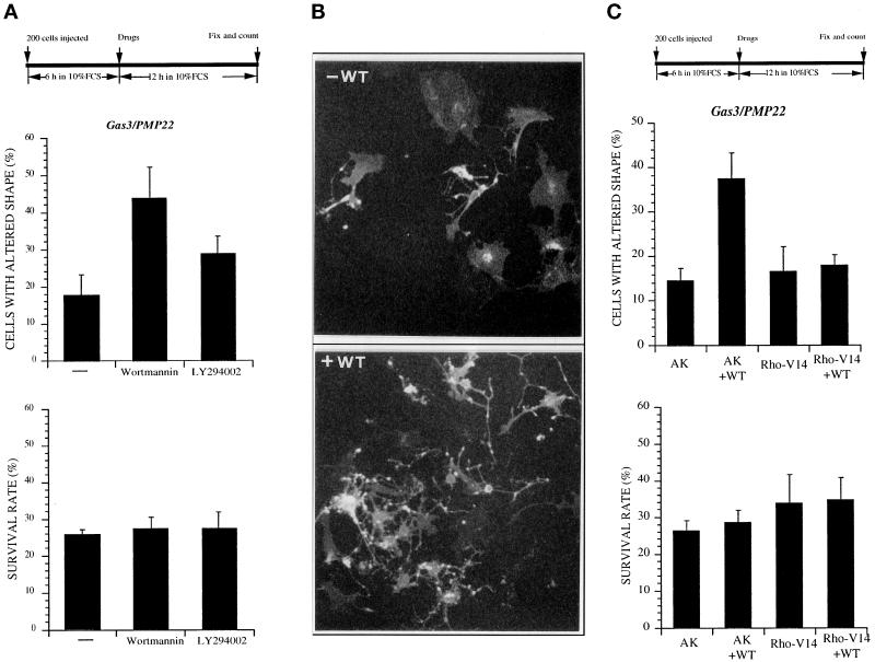
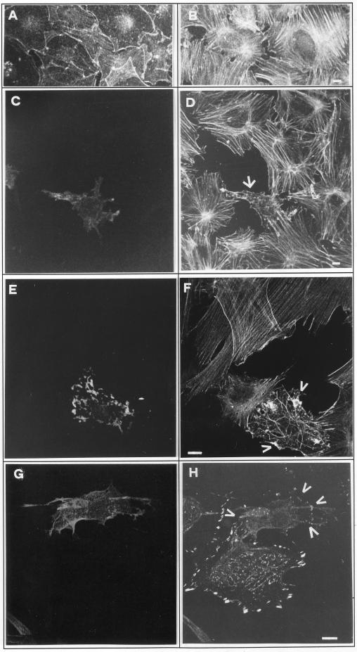
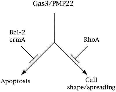
Gas3/PMP22 plays a crucial role in regulating myelin formation and maintenance, and different genetic alterations in gas3/PMP22 are responsible for a set of human peripheral neuropathies. We have previously demonstrated that Gas3/PMP22 could regulate susceptibility to apoptosis in NIH3T3 cells but not in REF 52 cells. In this report we demonstrate that when the apoptotic response triggered by gas3/PMP22 was counteracted by Bcl-2 coexpression, morphological changes were observed. Time-lapse analysis confirmed that Gas3/PMP22 can modulate cell spreading, and this effect was strengthened after inhibition of phosphoinositide 3-kinase. Using the active form of the small GTPase RhoA, we have been able to dissect the different Gas3/PMP22 biological activities. RhoA counteracted the Gas3/PMP22-dependent morphological response but was unable to neutralize the apoptotic response. Treatment of NIH3T3 cells with cytotoxic necrotizing factor 1, which activates endogenous Rho, also counteracted Gas3/PMP22-mediated cell shape and spreading changes. Treatment of REF 52 cells, which are unresponsive to Gas3/PMP22 overexpression, with the C3 exoenzyme, inhibiting Rho activity, renders REF 52 cells responsive to Gas3/PMP22 overexpression for cell shape and spreading changes. Finally, assembly of stress fibers and focal adhesions complexes, in response to lysophosphatidic acid-induced endogenous Rho activation, was impaired in Gas3/PMP22-overexpressing cells. We hypothesize that cell shape and spreading regulated by Gas3/PMP22 through the Rho GTPase might have an important role during Schwann cells differentiation and myelinization.


Figures

Figure 1
Figure 1
bcl-2 counteracts apoptosis induced by gas3/PMP22. (A) bcl-2, crmA, gas2, and hPLAP were coexpressed with h-TR in NIH3T3 cells. Six hours after microinjection apoptosis was induced by serum deprivation. Sixteen hours later cells were fixed and processed for immunofluorescence to detect h-TR. Survival was scored as described in the text. Data represent arithmetic means ± SD for five independent experiments (p < 0.001). (B) bcl-2, crmA, gas2, and hPLAP were coexpressed with gas3/PMP22 in NIH3T3 cells. After 24 h from microinjection cells were fixed and processed for immunofluorescence to detect Gas3/PMP22. Survival was scored as described in the text. Data represent arithmetic means ± SD for five independent experiments (p < 0.001). (C) gas3/PMP22 and bax killer activity. Different amounts of pGDSV7S containing gas3/PMP22, bax, or gas2 were comicroinjected with pGDSV7h-TR (25 ng/μl) in the nuclei of NIH3T3 cells. After 24 h from microinjection cells were fixed and processed for immunofluorescence to detect h-TR. Survival was scored as described in the text. Datarepresent arithmetic means ± SD for three independent experiments. (D) gas3/PMP22-dependent morphological changes: confocal generated overlay showing cellular and nuclear phenotypes in NIH3T3 cells coexpressing Gas3/PMP22 and Bcl-2 (A) or h-Tr and Bcl-2 (B) or P0 (C). NIH3T3 cells 24 h after seeding were microinjected with pGDSV3-hTR (100 ng/μl) and pGDSV7-bcl-2 (50 ng/μl), with pGDSV7-gas3/PMP22 (100 ng/μl) and pGDSV7-bcl-2 (50 ng/μl), or with pEXV-P0 (100 ng/μl). After 24 h cells were fixed and processed for immunofluorescence analysis to visualize Gas3/PMP22 (dA), h-TR (dB), or P0 (dC) (green) using the specific antibodies. Propidium iodide was used to visualize nuclei (red). Images were overlayered using a Zeiss confocal microscope and are displayed in pseudocolor. Bar, 5 μm.
Figure 2
Figure 2
gas3/PMP22-dependent biological activities in Schwann cells. (A) Confocal generated overlay showing nuclear morphology in rat Schwann cells overexpressing Gas3/PMP22 or h-TR. Schwann cells 24 h after seeding were microinjected with pGDSV7-gas3/PMP22 (100 ng/μl) or with pGDSV3-hTR (100 ng/μl); after 24 h cells were fixed and processed for immunofluorescence analysis to visualize Gas3/PMP22 or h-TR using the specific antibodies (green). Propidium iodide was used to visualize nuclei (red). Images were overlayered using a Zeiss confocal microscope and are displayed in pseudocolor. The arrow indicates a Gas3/PMP22-overexpressing cell. Bar, 5 μm. (B) bcl-2 and h-TR were coexpressed with gas3/PMP22 in Schwann cells. After 24 h from microinjection cells were fixed and processed for immunofluorescence to detect Gas3/PMP22. Survival was scored as described in the text. Data represent arithmetic means ± SD for five independent experiments (p < 0.001). (C) Confocal generated overlay showing actin architecture and cell morphology in Schwann cells cells coexpressing Gas3/PMP22 Bcl-2 and Gas2 or h-Tr Bcl-2 and Gas2. Schwann cells 24 h after seeding were microinjected with pGDSV3-hTR (50 ng/μl), pGDSV7-bcl-2 (50 ng/μl), and pGDSV7-gas2 (25 ng/μl) or with pGDSV7-gas3/PMP22 (50 ng/μl), pGDSV7-bcl-2 (50 ng/μl), and pGDSV7-gas2 (25 ng/μl). After 24 h cells were fixed and processed for immunofluorescence analysis to visualize Gas2 (green), using the specific antibody and actin filaments (red) and rhodamine-phalloidin. Images were overlayered using a Zeiss confocal microscope and are displayed in pseudocolor. Bar, 15 μm.
Figure 3
Figure 3
Time-lapse images of a NIH3T3 cell overexpressing gas3/PMP22. Representative cell (arrow) injected with pGDSV7-gas3/PMP22 (100 ng/μl). Pictures at selected times after microinjection (as indicated) show the morphological changes from 17 h and the appearance of membrane blebbing at 28 h. Bar, 20 μm.
Figure 4
Figure 4
Time-lapse images of an NIH3T3 cell overexpressing bax. Representative cell (arrow) injected with pGDSV7-bax (50 ng/μl). Pictures at selected times after microinjection (as indicated) show the appearance of membrane blebbing at 19 h. Bar 20 μm.
Figure 5
Figure 5
Coexpression of Gas3/PMP22 and RhoA-V14 in NIH3T3 cells. (A) AK and rhoA-V14 were coexpressed with gas3/PMP22 in NIH3T3 cells. After 24 h from microinjection cells were fixed and processed for immunofluorescence to detect Gas3/PMP22FLAG. Survival and morphological changes were scored as described in the text. Data represent arithmetic means ± SD for seven independent experiments (survival rate, p = 0.225; altered shape, p < 0.001). (B) Immunofluorescence analysis of NIH3T3 cells coexpressing gas3/PMP22 and AK or gas3/PMP22 and rhoA-V14 or gas3/PMP22. NIH3T3 cells 24 h after seeding were microinjected with pEXV-gas3/PMP22FLAG (100 ng/μl) and pEXV-AK (25 ng/μl) or with pEXV-gas3/PMP22FLAG (100 ng/μl) and pEXV-RhoA-V14 (25 ng/μl). After 24 h cells were fixed and processed for immunofluorescence to visualize Gas3/PMP22FLAG. Bar, 5 μm.
Figure 6
Figure 6
Coexpression of Gas3/PMP22, RhoA-V14 and Bcl-2 in NIH3T3 cells. (A) AK, rhoA-V14, and bcl-2 were coexpressed with gas3/PMP22 in NIH3T3 cells. After 24 h from microinjection cells were fixed and processed for immunofluorescence to detect Gas3/PMP22FLAG. Survival and morphological changes were scored as described in the text. Data represent arithmetic means ± SD for seven independent experiments (p < 0.001). (B) gas3/PMP22 and rhoA-V14 were coexpressed in NIH3T3NEO and in NIH3T3bcl-2 cells by nuclear microinjection. After 24 h from microinjection cells were fixed and processed for immunofluorescence to detect Gas3/PMP22FLAG. Survival and morphological changes were scored as described in the text. Data represent arithmetic means ± SD for three independent experiments.
Figure 7
Figure 7
Gas3/PMP22 effect on cell shape and spreading can be modulated by bacterial toxin regulating Rho activity. (A) gas3/PMP22 FLAG was overexpressed in NIH3T3 cells by nuclear microinjection. After microinjection cells were treated or not with CNF-1 in serum-free medium; 24 h later cells were fixed and processed for immunofluorescence to detect Gas3/PMP22FLAG. Morphological changes were scored as described in the text. Data represent arithmetic means ± SD for four independent experiments (p = 0.002). (B) Immunofluorescence
Figure 8
Figure 8
Overexpression of Gas3/PMP22 CMT1A point mutants in NIH3T3 cells. gas3/PMP22wt-FLAG, gas3/PMP22L16P-FLAG, gas3/PMP22S79C-FLAG, and gas3/PMP22G150D-FLAG were overexpressed in NIH3T3 cells by nuclear microinjection. After 24 h from microinjection cells were fixed and processed for immunofluorescence to detect Gas3/PMP22FLAG. Survival and morphological changes were scored as described in the text. Data represent arithmetic means ± SD for five independent experiments (p < 0.001).analysis of NIH3T3 cells expressing gas3/PMP22 in the presence of CNF-1. NIH3T3 cells 24 h after seeding were microinjected with pGDSV7-gas3/PMP22FLAG (100 ng/μl). After microinjection medium was changed to serum free, and cells were treated or not with CNF-1. Twenty-four hours later cells were fixed and processed for immunofluorescence to visualize Gas3/PMP22FLAG. Bar, 25 μm. (C) C3 exoenxyme was coexpressed with gas3/PMP22 or gas2 in REF 52 cells. After 24 h from microinjection cells were fixed and processed for immunofluorescence to detect Gas3/PMP22FLAG or Gas2. Morphological changes were scored as described in the text. Data represent arithmetic means ± SD for four independent experiments (p < 0.001). (D) Immunofluorescence analysis of REF 52 cells expressing gas3/PMP22. REF 52 cells 24 h after seeding were microinjected with pGDSV7-gas3/PMP22FLAG (100 ng/μl) and pcDNA3NEO (25 ng/μl), with pGDSV7-gas3/PMP22FLAG (100 ng/μl) and pcDNA3C3 (25 ng/μl), or with pGDSV7-gas2 (100 ng/μl) and pcDNA3C3 (25 ng/μl). Twenty-four hours later cells were fixed and processed for immunofluorescence to visualize Gas3/PMP22FLAG or Gas2. Bar, 25 μm.
Figure 9
Figure 9
Wortmannin (WT) can agument changes of cell shape and spreading induced by Gas3/PMP22. (A) gas3/PMP22 FLAG was overexpressed in NIH3T3 cells by nuclear microinjection. After 6 h cells were treated or not with WT and LY294002; 12 h later cells were fixed and processed for immunofluorescence to detect Gas3/PMP22FLAG. Survival and morphological changes were scored as described in the text. Data represent arithmetic means ± SD for five independent experiments (altered shape, p < 0.001). (B) Immunofluorescence analysis of NIH3T3 cells expressing gas3/PMP22 in the presence of WT. NIH3T3 cells 24 h after seeding were microinjected with pEXV-gas3/PMP22FLAG (50 ng/μl). After 6 h cells were treated or not with WT; 12 h later cells were fixed and processed for immunofluorescence to visualize Gas3/PMP22FLAG. Bar, 25 μm. (C) AK and rhoA-V14 were coexpressed with gas3/PMP22 in NIH3T3 cells. After 6 h cells were treated or not with WT; 12 h later cells were fixed and processed for immunofluorescence to detect Gas3/PMP22FLAG. Survival and morphological changes were scored as described in the text. Data represent arithmetic means ± SD for five independent experiments (altered shape, p < 0.001).
Figure 10
Figure 10
Gas3/PMP22 overexpression and stress fiber formation. Swiss 3T3 grown for 48 h in 0.1% FCS (A) and then treated for 30 min with LPA (B) were stained to visualize microfilaments. Serum-starved Swiss 3T3 cells were microinjected with pEXVhGas3/PMP22 (100 ng/μl). After 15 h from microinjection, LPA was added, and 30 min later cells were fixed and processed for immunofluorescence analysis to visualize actin filaments (D and F), Gas3/PMP22 (C, E, and G) and talin (H). Arrows indicate microinjected cells. Bar, 10 μm.
Figure 11
Figure 11
Schematic representation of Gas3/PMP22 biological activities. Gas3/PMP22 could regulate both susceptibility to apoptosis and morphological changes. Bcl-2 counteracts the apoptotic response, whereas Rho specifically counteracts morphological changes.

Similar articles

Cited by

References

    1. Adlkofer K, Martini R, Aguzzi A, Zielasek J, Toyka KV, Suter U. Hypermyelination and demyelinating peripheral neuropathy in PMP22-deficient mice. Nat Genet. 1995;11:274–280. - PubMed
    1. Amano M, Chiahara K, Kimura K, Fukuta Y, Nakamura N, Matsuura Y, Kaibuchi K. Formation of actin stress fibers and focal adhesions is enhanced by Rho kinase. Science. 1997;275:1308–1311. - PubMed
    1. Amano M, Ito M, Kimura K, Fukata Y, Chihara K, Nakano T, Matsuura Y, Kaibuchi K. Phosphorylation and activation of myosin by Rho-associated kinase (Rho-kinase) J Biol Chem. 1996;271:20246–20249. - PubMed
    1. Archelos JJ, Roggenbuck K, Schneider-Schaulies J, Linington C, Toyka KV, Hartung HP. Production and characterization of monoclonal antibodies to the extracellular domain of P0. J Neurosci Res. 1993;35:46–53. - PubMed
    1. Baechner D, Liehr T, Hameister H, Altenberger H, Grehl H, Suter U, Rautenstrauss B. Widespread expression of the peripheral myelin protein-22 gene (pmp22) in neural and nonneural tissue during murine development. J Neurosci Res. 1995;42:733–741. - PubMed

Publication types

MeSH terms

Grants and funding

LinkOut - more resources