Review
. 1999 Jul;12(3):405-28.
doi: 10.1128/CMR.12.3.405.

Molecular basis of the interaction of Salmonella with the intestinal mucosa

Affiliations
Review

Molecular basis of the interaction of Salmonella with the intestinal mucosa

K H Darwin et al. Clin Microbiol Rev. 1999 Jul.

Abstract

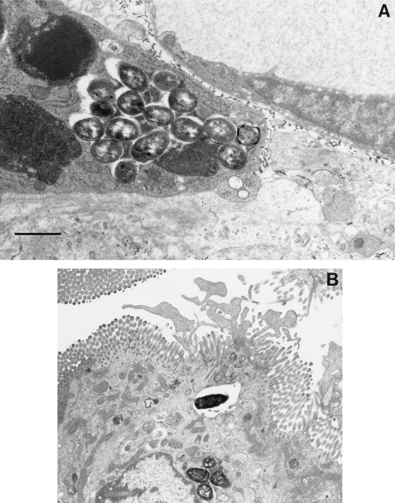
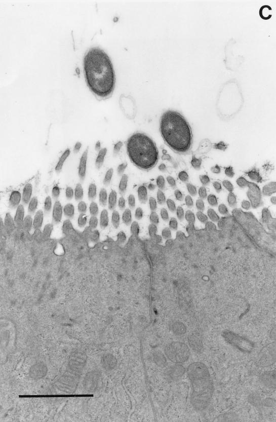
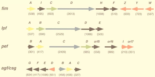
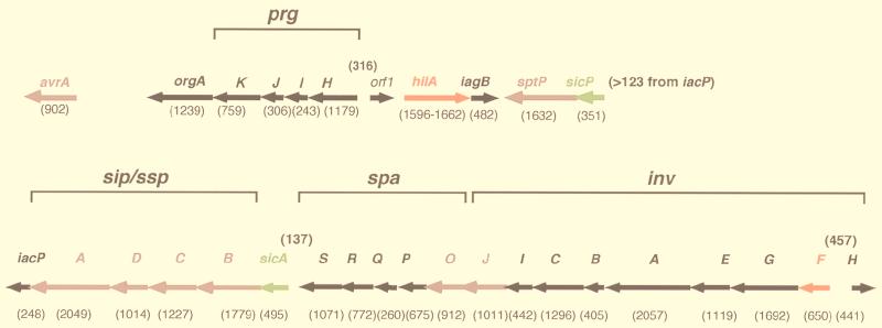
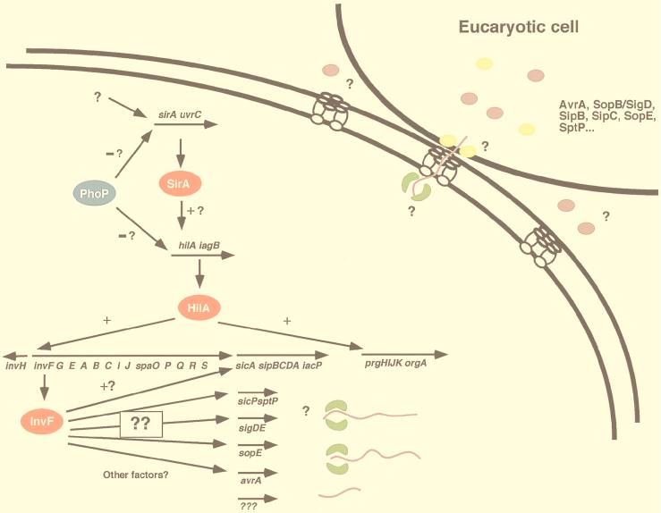
Salmonella is one of the most extensively characterized bacterial pathogens and is a leading cause of bacterial gastroenteritis. Despite this, we are only just beginning to understand at a molecular level how Salmonella interacts with its mammalian hosts to cause disease. Studies during the past decade on the genetic basis of virulence of Salmonella have significantly advanced our understanding of the molecular basis of the host-pathogen interaction, yet many questions remain. In this review, we focus on the interaction of enterocolitis-causing salmonellae with the intestinal mucosa, since this is the initiating step for most infections caused by Salmonella. Animal and in vitro cell culture models for the interaction of these bacteria with the intestinal epithelium are reviewed, along with the bacterial genes that are thought to affect this interaction. Lastly, recent studies on the response of epithelial cells to Salmonella infection and how this might promote diarrhea are discussed.


Figures

FIG. 1
FIG. 1
Interaction of Salmonella with intestinal mucosa. Transmission electron micrographs of non-follicle-associated epithelia from calves infected with either wild-type S. typhimurium ST4/74 (A and B) or with an isogenic invH mutant (C) are shown. (A) Macrophage from the laminae propriae of the absorptive villi with several internalized bacteria 3 h postinfection. (B) Enterocyte with an internalized bacterium. Note the membrane ruffling. (C) Enterocyte with attached bacteria (invH mutant). No ruffling was observed. Bars, 1 μm. Reprinted from reference with permission of the publisher.
FIG. 1
FIG. 1
Interaction of Salmonella with intestinal mucosa. Transmission electron micrographs of non-follicle-associated epithelia from calves infected with either wild-type S. typhimurium ST4/74 (A and B) or with an isogenic invH mutant (C) are shown. (A) Macrophage from the laminae propriae of the absorptive villi with several internalized bacteria 3 h postinfection. (B) Enterocyte with an internalized bacterium. Note the membrane ruffling. (C) Enterocyte with attached bacteria (invH mutant). No ruffling was observed. Bars, 1 μm. Reprinted from reference with permission of the publisher.
FIG. 2
FIG. 2
Organization of genes within the four fimbrial operons of S. typhimurium. Gene lengths (in base pairs) are indicated below each open reading frame. Genes encoding the major fimbrial subunits are indicated in yellow, genes encoding chaperones are indicated in green, genes encoding ushers are indicated in blue, genes encoding minor subunits are indicated in black, and genes encoding regulatory proteins are indicated in red.
FIG. 3
FIG. 3
Organization of genes in SPI1 at centisome 63. Gene lengths (in base pairs) are indicated below each open reading frame. Gaps longer than 30 bp between genes are indicated above intergenic spaces. Genes encoding regulatory proteins are indicated in red, genes encoding secreted proteins are indicated in mauve, genes encoding chaperones are indicated in green, and genes encoding apparatus proteins or proteins of uncharacterized function are indicated in black.
FIG. 4
FIG. 4
Model of expression of virulence genes and localization of their gene products. Putative activators are indicated in red, chaperones are indicated in green, and secreted proteins are indicated in mauve (effectors) and yellow (translocators).

References

    1. Reference deleted.
    1. Reference deleted.
    1. Abraham J M, Freitag C S, Clements J R, Eisenstein B I. An invertible element of DNA controls phase variation of type 1 fimbriae in Escherichia coli. Proc Natl Acad Sci USA. 1985;82:5724–5727. - PMC - PubMed
    1. Ager E A, Nelson K E, Galton M M, Boring III J, Jernigan J. Two outbreaks of egg-borne salmonellosis and implications for their prevention. JAMA. 1967;199:372–378. - PubMed
    1. Alfano J R, Collmer A. The type III (Hrp) secretion pathway of plant pathogenic bacteria: trafficking harpins, Avr protein, and death. J Bacteriol. 1997;179:5655–5662. - PMC - PubMed

MeSH terms

Substances

LinkOut - more resources