Review
. 1999 Jul;12(3):429-44.
doi: 10.1128/CMR.12.3.429.

Cellular biology of prion diseases

Affiliations
Review

Cellular biology of prion diseases

D A Harris. Clin Microbiol Rev. 1999 Jul.

Abstract

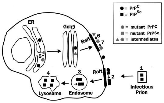
Prion diseases are fatal neurodegenerative disorders of humans and animals that are important because of their impact on public health and because they exemplify a novel mechanism of infectivity and biological information transfer. These diseases are caused by conformational conversion of a normal host glycoprotein (PrPC) into an infectious isoform (PrPSc) that is devoid of nucleic acid. This review focuses on the current understanding of prion diseases at the cell biological level. The characteristics of the diseases are introduced, and a brief history and description of the prion hypothesis are given. Information is then presented about the structure, expression, biosynthesis, and possible function of PrPC, as well as its posttranslational processing, cellular localization, and trafficking. The latest findings concerning PrPSc are then discussed, including cell culture systems used to generate this pathogenic isoform, the subcellular distribution of the protein, its membrane attachment, proteolytic processing, and its kinetics and sites of synthesis. Information is also provided on molecular models of the PrPC-->PrPSc conversion reaction and the possible role of cellular chaperones. The review concludes with suggestions of several important avenues for future investigation.


Figures

FIG. 1
FIG. 1
Structure and posttranslational processing of PrP. (Top) Structure of the primary translation product of mammalian PrP. The five proline- and glycine-rich repeats in mouse PrP have the sequence P(Q/H)GG(T/G/S)WGQ. (Bottom) Structure of the mature protein. The GPI anchor attaches the polypeptide chain to the membrane (see Fig. 7 for a schematic of the core anchor structure). Arrows A and B indicate the positions of cleavage sites in PrPC, and arrow C indicates a cleavage site in PrPSc. Site A lies within the GPI anchor, between the diacylglycerol moiety and the ethanolamine residue that is attached to the C-terminal amino acid. Site B lies near position 110, and site C lies near position 89.
FIG. 2
FIG. 2
Steps in the biosynthesis of PrPC. CHO, oligosaccharide; S-S, disulfide bond; Sig. pep., signal peptide.
FIG. 3
FIG. 3
Cellular trafficking and cleavage of PrP. After reaching the cell surface, PrPC is internalized into an endocytic compartment from which most of the molecules are recycled intact to the cell surface. A small percentage of the endocytosed molecules are proteolytically cleaved (site B in Fig. 1), and the N- and C-terminal cleavage products are then externalized. Some of the membrane-anchored protein is released into the extracellular medium by cleavage within the GPI anchor (site A in Fig. 1). Reprinted from reference with permission of the publisher.
FIG. 4
FIG. 4
Hypothetical model for a PrPC receptor. The cytoplasmic domain of the receptor contains signals for interacting with adapter molecules and clathrin, which are components of coated pits. The extracellular domain of the receptor binds to the N-terminal part of the PrPC molecule (solid rectangle), a region that is essential for efficient endocytosis. Reprinted from reference with permission of the publisher.
FIG. 5
FIG. 5
Cell culture systems for generation of PrPSc. (A) To model the infectious manifestation of prion diseases, several kinds of cultured cells have been infected with prions purified from rodent brain. (B) To model familial prion diseases, cultured cells have been transfected to express PrP molecules carrying disease-specific mutations. N2a and M17 are, respectively, mouse and human neuroblastoma cells; HaB are spontaneously immortalized hamster brain cells; PC12 are rat pheochromocytoma cells; GT1 are T-antigen-immortalized hypothalamic neurons; CHO are Chinese hamster ovary cells; and 3T3 are transformed mouse fibroblasts.
FIG. 6
FIG. 6
MoPrPs carrying disease-related mutations are detergent-insoluble and protease-resistant when expressed in cultured CHO cells. (A) CHO cells expressing wild-type (WT) and mutant moPrPs were labeled with [35S]methionine for 20 min and then chased for 3 h. Detergent lysates of the cells were centrifuged first at 16,000 × g for 5 min and then at 265,000 × g for 40 min. moPrP in the supernatants and pellets from the second centrifugation was immunoprecipitated and analyzed by SDS-polyacrylamide gel electrophoresis. PrP-specific bands were quantitated with a PhosphorImager, and the percentage of PrP in the pellet was calculated. Each bar represents the mean ± standard deviation of values from three experiments. Values that are significantly different from wild-type moPrP by Student’s t test (P < 0.001) are indicated by an asterisk. MoPrPs carrying disease-related mutations sediment (i.e., are detergent insoluble), while WT and M128V moPrPs remain largely in the supernatant. Human homologues of the mutant moPrPs analyzed here are associated with the following phenotypes: PG14 (9-octapeptide insertion), CJD variant; P101L, GSS; M128V, normal; D177N/Met128, FFI; D177N/Val128, CJD; F197S/Val128, GSS; E199K, CJD. (B) CHO cells expressing each moPrP were labeled for 3 h with [35S]methionine and chased for 4 h. Proteins in cell lysates were either digested at 37°C for 10 min with 3.3 μg of proteinase K per ml (+ lanes) or were untreated (− lanes) prior to recovery of moPrP by immunoprecipitation. Five times as many cell equivalents were loaded in the + lanes as in the − lanes. Molecular mass markers are in kilodaltons. moPrPs carrying pathogenic mutations yield a protease-resistant fragment of 27 to 30 kDa, while WT and M128V moPrPs are completely degraded. In separate experiments, we have shown that the PrP 27- to 30-kDa fragments are N-terminally truncated after the octapeptide repeats, the same region within which PrPSc from infected brain is cleaved (96). Modified from reference with permission of the publisher.
FIG. 7
FIG. 7
moPrPs carrying disease-related mutations are not released from the cell surface by PI-PLC. (A) CHO cells expressing wild-type (WT) and mutant PrPs were biotinylated with the membrane-impermeant reagent sulfo-biotin-X-NHS at 4°C and were then incubated with PI-PLC at 4°C prior to lysis. moPrP in the PI-PLC incubation media and cell lysates was immunoprecipitated, separated by SDS-polyacrylamide gel electrophoresis, and visualized by developing blots of the gel with horseradish peroxidase-streptavidin and enhanced chemiluminescence. PrP bands from three separate experiments were quantitated by densitometry, and the amount of PrP released by PI-PLC was plotted as a percentage of the total amount of PrP (medium plus cell lysate). Each bar represents the mean ± standard deviation. Values that are significantly different from wild-type moPrP by Student’s t test are indicated by single (P < 0.01) and double (P < 0.001) asterisks. All of the moPrPs carrying pathogenic mutations are less PI-PLC releasable than are wild-type and M128V moPrPs. E199K moPrP is more releasable than the other mutants, consistent with our observation that there are subtle biochemical differences among the mutant proteins. Phenotypes associated with the homologous human PrPs are given in the legend to Fig. 6. (B) Schematic of the membrane attachment of wild-type PrPC, which is can be completely released from cells by treatment with PI-PLC. The core structure of the GPI anchor, along with the site cleaved by PI-PLC, is indicated. (C to E) Schematics showing how PI-PLC is proposed to interact with mutant PrP. (C) On intact cells, the mutant protein adopts a PrPSc-like conformation that physically blocks access of PI-PLC to the GPI anchor. It is also possible that aggregation of mutant PrP molecules contributes to the inaccessibility of the anchor. (D) After extraction from the membrane by using nondenaturing detergents like Triton X-100 and deoxycholate (DOC), the abnormal conformation of the mutant protein is maintained and the anchor is still inaccessible to PI-PLC. (E) After denaturation in SDS, the conformation of the mutant protein is disrupted, and the anchor becomes susceptible to PI-PLC cleavage. Panel A is modified from reference with permission of the publisher.
FIG. 8
FIG. 8
Scheme for transformation of mutant PrPs to a PrPSc state. Mutant PrPs are initially synthesized in the PrPC state and acquire PrPSc properties in a stepwise fashion as they traverse different cellular compartments. PI-PLC resistance, which develops in the ER, reflects folding of the polypeptide chain into the PrPSc conformation. Detergent insolubility and protease resistance, which develop upon arrival at the plasma membrane or along an endocytic pathway, result from intermolecular aggregation (“maturation”). The times given underneath the boxes indicate when after pulse-labeling the corresponding property is detected. Addition of brefeldin A (BFA) to cells or incubation at 18°C, treatments which block the movement of proteins beyond the Golgi apparatus, inhibit the acquisition of detergent insolubility and protease resistance but not PI-PLC resistance. Adapted from reference with permission of the publisher.
FIG. 9
FIG. 9
Model of the cellular pathways involved in generation of PrPSc. In the infectious manifestation of prion diseases, extracellular PrPSc in the form of a prion particle (labeled 1) interacts with PrPC on the cell surface, possibly in detergent-resistant rafts, catalyzing its conversion to PrPSc (step 2). Conversion may also occur after uptake of the proteins into an endosomal compartment (step 3). Once formed, some PrPSc accumulates in lysosomes (step 4), although the protein is probably found in a number of other cellular locations as well. In familial prion disorders, mutant PrPC is converted spontaneously to the PrPSc state via a series of biochemical intermediates, the earliest of which is a PI-PLC-resistant form generated in the ER (step 5). Mutant PrP molecules are subsequently delivered to the cell surface, where they become detergent insoluble (step 6) and then protease resistant (step 7), possibly in raft domains. Steps 6 and 7 could also occur in endocytic organelles.

References

    1. Alper T, Cramp W A, Haig D A, Clarke M C. Does the agent of scrapie replicate without nucleic acid? Nature. 1967;214:764–766. - PubMed
    1. Anderson R G W. Plasmalemmal caveolae and GPI-anchored membrane proteins. Curr Opin Cell Biol. 1993;5:647–652. - PubMed
    1. Anderson R M, Donnelly C A, Ferguson N M, Woolhouse M E J, Watt C J, Udy H J, MaWhinney S, Dunstan S P, Southwood M E J, Wilesmith J W, Ryan J B M, Hoinville L J, Hillerton J E, Austin A R, Wells G A H. Transmission dynamics and epidemiology of BSE in British cattle. Nature. 1996;382:779–788. - PubMed
    1. Arnold J E, Tipler C, Laszlo L, Hope J, Landon M, Mayer R J. The abnormal isoform of the prion protein accumulates in late-endosome-like organelles in scrapie-infected mouse brain. J Pathol. 1995;176:403–411. - PubMed
    1. Bamborough P, Wille H, Telling G C, Yehiely F, Prusiner S B, Cohen F E. Prion protein structure and scrapie replication: theoretical, spectroscopic, and genetic investigations. Cold Spring Harbor Symp Quant Biol. 1996;61:495–509. - PubMed

Publication types

LinkOut - more resources