

doi: 10.1101/gad.13.13.1647.
NeuroD is required for differentiation of the granule cells in the cerebellum and hippocampus
Affiliations
- PMID: 10398678
- PMCID: PMC316850
- DOI: 10.1101/gad.13.13.1647
Item in Clipboard
NeuroD is required for differentiation of the granule cells in the cerebellum and hippocampus
Genes Dev.
.
Abstract
NeuroD, a bHLH transcription factor, is implicated in differentiation of neurons and pancreatic beta cells. NeuroD-null mice die shortly after birth due to severe neonatal diabetes. To examine if there is postnatal neuronal phenotype in these mice, we rescued them from neonatal lethality by introducing a transgene encoding the mouse neuroD gene under the insulin promoter. These mice survive to adulthood but display severe neurological phenotype due to neuronal deficit in the granule layers of the cerebellum and hippocampus. We show here that NeuroD is required for these postnatally generated microneurons to undergo proper differentiation, the absence of which results in cell death.
Figures
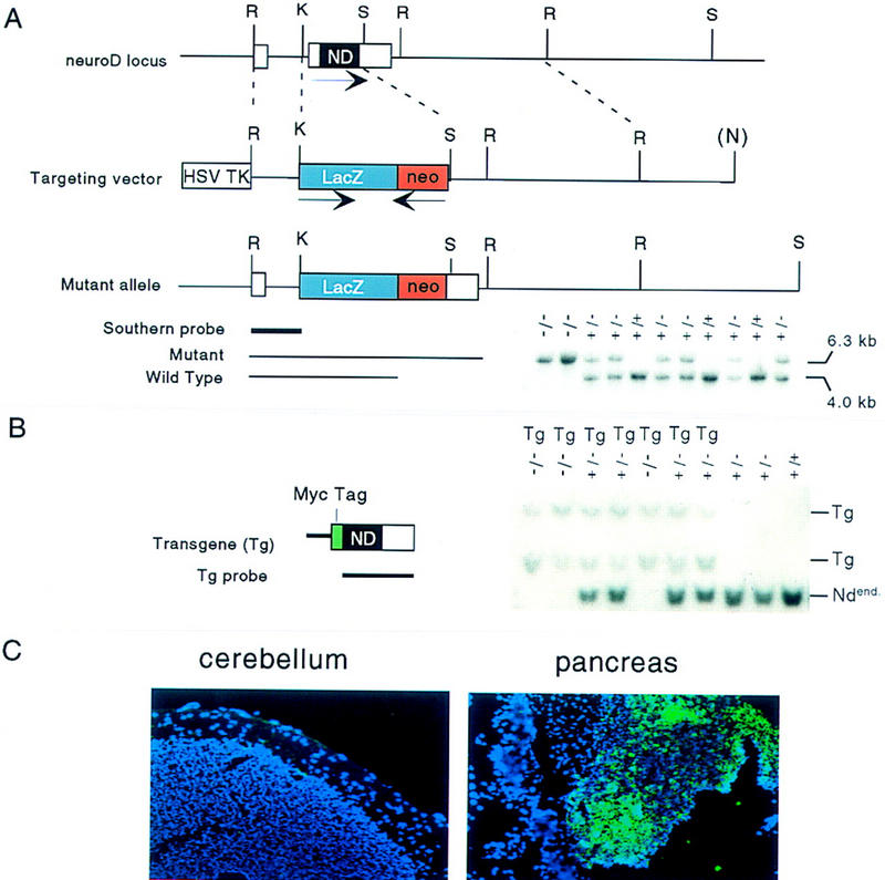
Gene targeting at the neuroD/beta-2 locus and introduction of transgene. (A). Schematic representation of the targeting vector, the wild-type neuroD locus, and the disrupted allele generated by homologous recombination. The orientations of the neuroD (noncoding regions in white; entire coding region in black), lacZ (in blue), and neoR gene (red) are indicated by arrows above below the genes. Insertion of the lacZ–neoR cassette deleted the second exon including the entire coding region. (R) EcoRI; (K) KpnI; (S) SpeI; (N) from the vector NotI. A sample of mouse genotyping by Southern blot analysis is shown below. Total mouse genomic DNA was digested with EcoRI; the blot was then hybridized with the Southern probe indicated; the presence of the mutant allele is confirmed by a 6.3-kb diagnotic band and the wild type by a 4.0-kb band. (B) Schematic representation of the transgene containing ∼700-bp RIP-1 and the 1.8-kb mouse neuroD coding region with its amino terminus fused to the Myc tag (green). A Southern blot analysis using the neuroD coding region as a probe is shown. Both the endogenous neuroD (Ndend) and Tg are detected. (C) Expression of transgene in ND−/− Tg mice. Transgene expression, as detected by immunostaining using anti-Myc tag antibody (fluorescent color), is detected in the pancreatic islets but not in the brains of P30 mice.
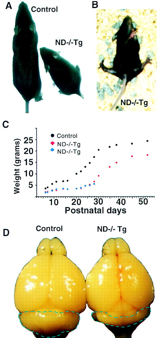
ND−/−Tg mice show retarded growth, an ataxic gait, and smaller cerebellar size. (A) Body size difference between ND+/−Tg and ND−/−Tg mice; (B) ND−/−Tg mice lose balance and fall over frequently; (C) a typical weight curve of littermates that include ND−/−Tg; (D) brains from ND+/−Tg and ND−/−Tg mice at P30.
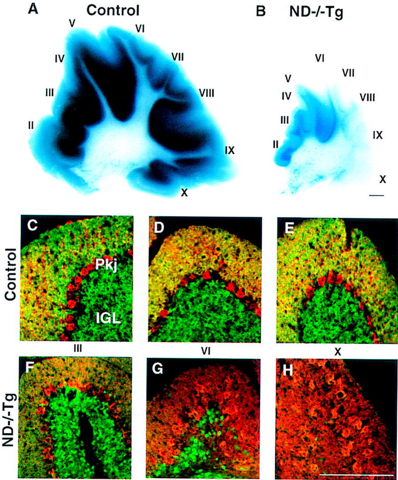
Cerebellar abnormalities in P30 ND−/−Tg mice. (A,B) X-gal-stained parasagittal sections showing a massive reduction in granule cells in ND−/−Tg mice. (C–H) β-Gal (green) and calbindin (red) double immunostaining showing the A–P difference in the degree of granule cell loss and the arrangement of Purkinje cells, respectively. (I–X) Individual cerebellar lobules. Bars, 100 μm.
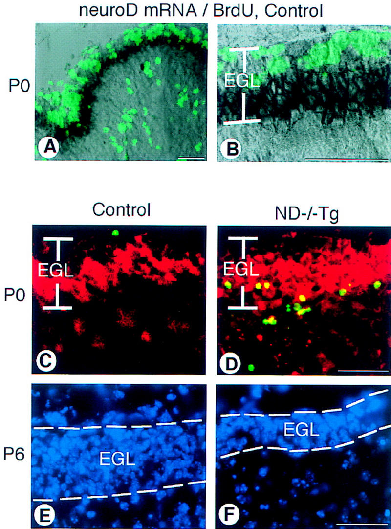
Cerebellar abnormalities in the ND−/−Tg mice. (A) neuroD in situ hybridization with BrdU labeling on control mice at P0. neuroD mRNA (black) is expressed in the inner zone of the EGL; the outer zone is cells incorporating BrdU (green). (B) A higher magnification of the EGL shown in A. (C,D) Apoptotic cells in the P0 cerebellar cortex. In the posterior lobules of ND−/−Tg mice, many TUNEL+ cells (green) are found in the inner EGL where immunostaining with antibody to β-galactosidase (red) is also found. (E,F) DAPI staining shows the reduced EGL thickness in the posterior lobules of ND−/−Tg cerebellum at P6. Bars, 20 μm.
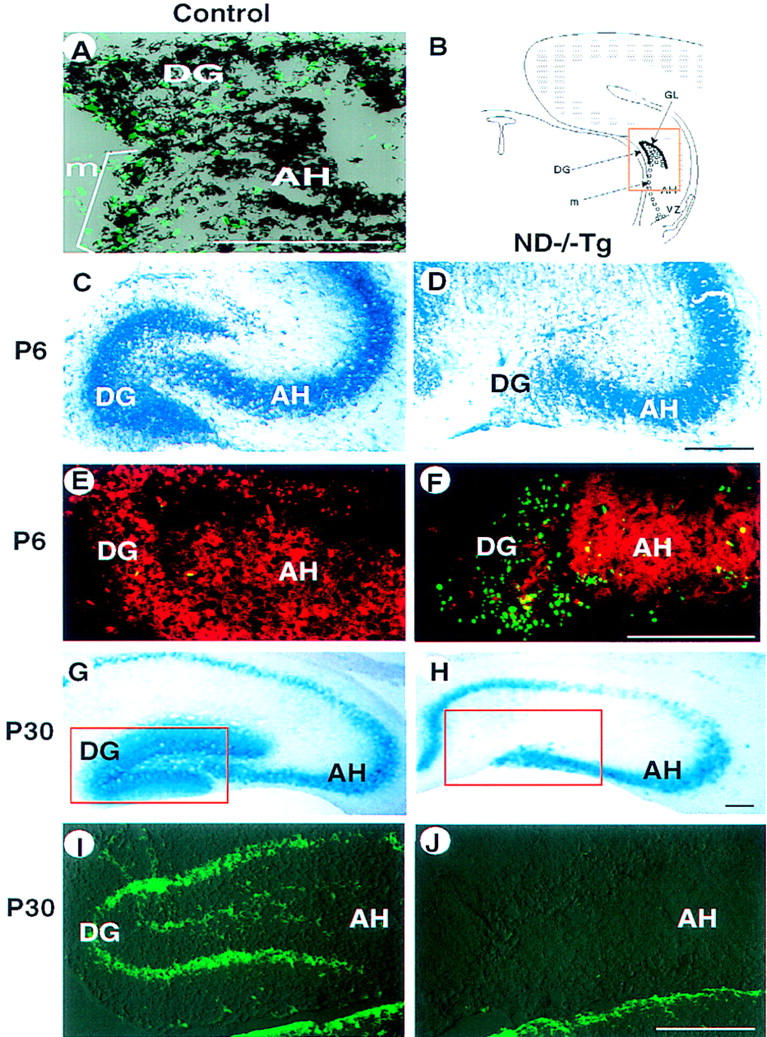
Hippocampal abnormalities in ND−/−Tg mice. (A) In situ hybridization at P0 showing that neuroD mRNA is expressed (black) in DG-forming granule cells and progenitor cells that are labeled with BrdU (green). (B) Schematic presentation of development of the DG in normal mice. The DG is formed by the granule neurons (●) generated by progenitor cells (○) after migration (m) from the VZ along the AH. (C–J) Degeneration of DG in ND−/−Tg mice as shown by staining with toluidine blue (C,D), anti-β-gal immunostaining (red in E,F), X-gal staining (G,H), or anti-calretinin immunostaining (I and J, corresponding to the squared areas in G and H, respectively; staining in the thalamic area below is also shown) at P6 (C–F) or P30 (G–J). TUNEL staining at P6 (E,F) shows many apoptotic cells (green) in the poorly formed DG in ND−/−Tg mice. (B–F) The squared region in B. Bars, 100 μm.
Similar articles
-
Atypical mouse cerebellar development is caused by ectopic expression of the forkhead box transcription factor HNF-3beta.Gene Expr. 2001;9(4-5):217-36. doi: 10.3727/000000001783992597. Gene Expr. 2001. PMID: 11444531 Free PMC article.
-
Neuronal basic helix-loop-helix proteins (NEX and BETA2/Neuro D) regulate terminal granule cell differentiation in the hippocampus.J Neurosci. 2000 May 15;20(10):3714-24. doi: 10.1523/JNEUROSCI.20-10-03714.2000. J Neurosci. 2000. PMID: 10804213 Free PMC article.
-
Neuronal basic helix-loop-helix proteins (NEX, neuroD, NDRF): spatiotemporal expression and targeted disruption of the NEX gene in transgenic mice.J Neurosci. 1998 Feb 15;18(4):1408-18. doi: 10.1523/JNEUROSCI.18-04-01408.1998. J Neurosci. 1998. PMID: 9454850 Free PMC article.
-
NeuroD: the predicted and the surprising.Mol Cells. 2004 Dec 31;18(3):271-88. Mol Cells. 2004. PMID: 15650322 Review.
-
NeuroD and neurogenesis.Dev Neurosci. 1997;19(1):27-32. doi: 10.1159/000111182. Dev Neurosci. 1997. PMID: 9078430 Review.
Cited by
-
The insulin regulatory network in adult hippocampus and pancreatic endocrine system.Stem Cells Int. 2012;2012:959737. doi: 10.1155/2012/959737. Epub 2012 Sep 4. Stem Cells Int. 2012. PMID: 22988465 Free PMC article.
-
Aryl hydrocarbon receptor deletion in cerebellar granule neuron precursors impairs neurogenesis.Dev Neurobiol. 2016 May;76(5):533-50. doi: 10.1002/dneu.22330. Epub 2015 Aug 17. Dev Neurobiol. 2016. PMID: 26243376 Free PMC article.
-
The neurogenic factor NeuroD1 is expressed in post-mitotic cells during juvenile and adult Xenopus neurogenesis and not in progenitor or radial glial cells.PLoS One. 2013 Jun 14;8(6):e66487. doi: 10.1371/journal.pone.0066487. Print 2013. PLoS One. 2013. PMID: 23799108 Free PMC article.
-
Regeneration of Xenopus laevis spinal cord requires Sox2/3 expressing cells.Dev Biol. 2015 Dec 15;408(2):229-43. doi: 10.1016/j.ydbio.2015.03.009. Epub 2015 Mar 19. Dev Biol. 2015. PMID: 25797152 Free PMC article.
-
Atypical mouse cerebellar development is caused by ectopic expression of the forkhead box transcription factor HNF-3beta.Gene Expr. 2001;9(4-5):217-36. doi: 10.3727/000000001783992597. Gene Expr. 2001. PMID: 11444531 Free PMC article.
References
-
- Alpert S, Hanahan D, Teitelman G. Hybrid insulin genes reveal a developmental lineage for pancreatic endocrine cells and imply a relationship with neurons. Cell. 1988;53:295–308. - PubMed
-
- Altman J. Postnatal growth and differentiation of the mammalian brain, with implications for a morphological theory of memory. In: Quarton GC, Melnechuk T, Schmitt FO, editors. The neurosciences. New York, NY: The Rockefeller University Press; 1967. pp. 723–743.
-
- Altman J, Bayer SA. Mosaic organization of the hippocampal neuroepithelium and the multiple germinal sources of dentate granule cells J. Comp Neurol. 1990;301:325–342. - PubMed
-
- Ben-Arie N, Bellen HJ, Armstrong DL, McCall AE, Gordadze PR, Guo Q, Matzuk MM, Zoghbi HY. Math1 is essential for genesis of cerebellar granule neurons. Nature. 1997;390:169–172. - PubMed
-
- Gage FH, Kempermann G, Palmer TD, Peterson DA, Ray J. Multipotent progenitor cells in the adult dentate gyrus. J Neurobiol. 1998;36:249–266. - PubMed
Publication types
MeSH terms
Substances
Grants and funding
LinkOut - more resources
Full Text Sources
Other Literature Sources
Molecular Biology Databases