. 1999 Jul;120(3):799-810.
doi: 10.1104/pp.120.3.799.

Molecular cloning and tissue-specific expression of an anionic peroxidase in zucchini

Affiliations

Molecular cloning and tissue-specific expression of an anionic peroxidase in zucchini

S Carpin et al. Plant Physiol. 1999 Jul.

Abstract

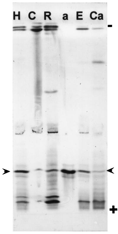
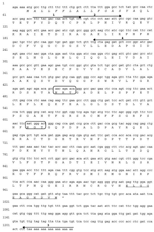
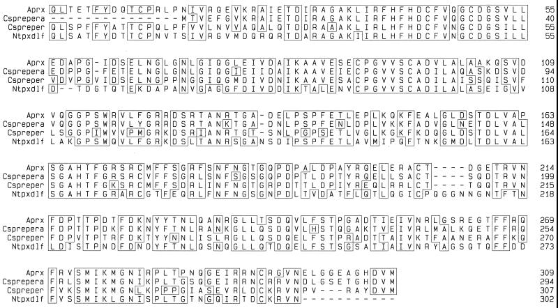
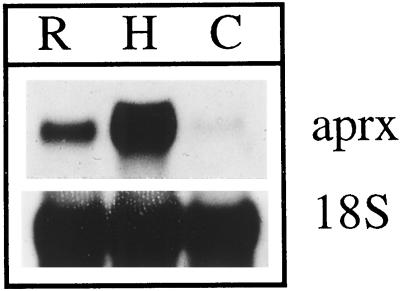
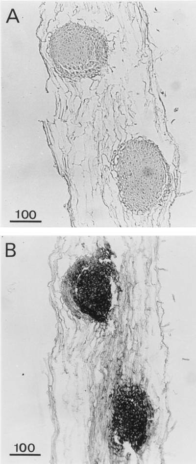
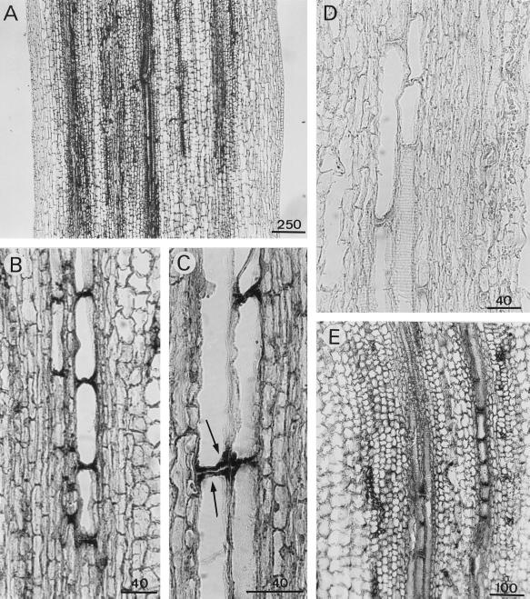
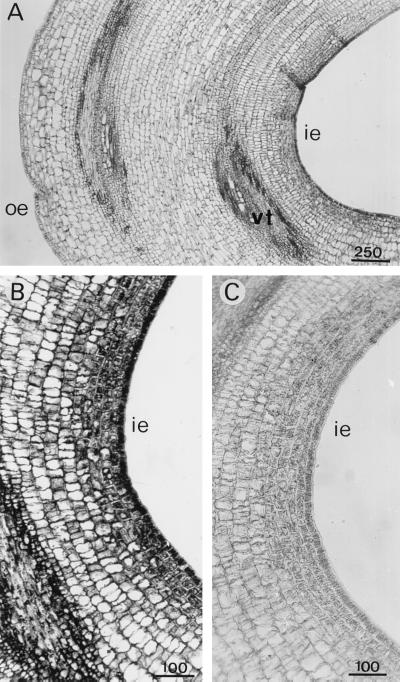
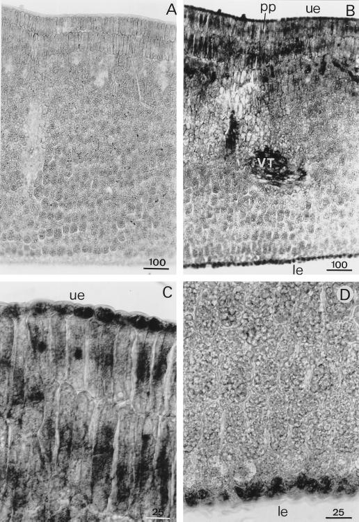
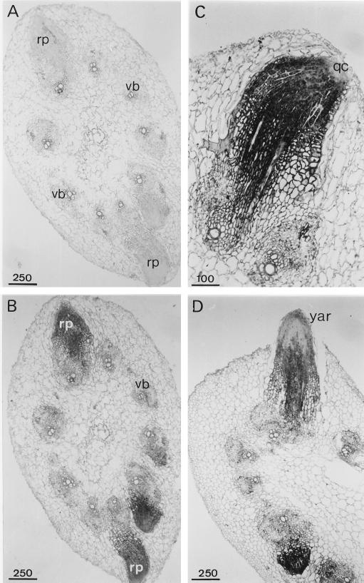
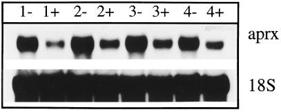
A calcium-pectate-binding anionic isoperoxidase (APRX) from zucchini (Cucurbita pepo) was purified and subjected to N-terminal amino acid microsequencing. The cDNA encoding this enzyme was obtained by reverse transcriptase polymerase chain reaction from a cDNA library. It encoded a mature protein of 309 amino acids exhibiting all of the sequence characteristics of a plant peroxidase. Despite the presence of a C-terminal propeptide, APRX was found in the apoplast. APRX protein and mRNA were found in the root, hypocotyls, and cotyledons. In situ hybridization showed that the APRX-encoding gene was expressed in many different tissues. The strongest expression was observed in root epidermis and in some cells of the stele, in differentiating tracheary elements of hypocotyl, in the lower and upper epidermis, in the palisade parenchyma of cotyledons, and in lateral and adventitious root primordia. In the hypocotyl hook there was an asymmetric expression, with the inner part containing more transcripts than the outer part. Treatment with 2,3,5-triiodobenzoic acid reduced the expression of the APRX-encoding gene in the lower part of the hypocotyl. Our observations suggest that APRX could be involved in lignin formation and that the transcription of its gene was related to auxin level.


Figures

Figure 1
Figure 1
Peroxidase isoenzymes from various fractions of zucchini seedlings separated on acrylamide gel by IEF. Extracts are from hypocotyls (H) (10 μg of proteins); cotyledons (C) (100 μg); roots (R) (10 μg); purified APRX (a) (28 ng); and extracellular fluid obtained in the presence of 5 mm EGTA (E) or 2 mm CaCl2 (C). Arrowheads indicate the positions of APRX on the gel.
Figure 2
Figure 2
The complete nucleotide and deduced amino acid sequences of APRX (accession no. Y17192). The signal peptide is shown in italics and the C-terminal propeptide is underlined. The four primers used to produce the full cDNA are shown. The two boxes designate the putative glycosylation sites. The stop codon is marked with an asterisk.
Figure 3
Figure 3
Mature peroxidase protein sequence comparison. The predicted protein sequences encoded by zucchini APRX clone (this study), two cucumber peroxidase cDNAs, Csprepera and Cspreper (Rasmussen et al., 1995), and a tobacco peroxidase cDNA, Ntpxdlf (Lagrimini et al., 1987), are shown. The alignment was created with the PileUp program and sequence homology was determined with the PrettyPlot program (both programs from Genetics Computer Group).
Figure 4
Figure 4
Northern-blot analysis using total RNA (10 μg) from root (R), hypocotyls (H), and cotyledons (C). APRX mRNA was detected with the 1145-bp cDNA as a specific probe. Detection of ribosomal 18S RNA was performed as a control.
Figure 5
Figure 5
Detection of APRX mRNA by in situ hybridization of root cross-sections. Sections were hybridized with sense (A) or antisense (B and C) probe. co, Cortex; ep, epidermis; st, stele; rc, root cap. Arrows indicate the accumulation of transcripts in epidermal cells. Scale bars are in μm.
Figure 6
Figure 6
Detection of APRX mRNA by in situ hybridization in root longitudinal sections showing lateral root primordia. Sections were hybridized with sense (A) or antisense (B) probes. Scale bars are in μm.
Figure 7
Figure 7
Detection of APRX mRNA by in situ hybridization in hypocotyl longitudinal sections taken 1 cm beneath the hook (A–D) or at the hook level (E). A, General view of a section hybridized with the antisense probe. B and C, Detailed views showing differentiating tracheary elements. D, Control section hybridized with the sense probe. Arrows in C indicate the accumulation of transcripts in differentiating xylem elements. Scale bars are in μm.
Figure 8
Figure 8
Detection of APRX mRNA by in situ hybridization in longitudinal sections of the hypocotyl hook. Sections were hybridized with antisense (A and B) or sense (C) probes. ie, Inner epidermis; oe, outer epidermis; vt, vascular tissues. Scale bars are in μm.
Figure 9
Figure 9
Detection of APRX mRNA by in situ hybridization in transversal sections of cotyledons. Sections were hybridized with sense (A) or antisense (B) probes. Upper (C) and lower (D) parts of the cotyledon were hybridized with the antisense probe. le, Lower epidermis; pp, palisade parenchyma; ue, upper epidermis; VT, vascular tissues. Scale bars are in μm.
Figure 10
Figure 10
Detection of APRX mRNA by in situ hybridization in cross-sections of hypocotyls. Shown are general views of adventitious root primordia in sections hybridized with sense (A) or antisense (B) probes, a young adventitious root (C), and an adventitious root emerging out of the hypocotyl (D) hybridized with the antisense probe. qc, Quiescent center; rp, root primordium; vb, vascular bundle; yar, young adventitious root. Scale bars are in μm.
Figure 11
Figure 11
Detection of mRNA encoding APRX in the lower part of hypocotyl of zucchini seedlings 1 to 4 d after excision of their root system. Rootless seedlings were kept without (−) or with (+) 10−4 m TIBA. Northern analysis was performed with 10 μg of total RNA. The detection of ribosomal 18S RNA was performed as a control.

References

    1. Blom TJM, Sierra M, van Vliet TB, Franke-van Dijk MEI, de Koning P, van Iren F, Verpoorte R, Libbenga KR. Uptake and accumulation of ajmalicine into isolated vacuoles of cultured cells of Catharanthus roseus (L.) G. Don. and its conversion into serpentine. Planta. 1991;183:170–177. - PubMed
    1. Brownleader M, Ahmed N, Trevan M, Chaplin MF, Dey PM. Purification and partial characterization of tomato extensin peroxidase. Plant Physiol. 1995;109:1115–1123. - PMC - PubMed
    1. Catesson A-M, Imberty A, Goldberg R, Czaninski Y (1986) Nature, localization and specificity of peroxidases involved in lignification processes. In H Greppin, C Penel, T Gaspar, eds, Molecular and Physiological Aspects of Plant Peroxidases. University of Geneva, Switzerland, pp 189–198
    1. Christensen JH, Bauw G, Welinder KG, Van Montagu M, Boerjan W. Purification and characterization of peroxidases correlated with lignification in poplar xylem. Plant Physiol. 1998;118:125–135. - PMC - PubMed
    1. Davies PJ. Plant Hormones and their Role in Plant Growth and Development. Dordrecht, The Netherlands: Martinus Nijhoff Publishers; 1987.

Publication types

Associated data

LinkOut - more resources