

The genes rubA and rubB for alkane degradation in Acinetobacter sp. strain ADP1 are in an operon with estB, encoding an esterase, and oxyR
- PMID: 10400587
- PMCID: PMC93931
- DOI: 10.1128/JB.181.14.4292-4298.1999
The genes rubA and rubB for alkane degradation in Acinetobacter sp. strain ADP1 are in an operon with estB, encoding an esterase, and oxyR
Abstract
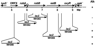
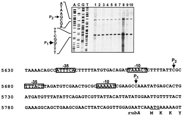
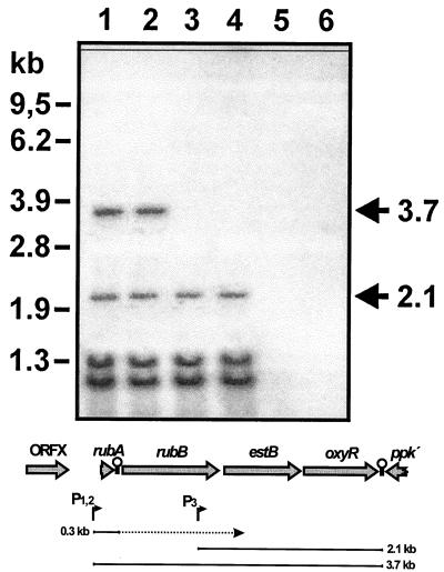
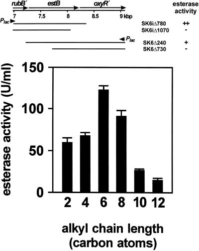
Alkanes are oxidized in Acinetobacter sp. strain ADP1 by a three-component alkane monooxygenase, composed of alkane hydroxylase, rubredoxin, and rubredoxin reductase. rubA and rubB encode rubredoxin and a NAD(P)H-dependent rubredoxin reductase. We demonstrate here that single base pair substitutions in rubA or rubB lead to defects in alkane degradation, showing that both genes are essential for alkane utilization. Differences in the degradation capacity for hexadecane and dodecane in these mutants are discussed. Two genes, estB and oxyR, are located downstream of rubB, but are not necessary for alkane degradation. estB encodes a functional esterase. oxyR encodes a LysR-type transcriptional regulator, conferring resistance to hydrogen peroxide. rubA, rubB, estB, and oxyR constitute an operon, which is constitutively transcribed from a sigma70 promoter, and an estB-oxyR containing message is also transcribed from an internal promoter.
Figures
Similar articles
-
Gene structures and regulation of the alkane hydroxylase complex in Acinetobacter sp. strain M-1.J Bacteriol. 2001 Mar;183(5):1819-23. doi: 10.1128/JB.183.5.1819-1823.2001. J Bacteriol. 2001. PMID: 11160120 Free PMC article.
-
Two genes encoding proteins with similarities to rubredoxin and rubredoxin reductase are required for conversion of dodecane to lauric acid in Acinetobacter calcoaceticus ADP1.Microbiology (Reading). 1995 Jun;141 ( Pt 6):1425-1432. doi: 10.1099/13500872-141-6-1425. Microbiology (Reading). 1995. PMID: 7670642
-
Involvement of an alkane hydroxylase system of Gordonia sp. strain SoCg in degradation of solid n-alkanes.Appl Environ Microbiol. 2011 Feb;77(4):1204-13. doi: 10.1128/AEM.02180-10. Epub 2010 Dec 23. Appl Environ Microbiol. 2011. PMID: 21183636 Free PMC article.
-
The OxyR regulon.Antonie Van Leeuwenhoek. 1990 Oct;58(3):157-61. doi: 10.1007/BF00548927. Antonie Van Leeuwenhoek. 1990. PMID: 2256675 Review.
-
OxyR: a regulator of antioxidant genes.J Nutr. 1992 Mar;122(3 Suppl):627-30. doi: 10.1093/jn/122.suppl_3.627. J Nutr. 1992. PMID: 1542022 Review.
Cited by
-
Transcriptomic analysis of the highly efficient oil-degrading bacterium Acinetobacter venetianus RAG-1 reveals genes important in dodecane uptake and utilization.FEMS Microbiol Lett. 2016 Oct;363(20):fnw224. doi: 10.1093/femsle/fnw224. Epub 2016 Sep 22. FEMS Microbiol Lett. 2016. PMID: 27664055 Free PMC article.
-
Identification of Brucella abortus OxyR and its role in control of catalase expression.J Bacteriol. 2000 Oct;182(19):5631-3. doi: 10.1128/JB.182.19.5631-5633.2000. J Bacteriol. 2000. PMID: 10986275 Free PMC article.
-
Functional Analysis of Novel alkB Genes Encoding Long-Chain n-Alkane Hydroxylases in Rhodococcus sp. Strain CH91.Microorganisms. 2023 Jun 9;11(6):1537. doi: 10.3390/microorganisms11061537. Microorganisms. 2023. PMID: 37375039 Free PMC article.
-
Characterization and Transcriptional Regulation of n-Alkane Hydroxylase Gene Cluster of Rhodococcus jostii RHA1.Microorganisms. 2019 Oct 23;7(11):479. doi: 10.3390/microorganisms7110479. Microorganisms. 2019. PMID: 31652785 Free PMC article.
-
Recent advances in petroleum microbiology.Microbiol Mol Biol Rev. 2003 Dec;67(4):503-49. doi: 10.1128/MMBR.67.4.503-549.2003. Microbiol Mol Biol Rev. 2003. PMID: 14665675 Free PMC article. Review.
References
-
- Asperger O, Kleber H-P. Metabolism of alkanes by Acinetobacter. In: Towner K J, Bergogne-Berezin E, Fewson C A, editors. The biology of Acinetobacter. New York, N.Y: Plenum Press; 1991. pp. 323–350.
-
- Asperger O, Naumann A, Kleber H-P. Occurrence of cytochrome P-450 in Acinetobacter strains after growth on n-hexadecane. FEMS Microbiol Lett. 1981;11:309–312.
-
- Aurich H, Sorger D, Asperger O. Isolation and characterization of rubredoxin from Acinetobacter calcoaceticus. Acta Biol Med Ger. 1976;35:443–451. - PubMed
-
- Ausubel F M, Brent R, Kingston R E, Moore D D, Seidman J G, Smith J A, Struhl K. Current protocols in molecular biology. I 1994. , II, and III. Greene Publishing Associates, New York, N.Y.
-
- Bagdasarian M, Lurz R, Rückert B, Franklin F C H, Bagdasarian M M, Frey J, Timmis K N. Specific-purpose plasmid cloning vectors. II. Broad host range, high copy number, RSF1010-derived vectors and a host-vector system for gene cloning in Pseudomonas. Gene. 1981;16:87–91. - PubMed
Publication types
MeSH terms
Substances
Associated data
- Actions
- Actions
LinkOut - more resources
Full Text Sources
Other Literature Sources
Molecular Biology Databases
