. 1999 Aug;73(8):6361-9.
doi: 10.1128/JVI.73.8.6361-6369.1999.

Reconstitution of human thymic implants is limited by human immunodeficiency virus breakthrough during antiretroviral therapy

Affiliations

Reconstitution of human thymic implants is limited by human immunodeficiency virus breakthrough during antiretroviral therapy

R G Amado et al. J Virol. 1999 Aug.

Abstract

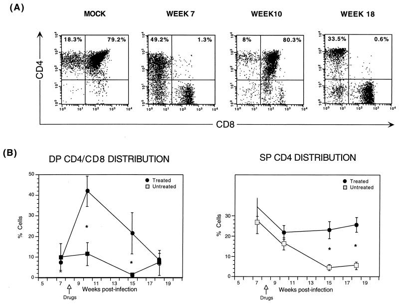
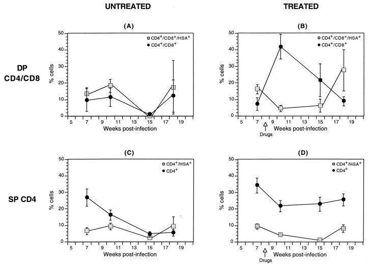
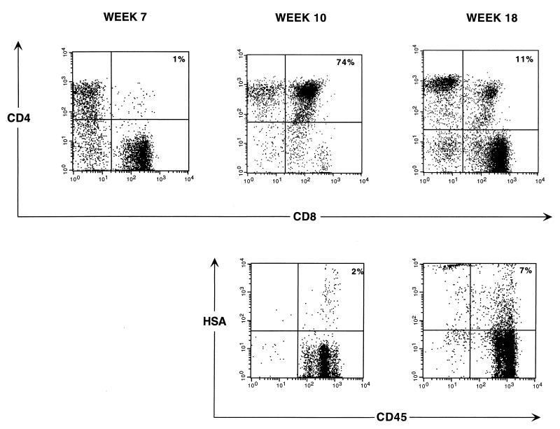
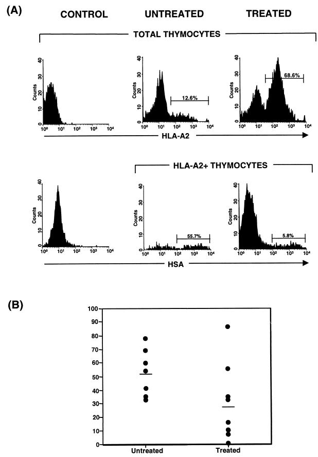
Human immunodeficiency virus type 1 (HIV-1)-infected SCID-hu thymic implants depleted of CD4(+) cells can support renewed thymopoiesis derived from both endogenous and exogenous T-cell progenitors after combination antiretroviral therapy. However, successful production of new thymocytes occurs transiently. Possible explanations for the temporary nature of this thymic reconstitution include cessation of the thymic stromal support function, exhaustion of T-cell progenitors, and viral resurgence. Distinguishing between these processes is important for the development of therapeutic strategies aimed at reconstituting the CD4(+) T-cell compartment in HIV-1 infection. Using an HIV-1 strain engineered to express the murine HSA heat-stable antigen surface marker, we explored the relationship between HIV-1 expression and CD4(+) cell resurgence kinetics in HIV-1-depleted SCID-hu implants following drug therapy. Antiviral therapy significantly suppressed HIV-1 expression in double-positive (DP) CD4/CD8 thymocytes, and the eventual secondary decline of DP thymocytes following therapy was associated with renewed viral expression in this cell subset. Thymocytes derived from exogenous T-cell progenitors induced to differentiate in HIV-1-depleted, drug-treated thymic implants also became infected. These results indicate that in this model, suppression of viral replication occurs transiently and that, in spite of drug therapy, virus resurgence contributes to the transient nature of the renewed thymic function.


Figures

FIG. 1
FIG. 1
Transient renewal of thymopoiesis in animals receiving antiretroviral therapy following infection with the reporter virus, NL-r-HSAL. Animals were infected with the reporter virus NL-r-HSAL, and biopsies were obtained at weeks 7, 10, 15, and 18 postinfection. Drug therapy was initiated at week 8 (arrows shown in panel B). Thymocytes were costained with CD4-PE and CD8-FITC. (A) Results of flow-cytometric analysis of CD4 and CD8 expression are shown for a representative animal that received antiretroviral therapy after DP CD4/CD8 thymocyte depletion, which was documented 7 weeks postinfection. In spite of continuing antiretroviral treatment, a secondary decline of DP CD/CD8 thymocytes occurred by week 18 postinfection. Results for a mock-infected animal are shown in the far-left plot to illustrate CD4/CD8 thymocyte distribution of an uninfected implant. The percentages of the subsets are indicated in the SP CD4 and DP CD4/CD8 quadrants. (B) Comparison of DP CD4/CD8 and SP CD4 thymocyte subset distributions of untreated and treated implants. Mean percentages of DP CD4/CD8 and SP CD4 cells and standard error bars are shown in each graph. An ∗ indicates a significant P value (see the text and Table 1). The numbers of animals analyzed at all time point are outlined in Table 1.
FIG. 2
FIG. 2
Distribution of total and HSA-expressing thymocyte subsets in treated and untreated implants. The distributions of DP CD4/CD8 (A and B) and SP CD4 (C and D) thymocytes are displayed in parallel with the percentages of the subsets that express the reporter virus (shaded squares) to contrast the trend of thymocyte kinetics with that of virus expression. Implants were infected with the reporter virus NL-r-HSAL, and biopsies were obtained at weeks 7, 10, 15, and 18 postinfection. The time of initiation of drug therapy is indicated in panels B and D. Thymocytes were costained with CD4-PE, CD8-FITC, and HSA-TC. Relevant isotype control antibodies were used to set quadrants. HSA-expressing subsets were determined by gating on DP CD4/CD8 or on SP CD4 thymocytes and by analyzing HSA expression. The numbers of animals analyzed at all time points are outlined in Table 1. Significance values are provided in the text and in Table 1.
FIG. 3
FIG. 3
Thymocyte subset distribution and HSA expression. Thymocytes obtained from biopsy samples of a representative animal at the indicated time points postinfection were costained for CD4-PE and CD8-FITC (top panels) and for CD45-FITC (human cells) and HSA-streptavidin-TC (lower panels). The percentage of each subset is indicated in each DP quadrant. Drugs were administered at week 8 postinfection.
FIG. 4
FIG. 4
Distributions of virus expression in thymocytes of donor origin in treated and untreated implants. Implants derived from HLA-A2 fetal tissue were injected with NL-r-HSAL virus. Infection was confirmed by measuring HSA expression at week 5. At week 6, nine animals were started on antiretroviral therapy and all implants were injected with 2.5 × 10⁵ CD34+ cells purified from an HLA-A2+ fetal liver. Costaining with HSA-FITC and HLA-A2 streptavidin-TC was performed at week 10 postinfection. (A) In the upper graphs HLA-A2 staining reveals chimeric engraftment in untreated and treated representative animals. In the lower graphs HSA expression in thymocytes derived from exogenous progenitors is shown by analyzing the HSA expression profile in the HLA-A2+ population (human cells of donor origin). Cells from a control, mock-infected, nontransplanted animal were stained in parallel with the same antibodies to set the relevant gates. (B) Distributions of HSA expression in donor-derived thymocytes of untreated and treated implants. Numeric values indicate the means of all data points for treated and untreated implants. Significance values are provided in the text and in Table 2.

Similar articles

Cited by

References

    1. Aldrovandi G M, Feuer G, Gao L, Jamieson B, Kristeva M, Chen I S Y, Zack J A. HIV-1 infection of the SCID-hu mouse: an animal model for virus pathogenesis. Nature. 1993;363:732–736. - PubMed
    1. Amado R G, Symonds G, Jamieson B D, Zhao G, Rosenblatt J D, Zack J A. Effects of megakaryocyte growth and development factor on survival and retroviral transduction of T lymphoid progenitor cells. Hum Gene Ther. 1998;9:173–183. - PubMed
    1. Angel J B, Kumar A, Parato K, Filion L G, Diaz-Mitoma F, Daftarian P, Pham B, Sun E, Leonard J M, Cameron D W. Improvement in cell-mediated immune function during potent anti-human immunodeficiency virus therapy with ritonavir plus saquinavir. J Infect Dis. 1998;177:898–904. - PubMed
    1. Autran B, Carcelain G, Li T S, Blanc C, Mathez D, Tubiana R, Katlama C, Debré P, Leibowitch J. Positive effects of combined antiretroviral therapy on CD4+ T cell homeostasis and function in advanced HIV disease. Science. 1997;277:112–116. - PubMed
    1. Bertho J M, Demarquay C, Moulian N, Van Der Meeren A, Berrih-Aknin S, Gourmelon P. Phenotypic and immunohistological analyses of the human adult thymus: evidence for an active thymus during adult life. Cell Immunol. 1997;179:30–40. - PubMed

Publication types

MeSH terms