. 1999 Aug;73(8):6670-9.
doi: 10.1128/JVI.73.8.6670-6679.1999.

Coupled integration of human immunodeficiency virus type 1 cDNA ends by purified integrase in vitro: stimulation by the viral nucleocapsid protein

Affiliations

Coupled integration of human immunodeficiency virus type 1 cDNA ends by purified integrase in vitro: stimulation by the viral nucleocapsid protein

S Carteau et al. J Virol. 1999 Aug.

Abstract

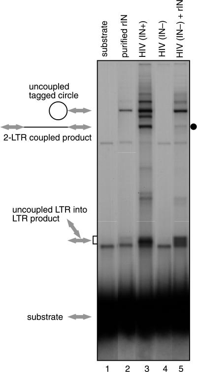
Integration of retroviral cDNA involves coupled joining of the two ends of the viral genome at precisely spaced positions in the host cell DNA. Correct coupled joining is essential for viral replication, as shown, for example, by the finding that viral mutants defective in coupled joining are defective in integration and replication. To date, reactions with purified human immunodeficiency virus type 1 (HIV-1) integrase protein in vitro have supported mainly uncoupled joining of single cDNA ends. We have analyzed an activity stimulating coupled joining present in HIV-1 virions, which led to the finding that the HIV-1 nucleocapsid (NC) protein can stimulate coupled joining more than 1,000-fold under some conditions. The requirements for stimulating coupled joining were investigated in assays with mutant NC proteins, revealing that mutations in the zinc finger domains can influence stimulation of integration. These findings (i) provide a means for assembling more authentic integrase complexes for mechanistic studies, (ii) reveal a new activity of NC protein in vitro, (iii) indicate a possible role for NC in vivo, and (iv) provide a possible method for identifying a new class of inhibitors that disrupt coupled joining.


Figures

FIG. 1
FIG. 1
Diagram of the integration reaction and reaction products.
FIG. 2
FIG. 2
Integration products generated in the presence of lysed HIV-1 virions or purified integrase. Lanes: 1, unreacted substrate; lane 2, 325 nM purified integrase protein; lane 3, lysed HIV-1 (HXB2) virions; lane 4, lysed HIV ΔIN; lane 5, mixture of R9 ΔIN and 325 nM recombinant integrase. Expected structures of integration products are indicated beside the gel; LTR DNAs are indicated by arrows, and target DNA is indicated by thin lines.
FIG. 3
FIG. 3
Characterization of products of integration reactions with lysed virions by digestion with restriction enzymes. Unintegrated HIV cDNA and solo LTR substrates have U3 sequences at one end and U5 sequences at the other end. (A) Autoradiogram of integration products analyzed by cleavage with restriction enzymes. Lane 1 displays the isolated integration product. Lane 2 presents the partially purified tagged circle form. Lane 3 presents the isolated two-LTR coupled form. Lanes 4 and 5 show these two forms cleaved with PpuMI. Lanes 6 to 8 contain the indicated markers. (B) Expected structure of the predominant tagged circle form. (C) Major coupled two-LTR products. The target DNA used was circular pCR2.1 (3.9 kb); the LTR DNA was 636 bp in length. The major products detected in panel A are as expected from the diagrams in panels B and C. The expected tagged circle form cleaved with PpuM1 runs between the uncleaved form and the relaxed circle form of the target DNA. Cleavage of the two-LTR coupled-joining product yielded two bands that migrated between the 4,822- and 4,361-bp markers; the expected sizes are 4,700 and 4,530 bp. Due to preferential joining of the U5 end, coupled products contained either two U5 ends or one U5 end and one U3 end joined to the target (C).
FIG. 4
FIG. 4
Stimulation of coupled joining by various nucleic acid binding proteins. (A) Products generated in the presence of 35 nM purified integrase and the indicated viral proteins. Expected structures of integration products are shown beside the gel. Maximum concentrations for each (right-most lane in each titration) are as follows: NC, 8 μg/ml (1 μM); MA, 16 μg/ml (0.94 μM); Rev, 4 μg/ml (0.2 μM); Tat, 4 μg/ml (0.28 μM); RT, 4 μg/ml (78 nM). Each protein was diluted 1:10 and 1:100 in the two left lanes. (B) Products generated in the presence of 35 nM purified integrase and the indicated cellular DNA binding proteins. Maximum concentrations for each (right-most lane in each titration) are as follows: NC, 8 μg/ml (1 μM); HMG I(Y), 16 μg/ml (1.4 μM); HMG-1, 4 μg/ml (0.16 μM); HMG-2, 8 μg/ml (0.64 μM); histone H1, 8 μg/ml (0.4 μM); Hu, 2.4 μg/ml (0.13 μM); BAF, 2 μg/ml (0.2 μM); RNase A, 4 μg/ml (0.3 μM); polylysine, 4 μg/ml (4 μM). Each protein was diluted 1:10 and 1:100 in the two left lanes.
FIG. 5
FIG. 5
Improvement of the yield of coupled-joining products by optimizing the concentrations of MgCl2 and NC. Concentrations of NC were 100 nM (lanes 3 and 8), 500 nM (lanes 4 and 9), 1 μM (lanes 5 and 10), and 2 μM (lanes 6 and 11). The integrase concentration was 35 nM. For this experiment, the LTR-supF donor was used. Other markings are as in Fig. 2.
FIG. 6
FIG. 6
Characterization of integration products generated in reactions with recombinant integrase and NC. The suspected tagged circle product (A) and coupled two-LTR product (B) were isolated from gels and characterized by digestion with restriction enzymes (diagrammed in panels C and D). The target DNA used was circular pZeo+. Cleavage of the putative tagged circle with HindIII (panel A, lane 2), which cleaves the LTR near the U5 terminus, yields predominantly a shortened labeled form that migrates near the target DNA relaxed circle form (C). Cleavage with PpuMI yielded a longer form, of a size consistent with cleavage near the U3 end (panel A, lane 3). Cleavage with XhoI, which cuts within the target, yields a smear of products (panel A, lane 4), as expected for Y-shaped cleavage products with heterogeneous branch points. The putative two-LTR coupled-joining product (panel B, lane 7) migrates at the molecular weight expected for a linearized target DNA joined in a coupled fashion to two LTRs (4,770 bp [D]). Cleavage with HindIII (panel B, lane 8) yielded a shortened labeled form of the size expected for the joining of two U5 ends (3,702 bp, diagrammed in panel D [top]). A longer form, of the size expected for joining of one U5 end and one U3 end, was also seen (4,134 bp, diagrammed in panel D [bottom]). Cleavage with PpuMI yielded predominantly a form of the size expected for the U5-U5 coupled two-LTR product (4,298 bp [panel B, lane 9]). The second expected PpuMI product, representing the U3-U5 form (expected size, 4,134 bp), migrated just below the U5-U5 form and is not clearly resolved in this gel. Cleavage with XhoI in the target DNA yielded a smear of labeled products, as expected since the points of integration in the target differ from molecule to molecule. The designation “hetero” indicates that this site is heterogeneously positioned in different product molecules due to the use of different integration sites.
FIG. 7
FIG. 7
Stimulation of integration in vitro by NC mutants. Mutants were tested at a concentration of 1.4 μM and integrase was present at 137.5 nM (His-tagged form), in otherwise standard reactions. The abundances of the two-LTR coupled-joining products (shaded bars) and the tagged circle products (open bars) were quantitated by PhosphorImager and normalized to wild-type NC. Each bar denotes the average of three reactions; half of the standard deviation is shown by the error bar.

Similar articles

Cited by

References

    1. Aiyar A, Hindmarsh P, Skalka A M, Leis J. Concerted integration of linear retroviral DNA by the avian sarcoma virus integrase in vitro: dependence on both long terminal repeat termini. J Virol. 1996;70:3571–3580. - PMC - PubMed
    1. Allen P, Worland S, Gold L. Isolation of high-affinity RNA ligands to HIV-1 integrase from a random pool. Virology. 1995;209:327–336. - PubMed
    1. Ausubel F M, Brent R, Kingston R E, Moore D D, Seidman J G, Smith J A, Struhl K. Current protocols in molecular biology. New York, N.Y: John Wiley and Sons, Inc.; 1987.
    1. Bess J W, Powell P J, Issaq H J, Schumack L J, Grimes M K, Henderson L E, Arthur L O. Tightly bound zinc in human immunodeficiency virus type 1, human T-cell leukemia virus type I, and other retroviruses. J Virol. 1992;66:840–847. - PMC - PubMed
    1. Bor Y-C, Bushman F, Orgel L. In vitro integration of human immunodeficiency virus type 1 cDNA into targets containing protein-induced bends. Proc Natl Acad Sci USA. 1995;92:10334–10338. - PMC - PubMed

Publication types

LinkOut - more resources