

Patterns of chemokine receptor fusion cofactor utilization by human immunodeficiency virus type 1 variants from the lungs and blood
- PMID: 10400765
- PMCID: PMC112752
- DOI: 10.1128/JVI.73.8.6680-6690.1999
Patterns of chemokine receptor fusion cofactor utilization by human immunodeficiency virus type 1 variants from the lungs and blood
Abstract
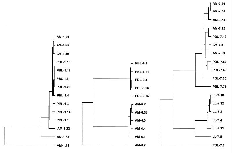
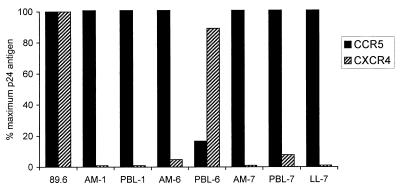
Human immunodeficiency virus type 1 (HIV-1) infection is highly compartmentalized, with distinct viral genotypes being found in the lungs, brain, and other organs compared with blood. CCR5 and CXCR4 are the principal HIV-1 coreceptors, and a number of other molecules support entry in vitro but their roles in vivo are uncertain. To address the relationship between tissue compartmentalization and the selective use of entry coreceptors, we generated functional env clones from primary isolates derived from the lungs and blood of three infected individuals and analyzed their use of the principal, secondary, orphan, and virus-encoded coreceptors for fusion. All Env proteins from lung viruses used CCR5 but not CXCR4, while those from blood viruses used CCR5 or CXCR4 or both. The orphan receptor APJ was widely used for fusion by Env proteins from both blood and lung viruses, but none used the cytomegalovirus-encoded receptor US28. Fusion mediated by the secondary coreceptors CCR2b, CCR3, CCR8, and CX3CR1 and orphan receptors GPR1, GPR15, and STRL33 was variable and heterogeneous, with relatively broad utilization by env clones from isolates of one subject but limited use by env clones from the other two subjects. However, there was no clear distinction between blood and lung viruses in secondary or orphan coreceptor fusion patterns. In contrast to fusion, none of the secondary or orphan receptors enabled efficient productive infection. These results confirm, at the level of cofactor utilization, previous observations that HIV-1 populations in the lungs and blood are biologically distinct and demonstrate diversity within lung-derived as well as blood-derived quasispecies. However, the heterogeneity in coreceptor utilization among clones from each isolate and the lack of clear distinction between lung- and blood-derived Env proteins argue against selective coreceptor utilization as a major determinant of compartmentalization.
Figures


References
-
- Agostini C, Trentin L, Zambello R, Semenzato G. HIV-1 and the lung. Infectivity, pathogenic mechanisms, and cellular immune responses taking place in the lower respiratory tract. Am Rev Respir Dis. 1993;147:1038–1049. - PubMed
-
- Alimohammadi A, Coker R, Miller R, Mitchell D, Williamson J, Clarke J. Genotypic variants of HIV-1 from peripheral blood and lungs of AIDS patients. AIDS. 1997;11:831–832. - PubMed
-
- Alkhatib G, Liao F, Berger E A, Farber J M, Peden K W C. A new SIV co-receptor, STRL33. Nature. 1997;388:238. - PubMed
-
- Ayehunie S, Garcia-Zepeda E A, Hoxie J A, Horuk R, Kupper T S, Luster A D, Ruprecht R M. Human immunodeficiency virus-1 entry into purified blood dendritic cells through CC and CXC chemokine coreceptors. Blood. 1997;90:1379–1386. - PubMed
Publication types
MeSH terms
Substances
Grants and funding
LinkOut - more resources
Full Text Sources
Molecular Biology Databases
