. 1999 Aug;73(8):6759-68.
doi: 10.1128/JVI.73.8.6759-6768.1999.

Structure of adenovirus complexed with its internalization receptor, alphavbeta5 integrin

Affiliations

Structure of adenovirus complexed with its internalization receptor, alphavbeta5 integrin

C Y Chiu et al. J Virol. 1999 Aug.

Abstract

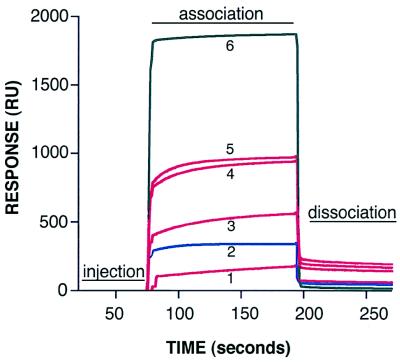
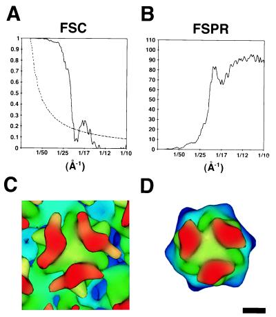
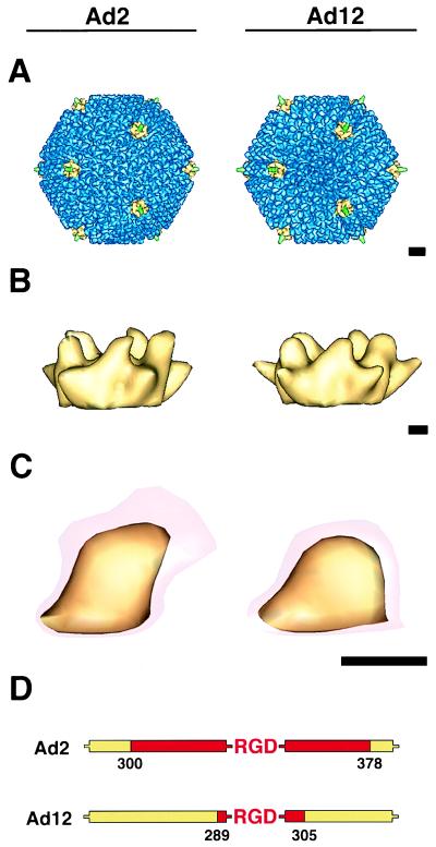
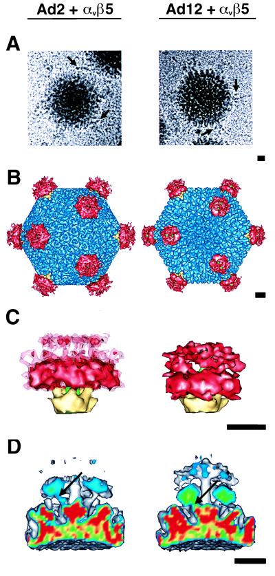
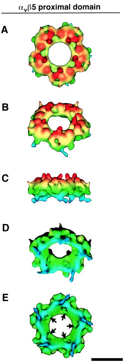
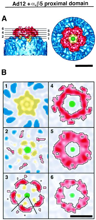
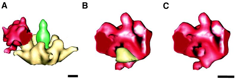
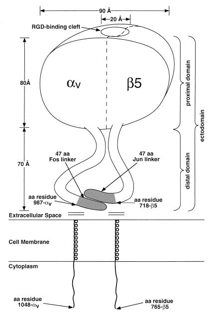
The three-dimensional structure of soluble recombinant integrin alphavbeta5 bound to human adenovirus types 2 and 12 (Ad2 and -12) has been determined at approximately 21-A resolution by cryoelectron microscopy (cryo-EM). The alphavbeta5 integrin is known to promote Ad cell entry. Cryo-EM has shown that the integrin-binding RGD (Arg-Gly-Asp) protrusion of the Ad2 penton base protein is highly mobile (P. L. Stewart, C. Y. Chiu, S. Huang, T. Muir, Y. Zhao, B. Chait, P. Mathias, and G. R. Nemerow, EMBO J. 16:1189-1198, 1997). Sequence analysis indicated that the Ad12 RGD surface loop is shorter than that of Ad2 and probably less flexible, hence more suitable for structural characterization of the Ad-integrin complex. The cryo-EM structures of the two virus-receptor complexes revealed a ring of integrin density above the penton base of each virus serotype. As expected, the integrin density in the Ad2 complex was diffuse while that in the Ad12 complex was better defined. The integrin consists of two discrete subdomains, a globular domain with an RGD-binding cleft approximately 20 A in diameter and a distal domain with extended, flexible tails. Kinetic analysis of Ad2 interactions with alphavbeta5 indicated approximately 4.2 integrin molecules bound per penton base at close to saturation. These results suggest that the precise spatial arrangement of five RGD protrusions on the penton base promotes integrin clustering and the signaling events required for virus internalization.


Figures

FIG. 1
FIG. 1
BIAcore sensorgrams of soluble αvβ5 integrin or DAV-1 MAb binding to Ad2 particles. Various amounts of soluble αvβ5 integrin or antibody (1, 500 nM integrin; 2, 50 nM DAV-1 MAb; 3, 2 μM integrin; 4, 4 μM integrin; 5, 8 μM integrin, 6, 500 nM integrin plus 20 mM EDTA) were passed over a biosensor chip containing immobilized Ad2 particles, and the association and dissociation rates were measured in real time with a BIAcore 2000 biosensor as described in Materials and Methods. For clarity, only the sensorgram of DAV-1 MAb binding (blue line) at saturating conditions is shown. The relatively large bulk-flow response seen in sensorgram 6 is due to the presence of 20 mM EDTA. This response does not represent specific integrin binding, as indicated by the absence of resonance units (RUs) following dissociation.
FIG. 2
FIG. 2
Resolution assessment of the Ad2 cryo-EM reconstruction. (A) Plot of the FSC (solid line) and the corresponding 23.2ς (3ς, adjusted for icosahedral symmetry) significance threshold curve (dashed line) (38). (B) Plot of the FSPR. Both the FSC and FSPR were calculated from soft masked reconstructions that only included the ordered protein capsid without the disordered fiber and core density. (C) One hexon from the Ad2 cryo-EM reconstruction filtered to 21 Å, the resolution indicated by the 0.5 correlation point of the FSC and the 45° criterion of the FSPR. (D) The crystallographic structure of the Ad2 hexon (6) filtered to 21-Å resolution. The density maps shown in panels C and D are color coded by height. The magnification of the cryo-EM structure was adjusted by ∼5% for the best match with the filtered crystallographic hexon. The scale bar is 25 Å.
FIG. 3
FIG. 3
Cryo-EM reconstructions of Ad2 (left) and Ad12 (right) at ∼21-Å resolution. (A) Ad capsids viewed along an icosahedral threefold axis. The penton base proteins at the icosahedral vertices are shown in yellow, the reconstructed portion of the flexible fibers are in green, and the remaining capsid density is in blue. (B) Side views of the external portion of the penton base contoured at a level corresponding to the strong capsid density. (C) Enlargements of a single penton base protrusion at two isosurface levels, one just above noise (transparent red) and the other showing well-defined density (yellow). (D) The lengths of the variable regions flanking the RGD sequence (red) in Ad2 and Ad12 are obtained from sequence alignment of five different Ad serotypes. The scale bars are 100 (A) and 25 (B and C) Å.
FIG. 4
FIG. 4
Cryo-EM reconstruction of the Ad2-integrin (left) and Ad12-integrin (right) complexes at ∼24-Å resolution. (A) Representative particle images with arrows indicating regions of density attributed to αvβ5 integrin. (B) Ad complexes viewed along an icosahedral threefold axis. The color scheme is the same as in Fig. 3A, with αvβ5 integrin density shown in red. (C) Side views of the penton base, fiber, and integrin. The Ad12-integrin density is contoured at a level corresponding to five bound αvβ5 heterodimers. The Ad2-integrin density is contoured at the same level (solid red) and just above noise (transparent red). (D) A cropped view of the vertex regions of the Ad complexes showing connections between the integrin and the penton base via the RGD protrusions (arrows). The slice plane is color coded on the basis of density, with the strongest density values shown in red and the weakest in blue. The scale bars are 100 Å.
FIG. 5
FIG. 5
Integrin ring from the Ad12-αvβ5 complex at ∼21-Å resolution. The ring is formed by associations between the RGD-binding proximal domains. (A to E) Five views of the height-color-coded ring from top to bottom. Note that the top surface has five columns of density (red) that connect the proximal domains to the more flexible distal domains (not shown). The bottom-surface view shows five clefts (arrows), each ∼20 Å in diameter, that bind the RGD-containing protrusions of the penton base protein. The scale bar is 100 Å.
FIG. 6
FIG. 6
Integrin ring in association with the Ad12 capsid. (A) Side- and top-surface views. (B) Slice planes through the integrin density perpendicular to an icosahedral fivefold symmetry axis. The heights of the slice planes are indicated by numbered lines in panel A. The color scheme for the individual proteins is as shown in Fig. 4B. Stronger density values are represented by darker shades, and weaker density values are represented by lighter shades. The black lines in slice 3 designate the boundary for the extracted model of one integrin proximal domain displayed in Fig. 7. The scale bars are 100 Å.
FIG. 7
FIG. 7
Model for the interaction between the integrin proximal domain and the Ad12 penton base protein. One-fifth of the integrin ring density is shown extracted along estimated boundaries to model the proximal domain of a single αvβ5 heterodimer. (A) The modeled proximal domain shown in association with the penton base protein. (B) The modeled proximal domain is rotated ∼90 degrees with respect to the view in panel A to show the interaction with a single penton base protrusion. (C) The same view as in panel B but with the protrusion removed to reveal the RGD-binding cleft on the inner surface. The scale bars are 25 Å.
FIG. 8
FIG. 8
Schematic diagram of the soluble αvβ5 integrin heterodimer. Approximate dimensions are derived from the cryo-EM reconstruction of the Ad12-integrin complex. Amino acid residue numbers are from Mathias et al. (34). The proximal and distal domains are defined with respect to the viral surface.

Similar articles

Cited by

References

    1. Acharya R, Fry E, Stuart D, Fox G, Rowlands D, Brown F. The three-dimensional structure of foot-and-mouth disease virus at 2.9 Å resolution. Nature. 1989;337:709–716. - PubMed
    1. Adler M, Lazarus R A, Dennis M S, Wagner G. Solution structure of kistrin, a potent platelet aggregation inhibitor and GP IIb-IIIa antagonist. Science. 1991;253:445–448. - PubMed
    1. Adrian M, Dubochet J, Lepault J, McDowall A W. Cryo-electron microscopy of viruses. Nature. 1984;308:32–36. - PubMed
    1. Akke M, Liu J, Cavanagh J, Erickson H P, Palmer A G I. Pervasive conformational fluctuations on microsecond time scales in a fibronectin type III domain. Nat Struct Biol. 1998;5:55–59. - PubMed
    1. Altschul S F, Gish W, Miller W, Myers E W, Lipman D J. Basic local alignment search tool. J Mol Biol. 1990;215:403–410. - PubMed

Publication types