. 1999 Aug;73(8):6852-61.
doi: 10.1128/JVI.73.8.6852-6861.1999.

In vivo monocyte tropism of pathogenic feline immunodeficiency viruses

Affiliations

In vivo monocyte tropism of pathogenic feline immunodeficiency viruses

S W Dow et al. J Virol. 1999 Aug.

Abstract

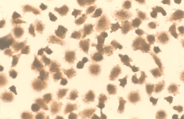
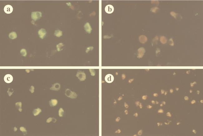
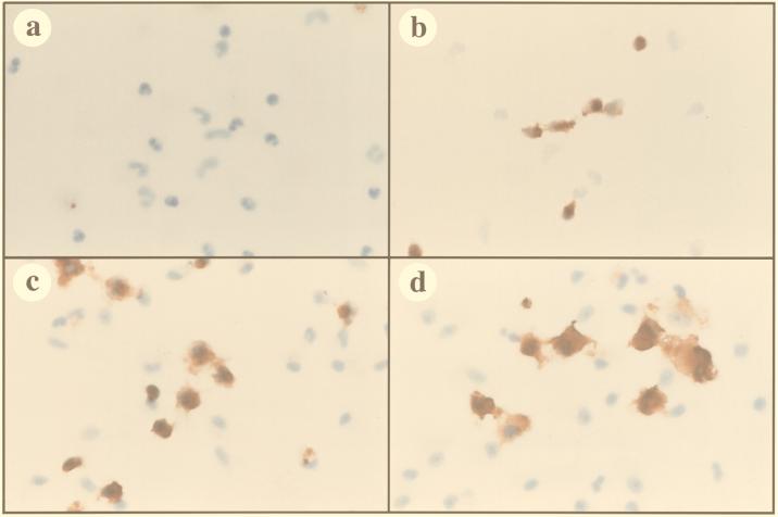
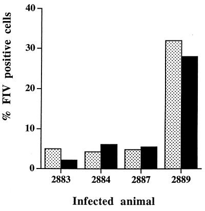
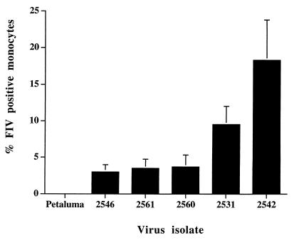
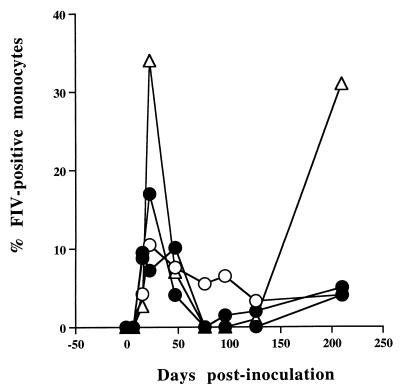
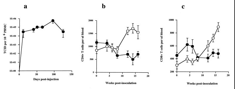
Virus-infected monocytes rarely are detected in the bloodstreams of animals or people infected with immunodeficiency-inducing lentiviruses, yet tissue macrophages are thought to be a major reservoir of virus-infected cells in vivo. We have identified feline immunodeficiency virus (FIV) clinical isolates that are pathogenic in cats and readily transmitted vertically. We report here that five of these FIV isolates are highly monocytotropic in vivo. However, while FIV-infected monocytes were numerous in the blood of experimentally infected cats, viral antigen was not detectable in freshly isolated cells. Only after a short-term (at least 12-h) in vitro monocyte culture were FIV antigens detectable (by immunocytochemical analysis or enzyme-linked immunosorbent assay). In vitro experiments suggested that monocyte adherence provided an important trigger for virus antigen expression. In the blood of cats infected with a prototype monocytotropic isolate (FIV subtype B strain 2542), infected monocytes appeared within 2 weeks, correlating with high blood mononuclear-cell-associated viral titers and CD4 cell depletion. By contrast, infected monocytes could not be detected in the blood of cats infected with a less pathogenic FIV strain (FIV subtype A strain Petaluma). We concluded that some strains of FIV are monocytotropic in vivo. Moreover, this property may relate to virus virulence, vertical transmission, and infection of tissue macrophages.


Figures

FIG. 1
FIG. 1
Enhancement of in vitro culture of feline monocytes by human rIL-4. Feline monocytes were obtained from PBMC by 1 h of adherence to plastic chamber slides that were precoated with purified feline IgG. After being washed to remove nonadherent cells, the monocytes were cultured in monocyte medium supplemented with human rIL-4 at 20 ng/ml (see Materials and Methods). Over the next 3 to 4 days of culture, the monocytes increased in size and assumed phenotypic characteristics of macrophages. Cells were fixed and stained for detection of nonspecific esterase activity, which revealed an essentially pure population of monocyte-derived macrophages (magnification, ×200).
FIG. 2
FIG. 2
Identification of in vivo FIV-infected monocytes by in vitro culture and immunofluorescence assay. Monocytes obtained from a cat infected with the FIV isotype B isolate 2542 were cultured in vitro for 3 days, fixed, and then immunostained for detection of FIV antigens. Cells were reacted first with FIV-immune cat serum (a), nonimmune cat serum (b), an anti-FIV p26 MAb (c), or an irrelevant isotype-matched MAb (d). The cells were then incubated with the appropriate FITC-conjugated secondary antibodies as described in Materials and Methods. Numerous FIV-positive monocytes were detected by staining with anti-FIV serum (a) or the anti-p26 MAb (c), whereas staining was minimal with nonimmune cat serum (b) or the irrelevant MAb (d). Monocytes from noninfected cats also did not stain with FIV-immune cat serum (data not shown) (magnification, ×200).
FIG. 3
FIG. 3
Kinetics of FIV antigen expression in in vivo FIV-infected monocytes during in vitro culture as assessed by immunocytochemical analysis. Monocytes were obtained from an FIV-infected cat by adherence to plastic for 1 h and then fixed either immediately after adherence (a) or after 24 h (b), 48 h (c), or 72 h (d) in culture. The monocytes were then immunostained to detect FIV antigens by using an immunoperoxidase technique as described in Materials and Methods. Expression of FIV antigens was not detected in monocytes immediately after adherence but then appeared and increased with time. However, the percentage of antigen-expressing monocytes decreased over a 3-day culture period, indicating that the infection did not spread to uninfected cells. (magnification, ×200).
FIG. 4
FIG. 4
Infection of bone marrow monocytes/macrophages with FIV in vivo. Bone marrow mononuclear cells were obtained by aspiration from the humerus of an FIV-infected cat. Bone marrow monocytes/macrophages were enriched by adherence for 1 h, washed, and then cultured for 3 days in monocyte medium. After the cells were fixed, FIV antigen expression was detected by immunocytochemical analysis. Immediately after adherence (a), FIV antigens could not be detected in bone marrow-derived monocytes. However, strong FIV expression could be detected in numerous bone marrow-derived monocytes/macrophages after 72 h in culture (b).
FIG. 5
FIG. 5
Similar levels of monocyte infection in blood and bone marrow. Monocytes were obtained from blood (black bars) and bone marrow (light bars) from four different cats 10 weeks after inoculation with in vivo-passaged FIV subtype B strain 2542. Monocytes were cultured for 72 h in monocyte medium, and then the percentage of FIV-infected monocytes in each culture was quantitated by immunohistochemical analysis. Similar results were obtained in one additional experiment using blood and bone marrow specimens from the same four cats.
FIG. 6
FIG. 6
FIV p26 Gag expression by in vivo-infected monocytes upon in vitro culture. A p26 (CA) capture ELISA was used to quantitate FIV expression by monocytes obtained from a cat infected with FIV subtype B strain 2542. Monocyte cultures were established from PBMC (as described in Materials and Methods) by using triplicate wells of a 24-well plate. At various time points during culture, supernatants were harvested from triplicate wells and the adherent cells in each well were then lysed in 1.0 ml of buffer containing 0.1% Triton X-100. The p26 concentrations in supernatants (□) and lysates (●) were determined by ELISA, and the mean (± the standard error) p26 concentration was plotted versus time in culture. Intracellular expression of p26 by in vivo-infected monocytes was first detectable at 12 h in culture, increased by 24 h in culture, and decreased thereafter, whereas p26 antigen was virtually undetectable in supernatants.
FIG. 7
FIG. 7
Triggering of FIV expression in in vivo-infected monocytes by adherence. To evaluate the influence of adherence on virus antigen expression by in vivo-infected monocytes, PBMC were obtained from the blood of an FIV-infected cat and cultured either under adherent conditions (adherence to tissue culture plastic) or under nonadherent conditions (culture on a feline fibroblast monolayer) for 3 days. During 3 days of culture on a fibroblast monolayer, monocytes remained fully viable but nonadherent and could be obtained by gentle washing and purified by 1 h of adherence to tissue culture plastic and fixed. The percentage of FIV-positive monocytes after culture under adherent or nonadherent conditions was then determined by immunocytochemical analysis. Virus antigen expression by nonadherent monocytes/macrophages was minimal (a), compared to that by monocytes cultured continuously under adherent conditions (Fig. 3). However, when the nonadherent monocytes/macrophages were cultured for 3 days in the presence of 10 nM PMA, virus expression was strongly upregulated (b). Other cytokines and macrophage stimulants were also evaluated for the ability to upregulate virus antigen expression by nonadherent monocytes/macrophages, including LPS at 10 μg/ml, IL-4 at 20 ng/ml, GM-CSF at 10 ng/ml, and TNF-α at 10 ng/ml (c). Only culture in the presence of 10 nM PMA stimulated virus expression by nonadherent monocytes/macrophages (c), although the level of expression was still less than that in monocytes cultured under continuously adherent conditions. Similar results were obtained in one additional experiment.
FIG. 7
FIG. 7
Triggering of FIV expression in in vivo-infected monocytes by adherence. To evaluate the influence of adherence on virus antigen expression by in vivo-infected monocytes, PBMC were obtained from the blood of an FIV-infected cat and cultured either under adherent conditions (adherence to tissue culture plastic) or under nonadherent conditions (culture on a feline fibroblast monolayer) for 3 days. During 3 days of culture on a fibroblast monolayer, monocytes remained fully viable but nonadherent and could be obtained by gentle washing and purified by 1 h of adherence to tissue culture plastic and fixed. The percentage of FIV-positive monocytes after culture under adherent or nonadherent conditions was then determined by immunocytochemical analysis. Virus antigen expression by nonadherent monocytes/macrophages was minimal (a), compared to that by monocytes cultured continuously under adherent conditions (Fig. 3). However, when the nonadherent monocytes/macrophages were cultured for 3 days in the presence of 10 nM PMA, virus expression was strongly upregulated (b). Other cytokines and macrophage stimulants were also evaluated for the ability to upregulate virus antigen expression by nonadherent monocytes/macrophages, including LPS at 10 μg/ml, IL-4 at 20 ng/ml, GM-CSF at 10 ng/ml, and TNF-α at 10 ng/ml (c). Only culture in the presence of 10 nM PMA stimulated virus expression by nonadherent monocytes/macrophages (c), although the level of expression was still less than that in monocytes cultured under continuously adherent conditions. Similar results were obtained in one additional experiment.
FIG. 8
FIG. 8
Identification of monocytotropic FIV strains by in vivo passage. Five groups of SPF cats (four per group) were each inoculated intravenously with 5.0 ml of whole blood obtained from an SPF cat that had been infected previously with one of five different clinical FIV isolates. Five additional SPF cats were inoculated with FIV subtype A strain Petaluma (provided by N. Pedersen, University of California, Davis). At 10 and again at 16 weeks p.i., the frequency of FIV infection in monocytes from each cat was evaluated by short-term culture and immunocytochemical analysis and the mean percentage of positive cells (± the standard error) was plotted. Two isolates (2531 and 2542) were identified as the most monocytotropic in vivo.
FIG. 9
FIG. 9
Kinetics of FIV subtype B strain 2542 monocyte infection after experimental infection. Four 8-week-old SPF cats were inoculated with whole blood from a cat infected with monocytotropic FIV subtype B strain 2542, four age-matched control cats were inoculated with blood from an uninfected control animal, and the level of monocyte infection was monitored over a 7-month period. The percentage of infected monocytes was determined as described in Materials and Methods, and the value for each cat was plotted versus time p.i. (cats: 2883 [●], 2884 [●], 2887 [○], and 2889 [▵]). In cats inoculated with blood from the FIV subtype B strain 2542-infected cat, peak monocyte infection occurred between 14 and 50 days p.i. The level of monocyte infection declined thereafter, remained intermittently detectable through 4 months, and then appeared to recrudesce at 7 months p.i. The cat with the highest level of monocyte infection (▵) was euthanized at 9 months p.i. due to progressive weight loss.
FIG. 10
FIG. 10
Viral replication and CD4 and CD8 T-cell kinetics in cats infected with FIV subtype B strain 2542. SPF cats (four per group) were inoculated with 5 ml of blood from a cat infected with FIV subtype B strain 2542 or sham inoculated with 5 ml of blood from an uninfected cat. The PBMC-associated virus titer was determined by coculture every 2 weeks. The mean viral titer (number of tissue culture-infective doses [TCID] per 106 PBMC ± the standard error) was plotted versus time p.i. (a). Also determined were the numbers of CD4+ (b) and CD8+ (c) cells. Symbols: ●, FIV-infected cats; ○, age-matched controls.

Similar articles

Cited by

References

    1. Alkhatib F, Combadiere C, Broder C C, Feng Y, Kennedy P E, Murphy P M, Berger E A. CC CKR5: a RANTES, MIP-I‡, MIP-1 · receptor as a fusion cofactor for macrophage-tropic HIV-1. Science. 1996;272:1955–1958. - PubMed
    1. Bagasra O, Hauptman S P, Lischner H W, Sachs M, Pomerantz R J. Detection of human immunodeficiency virus type 1 provirus in mononuclear cells by in situ polymerase chain reaction. N Engl J Med. 1992;326:1385–1391. - PubMed
    1. Beebe A M, Dua N, Faith T G, Moore P F, Pedersen N C, Dandekar S. Primary stage of feline immunodeficiency virus infection: viral dissemination and cellular target. J Virol. 1994;68:3080–3091. - PMC - PubMed
    1. Brunner D, Pedersen N C. Infection of peritoneal macrophages in vitro and in vivo with feline immunodeficiency virus. J Virol. 1989;63:5483–5488. - PMC - PubMed
    1. Choe H, Farzan M, Sun Y, Sullivan N, Rollins B, Ponath P D, Wu L, Mackay C R, LaRosa G, Newman W, Gerard N, Gerard C. The · -chemokine receptors CCR3 and CCR5 facilitate infection by primary HIV-1 isolates. Cell. 1996;85:1135–1148. - PubMed

Publication types