. 1999 Aug;73(8):6872-81.
doi: 10.1128/JVI.73.8.6872-6881.1999.

Association with the cellular export receptor CRM 1 mediates function and intracellular localization of Epstein-Barr virus SM protein, a regulator of gene expression

Affiliations

Association with the cellular export receptor CRM 1 mediates function and intracellular localization of Epstein-Barr virus SM protein, a regulator of gene expression

S M Boyle et al. J Virol. 1999 Aug.

Abstract

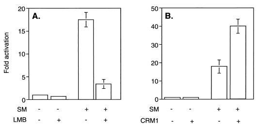
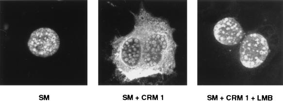
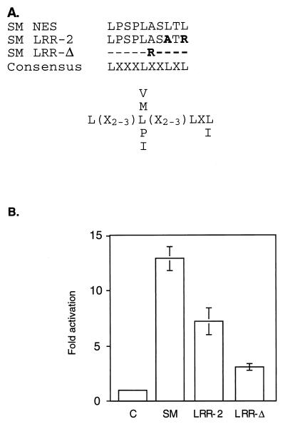
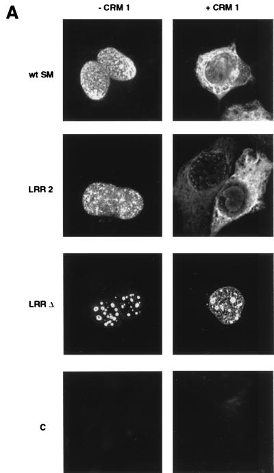
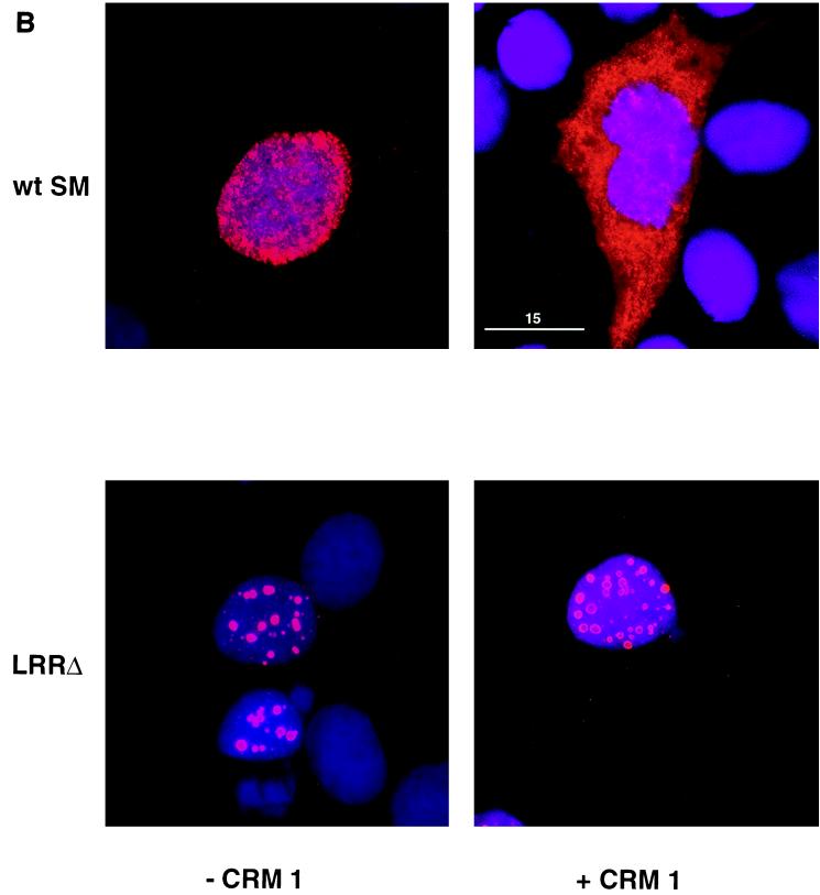
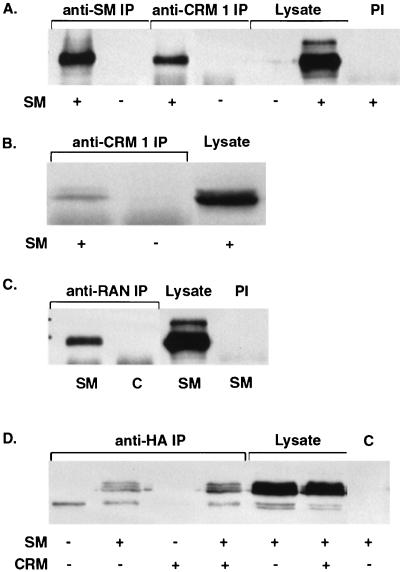
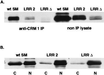
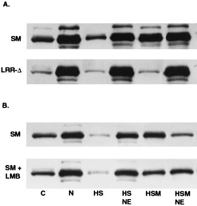
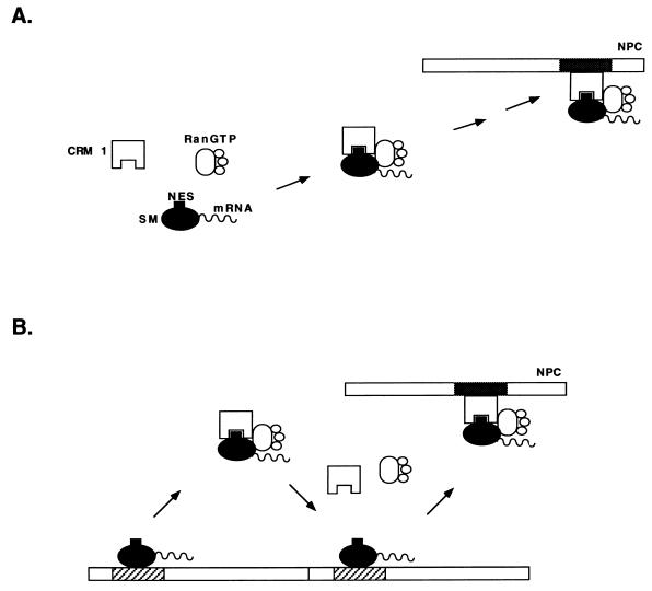
Splicing and posttranscriptional processing of eukaryotic gene transcripts are linked to their nuclear export and cytoplasmic expression. Unspliced pre-mRNAs and intronless transcripts are thus inherently poorly expressed. Nevertheless, human and animal viruses encode essential genes as single open reading frames or in the intervening sequences of other genes. Many retroviruses have evolved mechanisms to facilitate nuclear export of their unspliced mRNAs. For example, the human immunodeficiency virus RNA-binding protein Rev associates with the soluble cellular export receptor CRM 1 (exportin 1), which mediates nucleocytoplasmic translocation of Rev-HIV RNA complexes through the nuclear pore. The transforming human herpesvirus Epstein-Barr virus (EBV) expresses a nuclear protein, SM, early in its lytic cycle; SM binds RNA and posttranscriptionally activates expression of certain intronless lytic EBV genes. Here we show that both the trans-activation function and cytoplasmic translocation of SM are dependent on association with CRM 1 in vivo. SM is also shown to be associated in vivo with other components of the CRM 1 export pathway, including the small GTPase Ran and the nucleoporin CAN/Nup214. SM is shown to be present in the cytoplasm, nucleoplasm, and nuclear envelope of transfected cells. Mutation of a leucine-rich region (LRR) of SM inhibited CRM 1-mediated cytoplasmic translocation and SM activity, as did leptomycin B, an inhibitor of CRM 1 complex formation. Surprisingly, however, leptomycin B treatment and mutation of the LRR both led to SM becoming more tightly attached to intranuclear structures. These findings suggest a model in which SM is not merely a soluble carrier protein for RNA but rather is bound directly to intranuclear proteins, possibly including the nuclear pore complex.


Figures

FIG. 1
FIG. 1
Effects of modulating CRM 1 activity on SM function. (A) Effect of LMB on trans-activation by SM. CAT activity was measured in lysates of BJAB cells transfected with SM or antisense control plasmid and CAT reporter plasmid CMV-CAT. Cells were treated with either LMB (10 nM) or control medium immediately after transfection. Results are means of at least three independent transfections. (B) Effect of CRM 1 overexpression on trans-activation by SM. CAT activity was measured in lysates of BJAB cells transfected with SM or antisense control plasmid, CAT reporter plasmid CMV-CAT, and either control or CRM 1 expression vector.
FIG. 2
FIG. 2
Effects of CRM 1 overexpression and LMB treatment on cellular distribution of SM. Cos 7 cells were grown on coverslips prior to transfection, and immunofluorescence microscopy was performed with anti-SM antibodies. Cells were transfected with SM plasmid alone or with SM and CRM 1 expression plasmids. Cells were also transfected with SM and CRM 1 expression plasmid and treated with LMB immediately after transfection.
FIG. 3
FIG. 3
Effect of mutation of a leucine-rich putative SM NES on SM-mediated activation. (A) Amino acids 227 to 236 of SM, with four leucines separated by three, two, and one amino acid, fits the broad consensus sequence described for an NES (3). LRR-2 and LRR-Δ are SM mutants with two leucines altered or the entire LRR deleted and replaced with an arginine, respectively. Amino acid substitutions are shown in bold. “X” represents no selection for a particular amino acid at that site. Preferred amino acids at particular sites are shown by their one-letter codes. (B) BJAB cells were transfected with a CAT reporter plasmid and either SM or a mutant SM plasmid, and CAT activity was measured as described in the text.
FIG. 4
FIG. 4
Effects of CRM 1 overexpression on cellular distribution of SM and SM LRR mutants. (A) Immunofluorescence microscopy was performed on SM- or SM mutant-transfected cells with anti-SM antibodies as for Fig. 2. Cos 7 cells were transfected with either wild-type SM (wt SM), SM mutant plasmid LRR-2 or LRR-Δ, or vector plasmid (C), as indicated. Cells were also cotransfected with either control vector (−CRM 1) or CRM 1 expression plasmid (+CRM 1). (B) Cos 7 cells transfected with wt SM or LRR-Δ plasmid and cotransfected with either CRM 1 or control plasmid were stained with anti-SM antibodies. Nuclei were visualized by staining with DAPI. Immunofluorescence images were acquired with a deconvolution fluorescence microscope system. SM staining appears red, and nuclei are purple. Bar, 15 μm.
FIG. 4
FIG. 4
Effects of CRM 1 overexpression on cellular distribution of SM and SM LRR mutants. (A) Immunofluorescence microscopy was performed on SM- or SM mutant-transfected cells with anti-SM antibodies as for Fig. 2. Cos 7 cells were transfected with either wild-type SM (wt SM), SM mutant plasmid LRR-2 or LRR-Δ, or vector plasmid (C), as indicated. Cells were also cotransfected with either control vector (−CRM 1) or CRM 1 expression plasmid (+CRM 1). (B) Cos 7 cells transfected with wt SM or LRR-Δ plasmid and cotransfected with either CRM 1 or control plasmid were stained with anti-SM antibodies. Nuclei were visualized by staining with DAPI. Immunofluorescence images were acquired with a deconvolution fluorescence microscope system. SM staining appears red, and nuclei are purple. Bar, 15 μm.
FIG. 5
FIG. 5
Coimmunoprecipitation of CRM 1 and CRM 1-associated proteins with SM. (A) Lysates of Cos 7 cells transfected with SM or control vector (SM + and −) were immunoprecipitated with anti CRM 1 (anti-CRM 1 IP) or anti-SM (anti-SM IP) antibodies and immunoblotted to detect SM. A control immunoprecipitation of SM-transfected cell lysate with preimmune rabbit serum (PI) is also shown. (B) Lysates of BJAB cells transfected with SM or control vector were immunoprecipitated with anti-CRM 1 antibodies and immunoblotted as in panel A. (C) Lysates of Cos 7 cells transfected with SM and CRM 1 were immunoprecipitated with anti-Ran antibodies (anti-RAN IP) and immunoblotted to detect SM. Control immunoprecipitations with preimmune rabbit serum (PI) are also shown. (D) Cells were transfected with a plasmid expressing an HA-tagged carboxy-terminal fragment of CAN/Nup214 (HA-ΔCAN) and SM or control plasmid. Lysates were immunoprecipitated with anti-HA monoclonal antibody CA125 (anti-HA IP) and immunoblotted with anti-SM antibodies. A control immunoprecipitation (C) performed with an irrelevant monoclonal antibody (anti-FLAG) is also shown. In all panels, lanes containing an equivalent amount of unimmunoprecipitated lysate are indicated.
FIG. 6
FIG. 6
Effect of LRR mutation on intracellular compartmentalization of SM. (A) Detergent-solubilized lysates of cells transfected with SM, LRR-2, or LRR-Δ were immunoprecipitated with anti-CRM 1 antibodies (anti-CRM 1 IP) or electrophoresed directly (non IP lysate) and immunoblotted with anti-SM antibodies. (B) Distribution of SM between detergent-soluble and insoluble fractions. Detergent-soluble (C) and insoluble nuclear pellet (N) fractions of cells transfected with wt SM, LRR-2, or LRR-Δ were analyzed by immunoblotting with anti-SM.
FIG. 7
FIG. 7
Effects of LRR mutation and LMB treatment on intranuclear compartmentalization of SM. Cells were lysed and separated into soluble (C) and nuclear (N) fractions. Intact nuclei were nuclease treated and extracted with high salt (HS) or high salt plus 2-mercaptoethanol (HSM). Nuclear envelopes remaining after HS extraction (HS NE) or HSM extraction (HSM NE) were collected by centrifugation. Equivalent amounts of each fraction were analyzed by immunoblotting with anti-SM antibodies. (A) Cells were transfected with SM plasmid or LRR-Δ plasmid, as shown, and harvested for fractionation after 48 h. (B) Cells were transfected with SM plasmid and incubated in growth medium alone (SM) or growth medium with LMB (SM + LMB), harvested, and fractionated 16 h posttransfection.
FIG. 8
FIG. 8
Models for intranuclear translocation of SM. (A) Conventional model for CRM 1-mediated export of an NES-containing protein. CRM 1 is shown binding to soluble SM via its NES and transporting it to the NPC. CRM 1 docks at the NPC by binding to an FXFG nucleoporin-binding site (shown in gray). (B) Alternatively, CRM 1 binding detaches SM from its sites on the nuclear matrix or nuclear envelope (diagonal bars). Successive rounds of SM release and binding by CRM 1 constitute a possible mechanism of translocation along the nuclear matrix and through the NPC.

Similar articles

Cited by

References

    1. Agard D A, Hiraoka Y, Shaw P, Sedat J W. Fluorescence microscopy in three dimensions. Methods Cell Biol. 1989;30:353–377. - PubMed
    1. Baer R, Bankier A T, Biggin M D. DNA sequence and expression of the B95-8 Epstein-Barr virus genome. Nature. 1984;310:207–211. - PubMed
    1. Bogerd H P, Echarri A, Ross T M, Cullen B R. Inhibition of human immunodeficiency virus Rev and human T-cell leukemia virus Rex function, but not Mason-Pfizer monkey virus constitutive transport element activity, by a mutant human nucleoporin targeted to Crm1. J Virol. 1998;72:8627–8635. - PMC - PubMed
    1. Bogerd H P, Fridell R A, Benson R E, Hua J, Cullen B R. Protein sequence requirements for function of the human T-cell leukemia virus type 1 Rex nuclear export signal delineated by a novel in vivo randomization-selection assay. Mol Cell Biol. 1996;16:4207–4214. - PMC - PubMed
    1. Callis J, Fromm M, Walbot V. Introns increase gene expression in cultured maize cells. Genes Dev. 1987;1:1183–1200. - PubMed

Publication types

MeSH terms

LinkOut - more resources