. 1999 Jul 12;146(1):13-28.
doi: 10.1083/jcb.146.1.13.

Mutations in the essential spindle checkpoint gene bub1 cause chromosome missegregation and fail to block apoptosis in Drosophila

Affiliations

Mutations in the essential spindle checkpoint gene bub1 cause chromosome missegregation and fail to block apoptosis in Drosophila

J Basu et al. J Cell Biol. .

Abstract

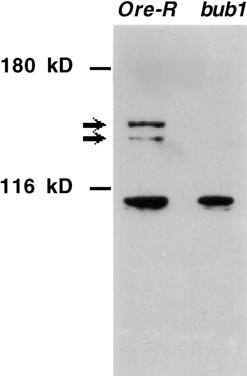
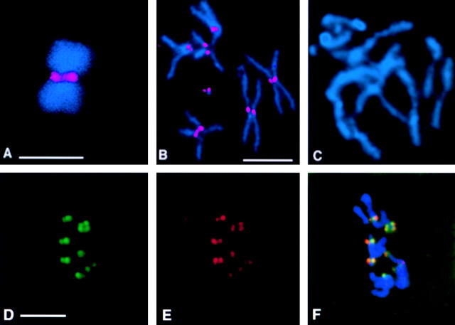
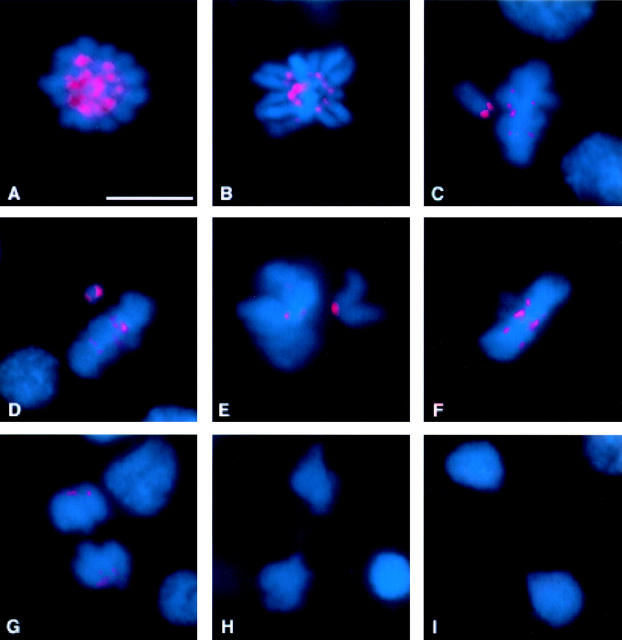
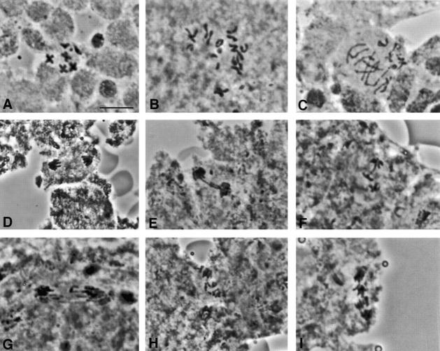
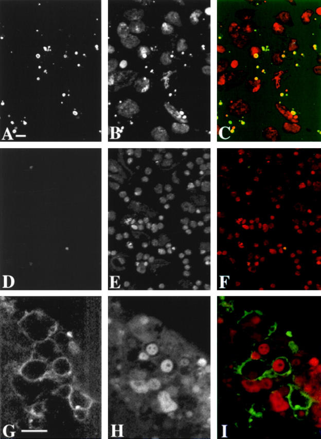
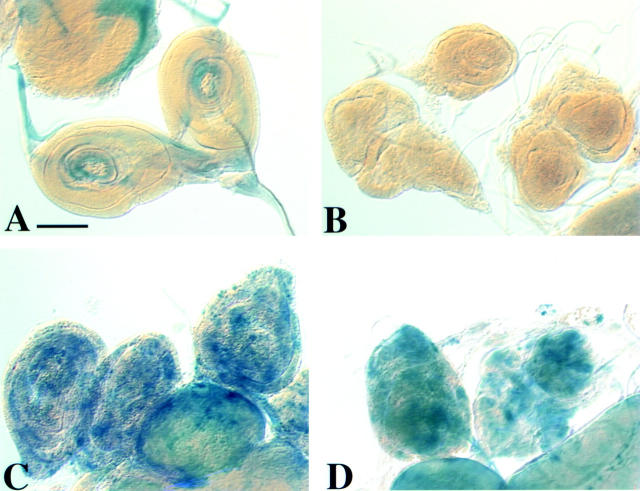
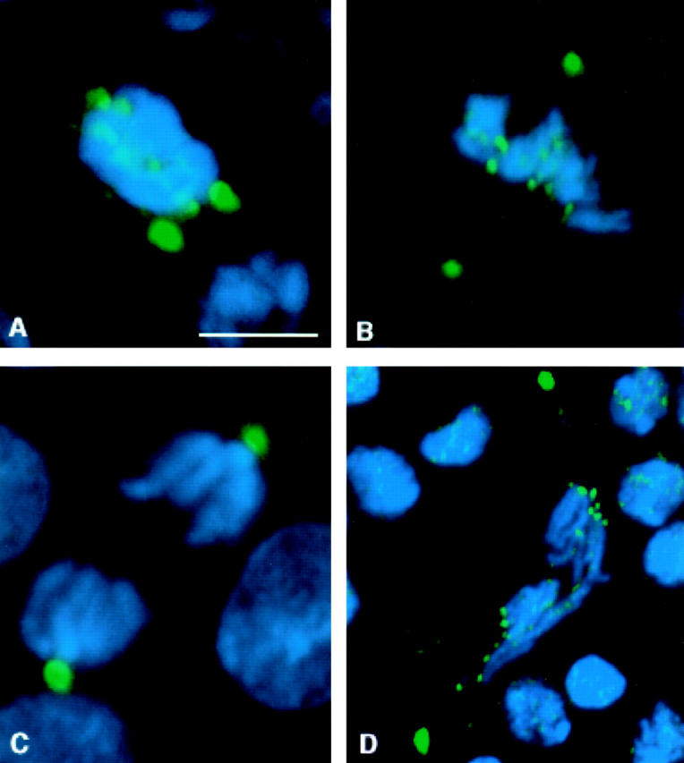
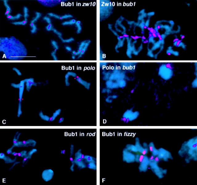
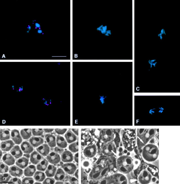
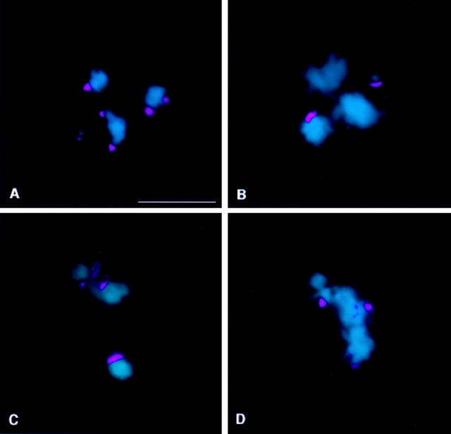
We have characterized the Drosophila mitotic checkpoint control protein Bub1 and obtained mutations in the bub1 gene. Drosophila Bub1 localizes strongly to the centromere/kinetochore of mitotic and meiotic chromosomes that have not yet reached the metaphase plate. Animals homozygous for P-element-induced, near-null mutations of bub1 die during late larval/pupal stages due to severe mitotic abnormalities indicative of a bypass of checkpoint function. These abnormalities include accelerated exit from metaphase and chromosome missegregation and fragmentation. Chromosome fragmentation possibly leads to the significantly elevated levels of apoptosis seen in mutants. We have also investigated the relationship between Bub1 and other kinetochore components. We show that Bub1 kinase activity is not required for phosphorylation of 3F3/2 epitopes at prophase/prometaphase, but is needed for 3F3/2 dephosphorylation at metaphase. Neither 3F3/2 dephosphorylation nor loss of Bub1 from the kinetochore is a prerequisite for anaphase entry. Bub1's localization to the kinetochore does not depend on the products of the genes zw10, rod, polo, or fizzy, indicating that the kinetochore is constructed from several independent subassemblies.


Figures

Figure 1
Figure 1
Specificity of affinity-purified anti-Drosophila Bub1 antibodies. Identical amounts of Drosophila third instar larval brain extracts, from either wild-type (Oregon R) or bub1 mutant homozygotes, were loaded onto the indicated lanes of a Western blot probed with affinity-purified anti-Drosophila Bub1 antibodies (see Materials and Methods). A doublet of ∼165 kD (the predicted size for Drosophila Bub1) is recognized in wild-type extracts. These two bands are almost completely absent in brain extracts made from bub1 mutant brains. The antibody also recognizes a band of ∼100 kD that is not Bub1-specific and that is also recognized by pre-immune IgY. This cross-reacting band serves as an internal loading control.
Figure 2
Figure 2
Drosophila Bub1 is recruited to kinetochores. DNA is shown in blue and Bub1 is in red. (A) A chromosome isolated from a Drosophila S2 cell arrested with colchicine, showing strong Bub1 staining at the kinetochores. (B) Bub1 is localized to the kinetochores in a wild-type (Oregon-R) neuroblast arrested with colchicine. (C) A neuroblast from a bub1 mutant (l(2)K06109/l(2)K06109) brain arrested with colchicine and stained with affinity-purified anti-Bub1 antibodies under identical conditions to those used in B. Note the complete absence of Bub1 staining in this figure; complete lack of Bub1 staining is also observed in all other bub1 allelic and deficiency combinations (not shown). D–F show that Bub1 (red) colocalizes with the kinetochore marker ZW10 (green) in wild-type neuroblasts, although the levels of staining of individual kinetochores with the two reagents are not always in concert. Bars, 5 μm. A–C are at the same magnification, as are D–F.
Figure 3
Figure 3
Bub1 distribution in cycling Drosophila S2 tissue culture cells. DNA is shown in blue and Bub1 is in red. (A) Prophase. Bub1 is strongly associated with the kinetochores of the condensing chromosomes. (B) Strong kinetochore staining continues to be observed into prometaphase. (C–E) As cells approach metaphase, chromosomes that are aligned along the metaphase plate show only weak Bub1 staining, while chromosomes that have not yet reached the metaphase plate continue to show intense Bub1 staining at one or both kinetochores. Occasionally, as in D, the two kinetochores of the lagging chromosome stain show different intensities of Bub1 signals. (F) At metaphase, all the chromosomes show weak Bub1 staining at the kinetochores, which continues to be detectable into early anaphase (G). (H) Later in anaphase, kinetochore staining is no longer detectable, although some staining of the spindle midzone is visible. In addition to the mitotic figures, interphase nuclei are also visible in G and H. (I) During telophase, no specific Bub1 staining pattern is observed. Bar, 5 μm.
Figure 4
Figure 4
Phenotype of bub1 mutants in Drosophila neuroblasts. (A–C) Orcein-stained mitotic figures from third instar larval brains treated with colchicine and hypotonic solution to perturb spindle assembly. Wild-type neuroblasts arrest in a prometaphase like configuration with attached sister chromatids as in A. In bub1 mutant neuroblasts treated in the same fashion (B and C), sister chromatids separate, and some evidence for aneuploidy or chromosome fragmentation is observed (diploid cells should contain 12 large chromatids and four dot-like 4th chromosome chromatids). (D–I) Orcein-stained anaphase figures from untreated brains. In contrast with a wild-type (D), bub1 mutant anaphases show several abnormalities including extensive chromatin bridging (E and F), chromosomes that lag in the vicinity of the metaphase plate (G), and apparent widespread chromosome fragmentation (H and I). Bar, 5 μm.
Figure 5
Figure 5
Larval brains of bub1 mutants contain many apoptotic nuclei. bub1 mutant (A–C) and wild-type (Oregon-R; D–F) brains were labeled by a TUNEL-based assay (A and D) for the presence of apoptotic nuclei, and stained with propidium iodide for DNA (B and E); a merged view with DNA in red and the TUNEL signal in green is shown in C and F. Many apoptotic nuclei are seen in bub1 mutant brains but not in wild-type; the majority of these TUNEL-positive nuclei are also pycnotic as seen by the abnormally condensed DNA signal. G–I show bub1 mutant larval brains stained with annexin V to reveal phosphatidylserine on the outside of the cell membrane (G), propidium iodide (H), and a merged view (I) with DNA staining in red and the annexin V signal in green. No annexin V staining is observed within wild-type brains (not shown). Bars: (A) 10 μm; (G) 5 μm. A–F are at the same magnification, as are G–I.
Figure 6
Figure 6
Expression of reaper in bub1 mutants. Imaginal discs were stained with X-gal to follow expression of a reaper-lacZ reporter construct (rpr-lacZ) in larvae of various genotypes. (A) rpr-lacZ/rpr-lacZ. (B) bub1/bub1 without the rpr reporter gene. (This control was necessitated by the fact that the P elements causing the bub1 mutations contain a lacZ gene.) (C and D) bub1/bub1; rpr-lacZ/rpr-lacZ imaginal discs showing very high levels of β-galactosidase expression, indicating that the cell death program is activated in many cells. Similar results were observed in the brains of these same animals (not shown). Bar, 100 μm.
Figure 7
Figure 7
Distribution of 3F3/2 phosphoepitopes in bub1 mutant neuroblasts. DNA is in blue, and 3F3/2 phosphoepitopes are in green. In all panels, the two strongest sites of 3F3/2 staining are the centrosomes. 3F3/2 distribution in prophase (A) and metaphase (B) neuroblasts from bub1 mutants. 3F3/2 epitopes at the centrosomes and kinetochores are strongly recognized, demonstrating that the Bub1 kinase is not a significant source of 3F3/2 phosphorylation activity in vivo. (C) 3F3/2 epitopes are completely dephosphorylated during anaphase in wild-type neuroblasts (see Bousbaa et al. 1997 for a detailed description of 3F3/2 distribution in wild-type Drosophila neuroblasts). (D) 3F3/2 distribution in an anaphase figure from a bub1 mutant neuroblast. 3F3/2 epitopes continue to remain phosphorylated in bub1 anaphases, though at reduced levels relative to those seen during prophase/prometaphase. Thus, total dephosphorylation of 3F3/2 phosphoepitopes cannot be a prerequisite for entry into anaphase. Bar, 5 μm.
Figure 8
Figure 8
Relationship of Bub1 to other kinetochore components. In all panels, DNA is in blue. Neuroblasts were treated with colchicine in all panels except D. (A) Bub1 (red) in a zw10S1/Y mutant neuroblast. Note the Bub1 staining at the kinetochores of separated sister chromatids. (B) ZW10 (red) stains the kinetochores in bub1 mutant neuroblasts. (C) In neuroblasts homozygous for the mutation polo1, Bub1 associates with the kinetochores. (D) In bub1 mutant neuroblasts (here shown at anaphase), Polo protein is bound to the kinetochores of the separating chromosomes, as in wild-type. (E) Null mutations in rough deal (rod) do not prevent the association of Bub1 with kinetochores. (F) Bub1 also binds to kinetochores in the neuroblasts of animals homozygous for the mutation fizzy6. Bar, 5 μm.
Figure 9
Figure 9
Role of Bub1 during Drosophila spermatogenesis. DNA is shown in blue and Bub1 is in red. (A–C) Localization of Bub1 during the first meiotic division. (A) Bub1 is strongly associated with the kinetochores at prometaphase I. (B) Kinetochore staining is significantly reduced by metaphase I and lost completely by anaphase I (C). (D–F) Localization of Bub1 during the second meiotic division parallels the behaviour of Bub1 during the first meiotic division. (G and H) Living spermatids from third instar larval testes viewed by phase contrast optics. A field of wild-type “onion stage” spermatids is shown in (G). Note that each spermatid contains a single phase light nucleus and a single phase dark Nebenkern (mitochondrial derivative), and that the volume of all nuclei are the same, indicating that chromosome segregation has occurred correctly. In contrast, a field of spermatids from a bub1 mutant testis (H) displays evidence of chromosome missegregation, in the form of spermatids with abnormal numbers of nuclei (arrows) or with micronuclei (arrowheads). Bars, 5 μm. A–F appear at the same magnification, as do G and H.
Figure 10
Figure 10
Bub1 responds to tension. DNA is shown in blue and Bub1 is in red. (A) Prometaphase I spermatocyte from a X^Y; C(4)RM stock, showing strong kinetochore staining of comparable intensity on both bivalents and univalents. (B–D) Once the bivalents align at the metaphase plate, the intensity of kinetochore staining is sharply decreased. Univalents continue to show strong kinetochore association with Bub1, indicating that Bub1 can discriminate between the presence and absence of bipolar tension at the kinetochores. Bar, 5 μm.

Similar articles

Cited by

References

    1. Ahmad K., Golic K.G. Telomere loss in somatic cells of Drosophila causes cell cycle arrest and apoptosis. Genetics. 1999;151:1041–1051. - PMC - PubMed
    1. Ault J.G., Lin H.P. Bivalent behaviour in Drosophila melanogaster males containing the In(1)sc4Lsc8RXchromosome. Chromosoma. 1984;90:222–228. - PubMed
    1. Ault J.G., Nicklas R.B. Tension, microtubule rearrangements, and the proper distribution of chromosomes in mitosis. Chromosoma. 1989;98:33–39. - PubMed
    1. Baker B.S., Smith D.A., Gatti M. Region specific effects on chromosome integrity of mutations at essential loci in Drosophila melanogaster . Proc. Natl. Acad. Sci. USA. 1982;79:1205–1209. - PMC - PubMed
    1. Basu, J. 1999. The spindle assembly checkpoint in Drosophila. Ph.D. thesis. Cornell University, Ithaca, NY.

Publication types

MeSH terms

Associated data