. 1999 Jul 12;146(1):29-44.
doi: 10.1083/jcb.146.1.29.

A visual screen of a GFP-fusion library identifies a new type of nuclear envelope membrane protein

Affiliations

A visual screen of a GFP-fusion library identifies a new type of nuclear envelope membrane protein

M M Rolls et al. J Cell Biol. .

Abstract

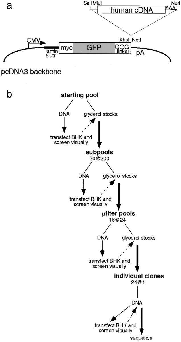
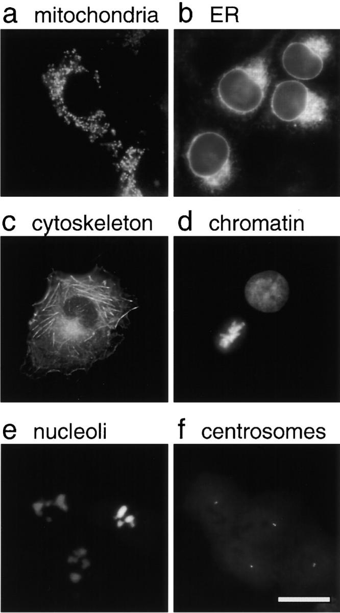
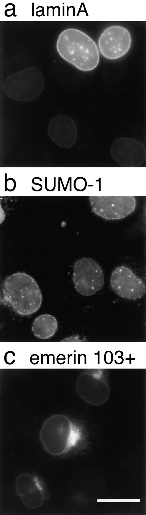
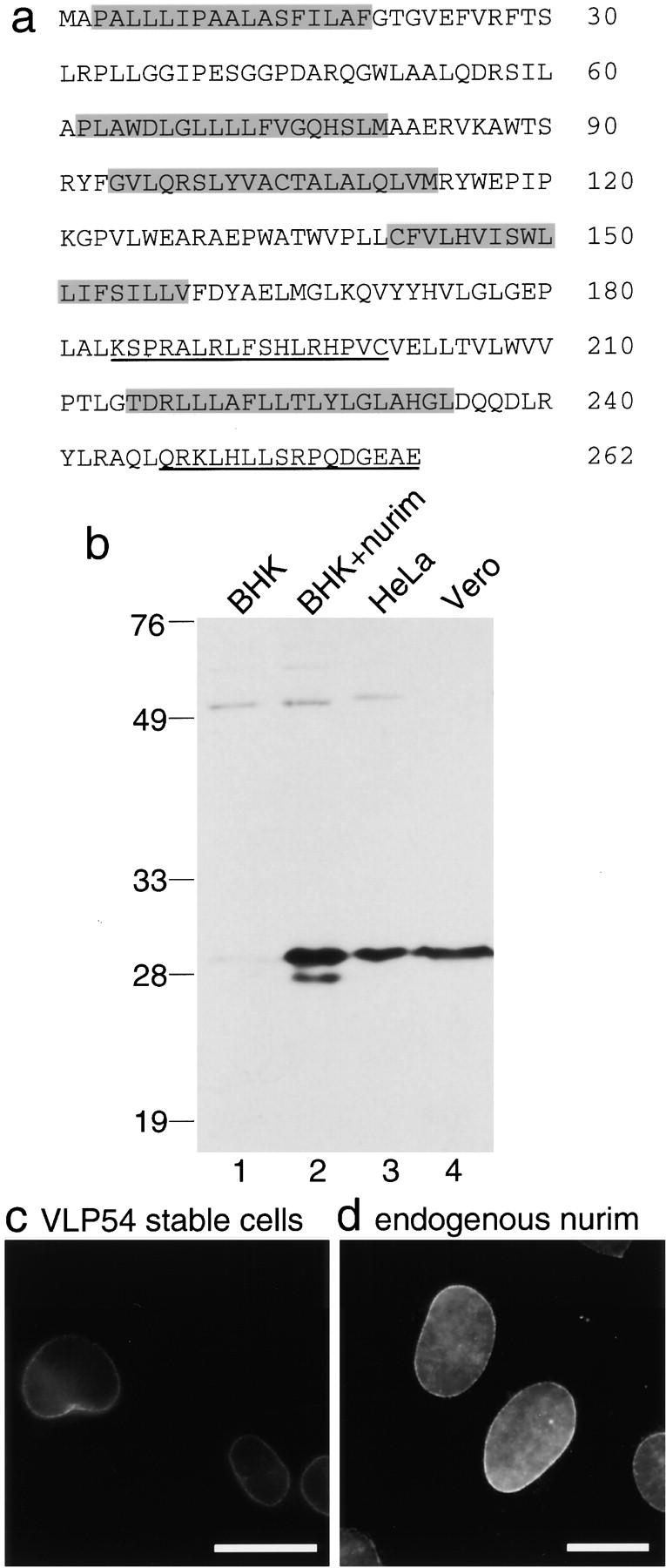
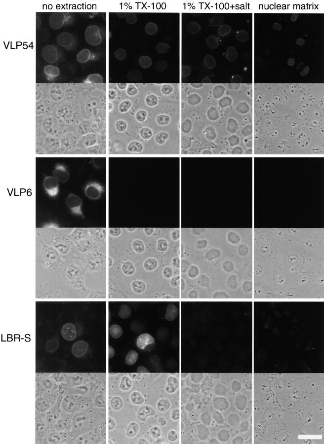
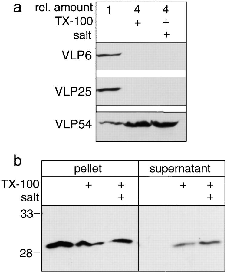
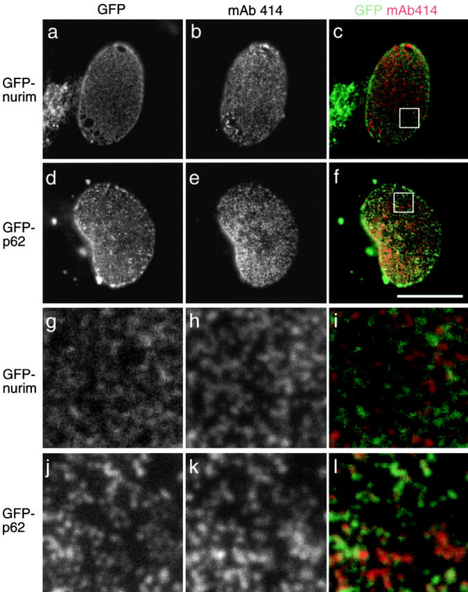
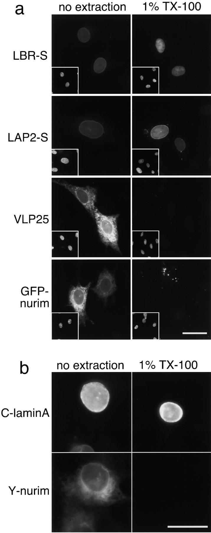
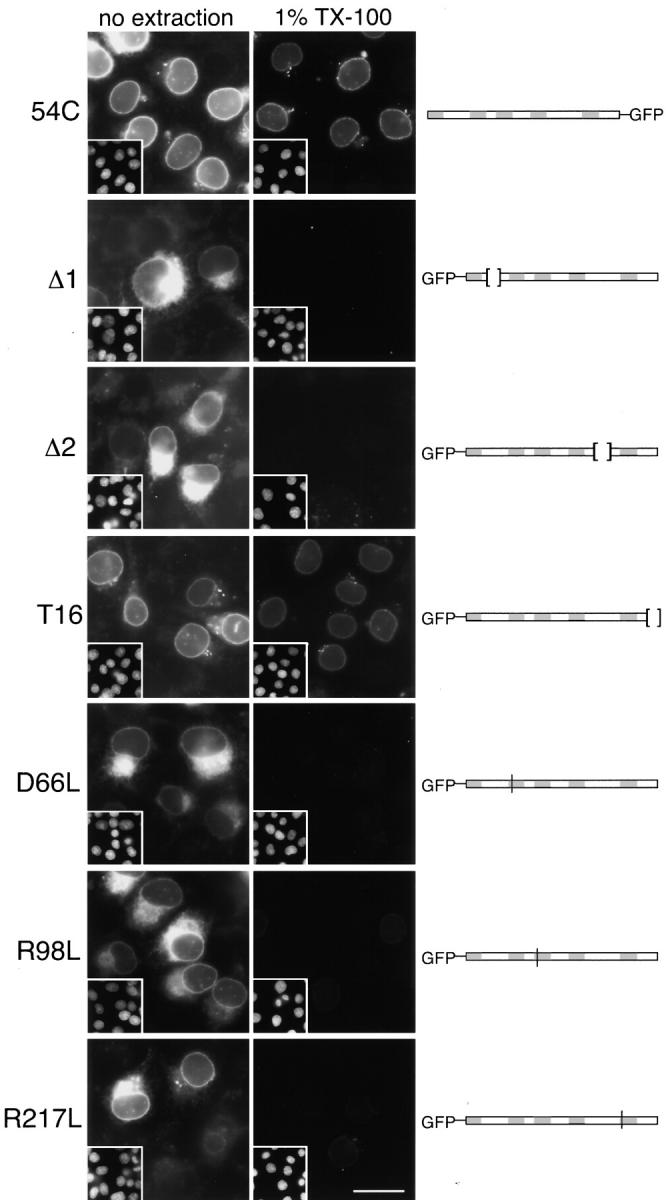
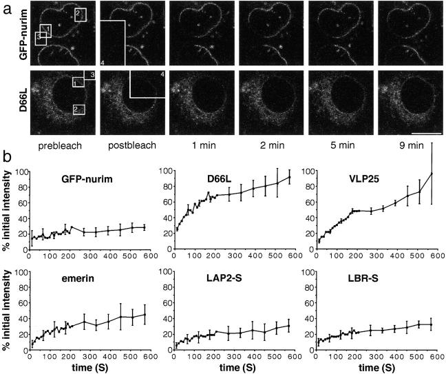
The nuclear envelope (NE) is a distinct subdomain of the ER, but few membrane components have been described that are specific to it. We performed a visual screen in tissue culture cells to identify proteins targeted to the NE. This approach does not require assumptions about the nature of the association with the NE or the physical separation of NE and ER. We confirmed that screening a library of fusions to the green fluorescent protein can be used to identify proteins targeted to various subcompartments of mammalian cells, including the NE. With this approach, we identified a new NE membrane protein, named nurim. Nurim is a multispanning membrane protein without large hydrophilic domains that is very tightly associated with the nucleus. Unlike the known NE membrane proteins, it is neither associated with nuclear pores, nor targeted like lamin-associated membrane proteins. Thus, nurim is a new type of NE membrane protein that is localized to the NE by a distinct mechanism.


Figures

Figure 1
Figure 1
Overview of the visual screen. (a) Construction of the expression library. Human cDNAs from an osteosarcoma cell line were inserted into a mammalian expression vector derived from pcDNA3. The vector was modified by the addition of part of the 5′ untranslated region of lamin A (lamin 5′ utr), as well as the coding sequences of a myc tag, a GFP variant (S65T and V163A), and a flexible linker (GGGLDPRVR). The library was initially generated as 20 master pools of 25,000 clones each. (b) Screening the expression library. Starting pools were generated by subdividing each master pool into 20 pools of 2,000 clones. These pools were screened for localized proteins by transfecting each pool into ∼2 × 10⁴ cells grown on a coverslip. The transfection efficiency varied from 20–50% meaning that each clone in the pool was expressed in several cells. Transfected pools were screened visually by epifluorescence microscopy. Pools that gave rise to interesting patterns were retrieved from glycerol stocks and further subdivided and screened until single clones were isolated.
Figure 2
Figure 2
Examples of fluorescence patterns generated by expression of individual clones isolated in the visual screen. Individual clones were transiently transfected into BHK cells. (a) A mitochondrial pattern is shown by VLP32, a GFP fusion to ADP/ATP translocase; (b) an ER pattern by VLP16, a fusion to the signal peptidase 25 kD subunit; (c) a cytoskeletal pattern by VLP11, a fusion to β-actin; (d) a chromatin pattern by VLP51, a fusion to histone H1; (e) a nucleolar pattern by VLP56, a fusion to ribosomal protein L27; and (f) a centrosomal pattern by VLP31, a fusion to the ATCase domain of CAD. Bar, 20 μm.
Figure 3
Figure 3
Examples of fluorescence patterns generated by GFP fusions to known NE proteins. BHK cells were transiently transfected with isolated clones (a) VLP4, a fusion to lamin A; (b) VLP23, a fusion to SUMO-1; and (c) VLP33, a fusion to the COOH-terminal half of emerin (starting at amino acid 103). Bar, 20 μm.
Figure 4
Figure 4
Nurim, a new NE membrane protein isolated in the visual screen. (a) Predicted amino acid sequence of nurim based on the coding sequence included in VLP54. Predicted transmembrane domains are shaded gray and peptide sequences 54.1 and 54.2 used to generate antibodies are underlined. These sequence data are available from GenBank/EMBL/DDBJ under accession number HSNRM29 AF143676. (b) An immunoblot with affinity-purified antibody 254 to peptide 54.2. Lane 1 was loaded with protein from 8 × 10⁵ BHK cells; lane 2 with 1 × 10⁵ BHK cells transiently transfected with an untagged version of VLP54; lane 3 with 8 × 10⁵ HeLa nuclei; and lane 4 with 8 × 10⁵ Vero nuclei. The position of molecular mass markers is shown at the left and their size is in kD. (c) Nuclear rim fluorescence is shown with a BHK stable cell line expressing VLP54 and (d) Vero cells stained with affinity-purified antibody 253 to peptide 54.2. Bars, 20 μm.
Figure 5
Figure 5
Extractions of cells expressing VLP54, VLP6 (a GFP fusion to the ER protein HO-2), and LBR-S (a GFP fusion to a truncated LBR). BHK cells were transiently transfected with expression constructs and fixed (no extraction) or extracted on ice with 1% TX-100 in PBS (1% TX-100), or 1% TX-100 in PBS supplemented with 350 mM NaCl (1% TX-100 + salt), or subjected to a nuclear matrix preparation (nuclear matrix) before fixation. Upper panels show GFP fluorescence and lower panels show corresponding phase-contrast images. All GFP images were taken at the same exposure and subsequently scaled identically. Bar, 20 μm.
Figure 6
Figure 6
Association of VLP54 and endogenous nurim with extracted nuclear pellets. (a) BHK cells stably expressing either VLP54 or a cyan version of VLP6 (HO-2) and yellow version of VLP25 (Sec61β) were treated with PBS, PBS with 1% TX-100, or PBS with 1% TX-100 and high salt. Nuclei were pelleted, washed, and analyzed by SDS-PAGE followed by immunoblotting with an anti-GFP antibody. Four times the amount of extracted nuclei compared with unextracted nuclei (1 × 10⁶) were analyzed. (b) HeLa cell nuclei were treated as in a, and analyzed by immunoblotting with affinity-purified antinurim antibody 254. In this experiment supernatants were also collected and the amount from equivalent numbers of cells (3 × 10⁵) were loaded in all lanes.
Figure 7
Figure 7
Localization of GFP-p62 and GFP-nurim compared with that of nuclear pores. Vero cells were transiently transfected with either GFP-nurim (a–c and g–i) or GFP-p62 (d–f and j–l). The bottom surfaces of nuclei are shown with enlargements of the regions indicated in c and f shown in g–i and j–l, respectively. (a, d, g, and j) GFP fluorescence and (b, e, h, and k) nuclear pore labeling by mAb 414 followed by a rhodamine secondary antibody. (c, f, i, and l) An overlay with GFP in green and rhodamine in red. Bar, 20 μm.
Figure 8
Figure 8
Localization and detergent sensitivity of GFP-fusions in chicken cells. (a) Chicken fibroblasts were transiently transfected with LBR-S and LAP2-S (GFP fusions to NE proteins), VLP25 (a GFP fusion to the ER protein Sec61β), or GFP-nurim. Cells were either fixed immediately (no extraction) or extracted with 1% TX-100 before fixation (1% TX-100). GFP fluorescence is shown in large images and Hoechst staining in insets. GFP images were taken at the same exposure and scaled identically. (b) Chicken cells were transiently transfected with both CFP-lamin A and YFP-nurim and treated as in a. The cyan and yellow images of the same cells are shown in the top and bottom frames, respectively. All images were taken at the same exposure and subsequently scaled identically. Bars, 20 μm.
Figure 9
Figure 9
Localization and detergent sensitivity of GFP-nurim deletions and point mutants. BHK cells were transiently transfected with nurim-tagged with GFP at the COOH terminus rather than the NH2 terminus (54C) or loop deletions (Δ1 and Δ2), truncation (T16), or point mutations (D66L, R98L, and R217L); diagrams of the mutants are included to the right with shading representing predicted transmembrane domains. Cells were either fixed immediately (no extraction) or extracted with 1% TX-100 before fixation (1% TX-100). GFP fluorescence is shown in large images and Hoechst staining to show nuclei in insets. All GFP images were taken at the same exposure and scaled identically. Bar, 20 μm.
Figure 10
Figure 10
FRAP of GFP-fusions to NE and ER proteins. (a) BHK cells transiently transfected with GFP-nurim or point mutant D66L were imaged with a confocal microscope. A portion of the cell (box 4) was subjected to photobleaching and fluorescence recovery monitored by imaging every 11s for 220 s, and then every minute for 5 min. Examples of images of cells at various times after recovery are shown. (b) FRAP experiments were performed as in a for GFP-nurim, D66L, VLP25 (a GFP fusion to Sec61β), YFP-emerin, LAP2-S, and LBR-S. The results from three bleached cells were quantitated and combined and the SD indicated with a bar (some of the SDs for early time points are not shown in the plots, but were similar to those shown). For quantitation, the total pixel intensity in a region of the cell that included the NE (a, box 1) was calculated. The intensity of an equivalent sized background area (box 3) was subtracted and, finally, the images were normalized for brightness using an unbleached region of the cell (box 2). Bar, 10 μm.

Similar articles

Cited by

References

    1. Altschul S.F., Madden T.L., Schaffer A.A., Zhang J., Zhang Z., Miller W., Lipman D.J. Gapped BLAST and PSI-BLASTa new generation of protein database search programs. Nucleic Acids Res. 1997;25:3389–3402. - PMC - PubMed
    1. Bailer S.M., Eppenberger H.M., Griffiths G., Nigg E.A. Characterization of a 54-kD protein of the inner nuclear membraneevidence for a cell cycle-dependent interaction with the nuclear lamina. J. Cell Biol. 1991;114:389–400. - PMC - PubMed
    1. Bastos R., Panté N., Burke B. Nuclear pore complex proteins. Int. Rev. Cytol. 1995;162B:257–302. - PubMed
    1. Bergmann J.E., Singer S.J. Immunoelectron microscopic studies of the intracellular transport of the membrane glycoprotein (G) of vesicular stomatitis virus in infected Chinese hamster ovary cells. J. Cell Biol. 1983;97:1777–1787. - PMC - PubMed
    1. Bione S., Maestrini E., Rivella S., Mancini M., Regis S., Romeo G., Toniolo D. Identification of a novel X-linked gene responsible for Emery-Dreifuss muscular dystrophy. Nat. Genet. 1994;8:323–327. - PubMed

Publication types

MeSH terms

Associated data