. 1999 Jul 12;146(1):57-70.
doi: 10.1083/jcb.146.1.57.

A role for the vesicle tethering protein, p115, in the post-mitotic stacking of reassembling Golgi cisternae in a cell-free system

Affiliations

A role for the vesicle tethering protein, p115, in the post-mitotic stacking of reassembling Golgi cisternae in a cell-free system

J Shorter et al. J Cell Biol. .

Abstract

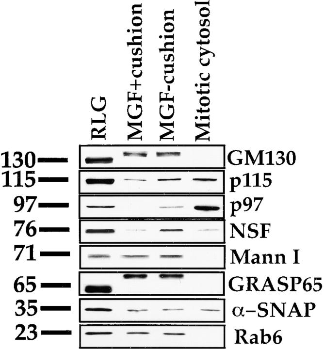
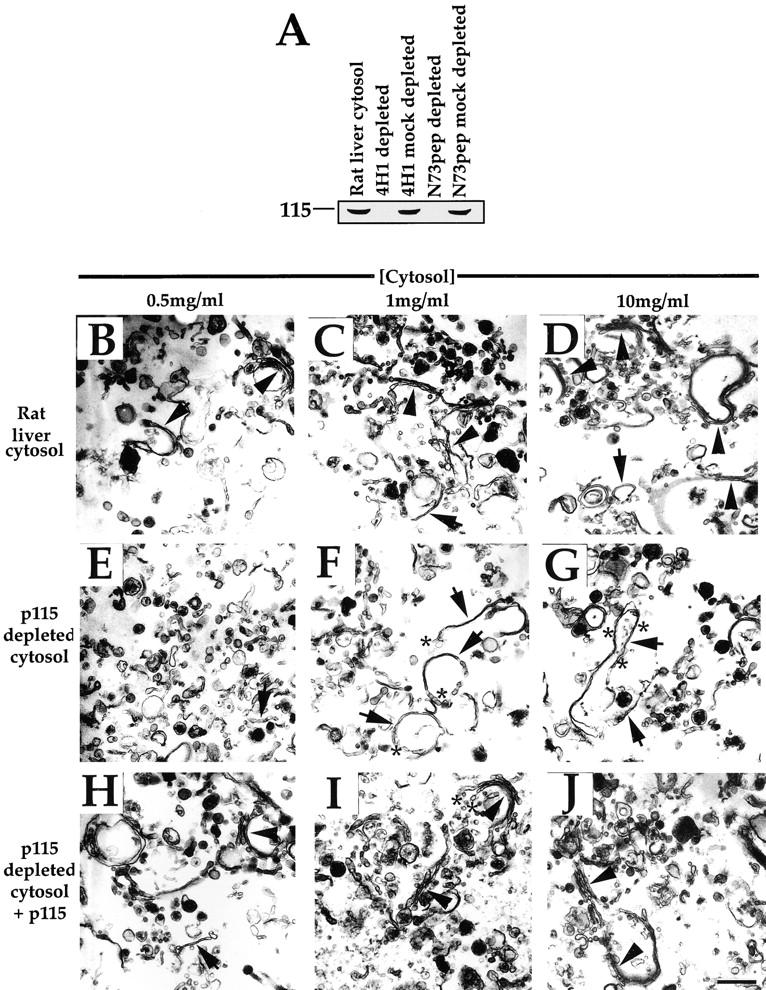
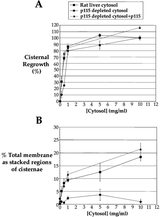
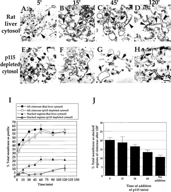
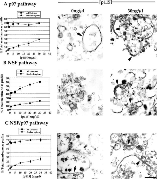
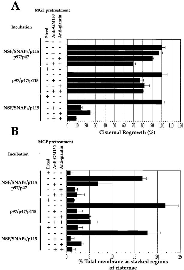
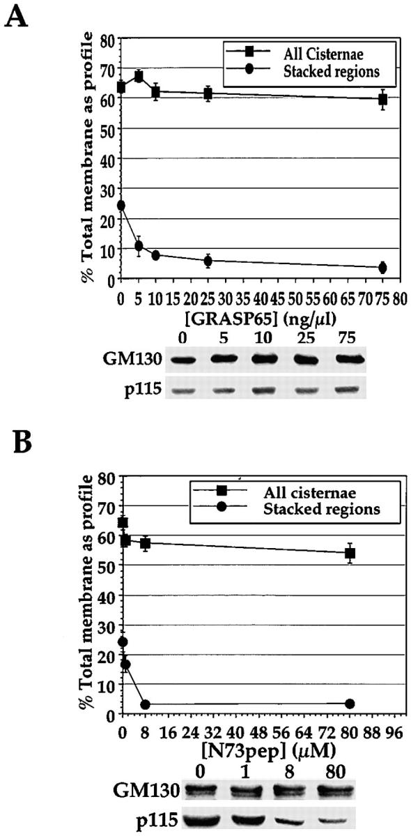
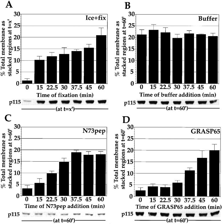
During telophase, Golgi cisternae are regenerated and stacked from a heterogeneous population of tubulovesicular clusters. A cell-free system that reconstructs these events has revealed that cisternal regrowth requires interplay between soluble factors and soluble N-ethylmaleimide (NEM)-sensitive fusion protein (NSF) attachment protein receptors (SNAREs) via two intersecting pathways controlled by the ATPases, p97 and NSF. Golgi reassembly stacking protein 65 (GRASP65), an NEM-sensitive membrane-bound component, is required for the stacking process. NSF-mediated cisternal regrowth requires a vesicle tethering protein, p115, which we now show operates through its two Golgi receptors, GM130 and giantin. p97-mediated cisternal regrowth is p115-independent, but we now demonstrate a role for p115, in conjunction with its receptors, in stacking p97 generated cisternae. Temporal analysis suggests that p115 plays a transient role in stacking that may be upstream of GRASP65-mediated stacking. These results implicate p115 and its receptors in the initial alignment and docking of single cisternae that may be an important prerequisite for stack formation.


Figures

Figure 1
Figure 1
Western analysis of MGF isolated with or without a 0.5-M sucrose cushion. 10 μg RLG, MGF derived from 10 μg RLG isolated with or without a 0.5-M sucrose cushion, and 10 μg sHeLa mitotic cytosol (1% of input) were fractionated by SDS-PAGE, transferred to nitrocellulose, and probed for GM130, p115, p97, NSF, Mann I, GRASP65, α-SNAP, and rab6 using specific antibodies. Molecular weights in kD are shown on the left. Note the shift in molecular weight of GM130 and GRASP65 in the MGF owing to mitotic phosphorylation of these two proteins.
Figure 2
Figure 2
Effect of interphase cytosol depleted of p115 on the reassembly process. (A) Rat liver cytosol was depleted of p115, as described in Materials and Methods, using either the anti-p115 mAb antibody 4H1 or the N73pep. 20 μg of cytosol was fractionated by SDS-PAGE using a 7.5% gel, transferred to nitrocellulose, and probed with the anti-p115 mAb 8A6. (B–J) MGF isolated through a 0.5-M sucrose cushion were incubated for 60 min at 37°C with increasing concentrations of either rat liver cytosol (B–D), p115-depleted cytosol (E–G), or p115-depleted cytosol supplemented with purified rat liver p115 (H–J), fixed, and processed for EM. Representative fields are shown. Note the presence of stacks (arrowheads) in B–D and H–J, but only single cisternae (arrows) in E–G. The reduced number of cisternae in E suggests poor reassembly at this concentration of p115-depleted cytosol. Note that the cisternae formed often have a wrinkled appearance (asterisks in G) in p115-depleted cytosol and are often blunt-ended with few associated vesicles (compare asterisks in F and I), in contrast to when p115 is present. Bar, 0.5 μm.
Figure 3
Figure 3
Quantitation of Golgi membrane reassembly in rat liver cytosol, p115-depleted cytosol, and p115-depleted cytosol supplemented with p115. MGF isolated through a 0.5-M sucrose cushion were incubated for 60 min at 37°C with increasing concentrations of either rat liver cytosol, p115-depleted cytosol, or p115-depleted cytosol supplemented with purified rat liver p115, fixed and processed for EM, and quantitated as described in Materials and Methods. (A) The percentage cisternal regrowth ± SEM formula image for each cytosol concentration tested. (B) The percentage total membrane present as stacked regions of cisternae ± SEM formula image for each cytosol concentration tested.
Figure 4
Figure 4
Kinetic analysis of Golgi membrane reassembly in rat liver cytosol and p115-depleted cytosol. (A–H) MGF isolated through a 0.5-M sucrose cushion were incubated for increasing time at 37°C with either rat liver cytosol (A–D) or p115-depleted cytosol (E–H) with the cytosol concentration set at 10 mg/ml, fixed and processed for EM, and quantitated as described in Materials and Methods. Representative fields are shown. In rat liver cytosol, note that the first intermediate formed is the single cisterna after 5 min (arrows in A). By 15 min, these single cisternae had grown in length, had tubular networks associated with their rims (asterisks in B), and had begun to align and dock to form stacks (arrowheads in B). Many discrete stacks had formed by 45 min (arrowheads in C), which had joined up by 120 min (arrowheads in D). In p115-depleted cytosol, single cisternae were again present after 5 min (arrows in E), and had increased in length by 15 min (arrows in F), but were often blunt-ended (asterisk in F) and were not stacked. At 45 min, these cisternae remained blunt-ended (asterisk in G) and unstacked. By 120 min, some stacks of blunt-ended cisternae had begun to form (arrowhead in H), but many single cisternae remained (arrows in H). Bar, 0.5 μm. (I) Quantitation of the time course. The percentage total membrane present as cisternae ± SEM formula image and stacked regions of cisternae ± SEM formula image are presented for each cytosol at every time point tested. (J) MGF were reassembled in p115-depleted cytosol and supplemented with p115 at different times (time of addition of p115). The reaction was allowed to proceed for a total time of 120 min at 37°C. Samples were then fixed and processed for EM, and the percentage total membrane as stacked regions of cisternae ± SEM formula image are presented for each time point tested.
Figure 5
Figure 5
Titration of p115 into the p97, NSF, and NSF/p97 catalyzed reassembly reactions. MGF isolated through a 0.5-M sucrose cushion were incubated for 60 min at 37°C with: A, p97 (70 ng/μl) and p47 (37.5 ng/μl); B, NSF (100 ng/μl), α-SNAP (25 ng/μl) and γ-SNAP (25 ng/μl); or C, all these components combined with from 0–30 ng/μl p115. Samples were fixed and processed for EM, and then quantitated for percentage total membrane as cisternae ± SEM formula image and percentage total membrane present as stacked regions of cisternae ± SEM formula image. Results are displayed in graphic form at the left and representative fields of the two extreme p115 concentrations are positioned adjacent. Note the long single, wrinkled cisternae formed by the p97 pathway in the absence of p115 (arrow in A) and the long stacks of two cisternae with maximum p115 (arrowheads in A). Note the virtual absence of cisternae for the NSF pathway in the absence of p115 (B) and the short stacks of three or more cisternae with maximum p115 (arrowheads in B). Note the long, wrinkled single cisternae formed by the NSF/p97 pathway in the absence of p115 (arrow in C) and the stacks of two or three cisternae with maximum p115 (arrowheads in C). Bar, 0.5 μm.
Figure 6
Figure 6
Effect of antibodies against giantin and GM130 on the reassembly process using pure components. MGF isolated through a 0.5-M sucrose cushion were either fixed and processed for EM, held on ice for 15 min, or preincubated on ice for 15 min with 1 μl of anti-GM130 NN15 serum, antigiantin serum, or a combination of both. The pretreated MGF were then incubated for 60 min at 37°C with: p97 (70 ng/μl); p47 (37.5 ng/μl) and p115 (30 ng/μl); or NSF (100 ng/μl), α-SNAP (25 ng/μl), γ-SNAP (25 ng/μl) and p115 (30 ng/μl); or all these components combined. Samples were fixed and processed for EM, and the percentage cisternal regrowth ± SEM formula image and the percentage total membrane present as stacked regions of cisternae ± SEM formula image was determined.
Figure 7
Figure 7
Effect of soluble GRASP65 and N73pep on the reassembly process using pure components. MGF isolated through a 0.5-M sucrose cushion were preincubated for 15 min on ice with increasing amounts of soluble GRASP65 (A) or N73pep (B). The pretreated MGF were then incubated for 60 min at 37°C with p97 (70 ng/μl), p47 (37.5 ng/μl), p115 (30 ng/μl), NSF (100 ng/μl), α-SNAP (25 ng/μl), and γ-SNAP (25 ng/μl). Samples were either fixed and processed for EM and quantitated, with the percentage total membrane present as cisternae ± SEM formula image and as stacked regions of cisternae ± SEM formula image shown; or the membranes were recovered and fractionated by SDS-PAGE using a 7.5% gel, transferred to nitrocellulose, and probed for GM130 and p115 using specific antibodies (shown below graphs in A and B).
Figure 8
Figure 8
Temporal sensitivity of stacking reaction to soluble GRASP65 and N73pep. MGF isolated through a 0.5-M sucrose cushion were incubated for the indicated time (time of addition) at 37°C with p97 (70 ng/μl), p47 (37.5 ng/μl), p115 (30 ng/μl), NSF (100 ng/μl), α-SNAP (25 ng/μl), and γ-SNAP (25 ng/μl). At this time, the reactions were transferred to ice and either fixed with 2% glutaraldehyde and processed for EM (A) and the percentage total membrane present as stacked regions of cisternae ± SEM formula image was determined, or the amount of p115 bound to the membranes at each time point was determined (shown below graph in A). Alternatively, reactions were transferred to ice and supplemented with buffer (B), 80 μM N73pep (C), or 75 ng/μl GRASP65 (D). After 15 min on ice, buffer-, N73pep-, and GRASP65-treated samples were transferred to 37°C and incubated for a total time of 60 min. Samples were fixed and processed for EM, quantitated, and the percentage total membrane present at the end of the incubation as stacked regions of cisternae ± SEM formula image was determined. The amount of p115 bound to Golgi membranes at the end of the incubation was also determined by Western blotting (shown below each graph).

References

    1. Acharya U., Jacobs R., Peters J.M., Watson N., Farquhar M.G., Malhotra V. The formation of Golgi stacks from vesiculated Golgi membranes requires two distinct fusion events. Cell. 1995;82:895–904. - PubMed
    1. Acharya U., Mallabiabarrena A., Acharya J.K., Malhotra V. Signaling via mitogen-activated protein-kinase (MEK 1) is required for Golgi fragmentation during mitosis. Cell. 1998;92:183–192. - PubMed
    1. Barr F.A., Puype M., Vandekerckhove J., Warren G. GRASP65, a protein involved in the stacking of Golgi cisternae. Cell. 1997;91:253–262. - PubMed
    1. Barr F.A., Nakamura N., Warren G. Mapping the interaction between GRASP65 and GM130, components of a protein complex involved in the stacking of Golgi cisternae. EMBO (Eur. Mol. Biol. Organ.) J. 1998;17:3258–3268. - PMC - PubMed
    1. Cao X., Ballew N., Barlowe C. Initial docking of ER-derived vesicles requires Uso1p and Ypt1p but is independent of SNARE proteins. EMBO (Eur. Mol. Biol. Organ.) J. 1998;17:2156–2165. - PMC - PubMed

Publication types

MeSH terms