. 1999 Jul 12;146(1):141-8.
doi: 10.1083/jcb.146.1.141.

DAP-kinase participates in TNF-alpha- and Fas-induced apoptosis and its function requires the death domain

Affiliations

DAP-kinase participates in TNF-alpha- and Fas-induced apoptosis and its function requires the death domain

O Cohen et al. J Cell Biol. .

Abstract

Death-associated protein (DAP)-kinase is a calcium/calmodulin regulated serine/threonine kinase that carries ankyrin repeats, a death domain, and is localized to the cytoskeleton. Here, we report that this kinase is involved in tumor necrosis factor (TNF)-alpha and Fas-induced apoptosis. Expression of DAP-kinase antisense RNA protected cells from killing by anti-Fas/APO-1 agonistic antibodies. Deletion of the death domain abrogated the apoptotic functions of the kinase, thus, documenting for the first time the importance of this protein domain. Overexpression of a fragment encompassing the death domain of DAP-kinase acted as a specific dominant negative mutant that protected cells from TNF-alpha, Fas, and FADD/MORT1-induced cell death. DAP-kinase apoptotic function was blocked by bcl-2 as well as by crmA and p35 inhibitors of caspases, but not by the dominant negative mutants of FADD/MORT1 or of caspase 8. Thus, it functions downstream to the receptor complex and upstream to other caspases. The multidomain structure of this serine/threonine kinase, combined with its involvement in cell death induced by several different triggers, place DAP-kinase at one of the central molecular pathways leading to apoptosis.


Figures

Figure 1
Figure 1
DAP-kinase antisense RNA expression protects HeLa cells from Fas-induced cell death. (a) HeLa cells were stably transfected with DAP kinase antisense cDNA fragment or with DHFR cDNA as a nonrelevant control. Viability was measured by neutral red dye uptake assay and was calculated as described in Materials and Methods either after 40 h of exposure to anti–Fas/APO-1 agonistic antibodies (50 ng/ml) or after 8 d of IFN-γ treatment (1,000 U/ml). The results represent an average of three independent experiments performed in quadruplet. (b) PARP cleavage analysis of the transfected HeLa cells treated with the indicated amount of anti–Fas/APO-1 antibodies. The upper band is the 116-kD noncleaved PARP and the lower band is the typical 85-kD cleaved product.
Figure 1
Figure 1
DAP-kinase antisense RNA expression protects HeLa cells from Fas-induced cell death. (a) HeLa cells were stably transfected with DAP kinase antisense cDNA fragment or with DHFR cDNA as a nonrelevant control. Viability was measured by neutral red dye uptake assay and was calculated as described in Materials and Methods either after 40 h of exposure to anti–Fas/APO-1 agonistic antibodies (50 ng/ml) or after 8 d of IFN-γ treatment (1,000 U/ml). The results represent an average of three independent experiments performed in quadruplet. (b) PARP cleavage analysis of the transfected HeLa cells treated with the indicated amount of anti–Fas/APO-1 antibodies. The upper band is the 116-kD noncleaved PARP and the lower band is the typical 85-kD cleaved product.
Figure 2
Figure 2
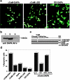
The death domain of DAP-kinase is important for its function in apoptosis. (a) Transient transfections of 293 cells with ΔCaM mutant, ΔCaM/ΔDD mutant, or with the DD-DAPk. Photographs were taken 24 h after transfection under fluorescent light microscope to visualize GFP positive cells. (b) Expression of ΔCaM and ΔCaM/ΔDD mutants in the transiently transfected 293 cells. Lane 1, expression of ΔCaM-DAPk; lane 2, expression of ΔCaM/ΔDD-DAPk; and lane 3, endogenous DAPk in cells transfected with a nonrelevant vector. Western blotting analysis was done with anti–DAP-kinase antibodies. (c) Schematic presentation of DAP-kinase mutant proteins used in these experiments. Kinase, kinase domain; CaM, calmodulin binding and regulatory domain; ankyrin, ankyrin repeats; and DD, death domain. Asterisks delineate the region that by deletion mapping was shown to be responsible for cytoskeletal binding. (d) Transfections as shown in a, with the indicated constructs or double transfections of ΔCaM mutant with the indicated constructs. The percentage of apoptotic cells was calculated as described in Materials and Methods.
Figure 3
Figure 3
Expression of DD-DAPk protects from TNF-α– and Fas-induced cell death. (a) Expression of endogenous DAP-kinase in cell lines. Western blotting analysis of extracts of indicated cells, using anti–DAP-kinase antibodies. (b) Expression of recombinant DD-DAPk. Extracts from cells transfected with DD-DAPk construct were immunoprecipitated by anti-Flag antibodies and analyzed by Western blotting using anti-Flag. (c) Transient transfection of 293, HeLa, or MCF7 cells with vectors encoding p55-TNF-R, GFP, and either luciferase (Luc), death domain of DAP-kinase (DD-DAPk), or dominant negative mutant of FADD/MORT1 (DN-MORT). The percentage of apoptotic cells was calculated as described in Materials and Methods. (d) Same as c, except that p55/Fas chimera was used instead of p55-TNF-R. (e) Transient transfection of HeLa cells with vectors encoding GFP and either luciferase, DD-DAPk, or DN-MORT. Cells were treated 24 h after transfection with a combination of TNF-α and cycloheximide. The number of apoptotic cells was scored under fluorescent microscopy 3 h after treatment.
Figure 4
Figure 4
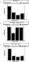
Functional positioning of DAP-kinase along TNF/Fas cell death pathways. Schematic representation of the results is depicted above each bar graph. (a) Transient transfections of 293 cells with vectors encoding FADD/MORT1 together with vectors encoding the indicated proteins. GFP plasmid is present in each of the transfections. (b and c) Transfections as in a, except that the ΔCaM mutant of DAP-kinase was used instead of FADD/MORT1 together with vectors encoding the indicated proteins.

Similar articles

Cited by

References

    1. Beidler D.R., Tewari M., Friesen P.D., Poirier G., Dixit V.M. The baculovirus p35 protein inhibits Fas- and tumor necrosis factor–induced apoptosis. J. Biol. Chem. 1995;270:16526–16528. - PubMed
    1. Boldin M.P., Varfolomeev E.E., Pancer Z., Mett I.L., Camonis J.H., Wallach D. A novel protein that interacts with the death domain of Fas/APO1 contains a sequence motif related to the death domain. J. Biol. Chem. 1995;270:7795–7798. - PubMed
    1. Boldin M.P., Goncharov T.M., Goltsev Y.V., Wallach D. Involvement of MACH, a novel Mort1/FADD–interacting protease, in Fas/APO-1 and TNF receptor-induced cell death. Cell. 1996;85:803–815. - PubMed
    1. Brancolini C., Benedetti M., Schneider C. Microfilament reorganization during apoptosisthe role of Gas2, a possible substrate for ICE-like proteases. EMBO (Eur. Mol. Biol. Organ.) J. 1995;14:5179–5190. - PMC - PubMed
    1. Chinnaiyan A.M., O'Rourke K., Tewari M., Dixit V.M. FADD, a novel death domain-containing protein, interacts with the death domain of Fas and initiates apoptosis. Cell. 1995;81:505–512. - PubMed

Publication types

MeSH terms

Substances