. 1999 Jul 12;146(1):149-64.
doi: 10.1083/jcb.146.1.149.

Myosin light chain kinase functions downstream of Ras/ERK to promote migration of urokinase-type plasminogen activator-stimulated cells in an integrin-selective manner

Affiliations

Myosin light chain kinase functions downstream of Ras/ERK to promote migration of urokinase-type plasminogen activator-stimulated cells in an integrin-selective manner

D H Nguyen et al. J Cell Biol. .

Abstract

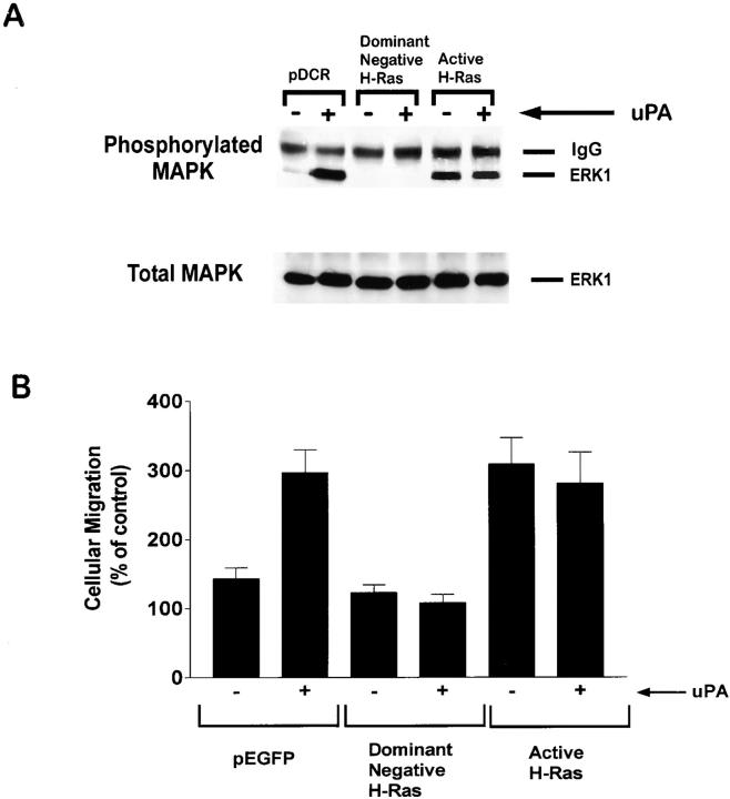
Urokinase-type plasminogen activator (uPA) activates the mitogen activated protein (MAP) kinases, extracellular signal-regulated kinase (ERK) 1 and 2, in diverse cell types. In this study, we demonstrate that uPA stimulates migration of MCF-7 breast cancer cells, HT 1080 fibrosarcoma cells, and uPAR-overexpressing MCF-7 cells by a mechanism that depends on uPA receptor (uPAR)-ligation and ERK activation. Ras and MAP kinase kinase (MEK) were necessary and sufficient for uPA-induced ERK activation and stimulation of cellular migration, as demonstrated in experiments with dominant-negative and constitutively active mutants of these signaling proteins. Myosin light chain kinase (MLCK) was also required for uPA-stimulated cellular migration, as determined in experiments with three separate MLCK inhibitors. When MCF-7 cells were treated with uPA, MLCK was phosphorylated by a MEK-dependent pathway and apparently activated, since serine-phosphorylation of myosin II regulatory light chain (RLC) was also increased. Despite the transient nature of ERK phosphorylation, MLCK remained phosphorylated for at least 6 h. The uPA-induced increase in MCF-7 cell migration was observed selectively on vitronectin-coated surfaces and was mediated by a beta1-integrin (probably alphaVbeta1) and alphaVbeta5. When MCF-7 cells were transfected to express alphaVbeta3 and treated with uPA, ERK was still phosphorylated; however, the cells did not demonstrate increased migration. Neutralizing the function of alphaVbeta3, with blocking antibody, restored the ability of uPA to promote cellular migration. Thus, we have demonstrated that uPA promotes cellular migration, in an integrin-selective manner, by initiating a uPAR-dependent signaling cascade in which Ras, MEK, ERK, and MLCK serve as essential downstream effectors.


Figures

Figure 1
Figure 1
uPA promotes cellular migration by a uPAR-dependent mechanism which is inhibited by PD098059 in parent and uPAR-overexpressing MCF-7 cells. Each of the designated cell types was treated with 10 nM DIP-uPA (+) or with vehicle (−) and allowed to migrate for 6 h on serum-coated Transwell membranes. Some cells were treated with uPA-specific antibody, uPAR-specific antibody, nonimmune IgG (each at 25 μg/ml), or with PD098059 (50 μM). Cellular migration is expressed as a percentage of that observed with control cells, which were not treated with DIP-uPA, antibodies, or PD098059 formula image.
Figure 2
Figure 2
uPA-stimulated HT 1080 cell migration requires ERK activation and uPAR ligation. (A) HT 1080 cells were treated with 10 nM DIP-uPA (+) or with vehicle (−) and allowed to migrate for 6 h on serum-coated Transwell membranes. Some cells were treated with uPA-specific antibody, uPAR-specific antibody (each at 25 μg/ml), or PD098059 (50 μM). Cellular migration is expressed as a percentage of that observed with control cells that were not treated with DIP-uPA, antibodies, or inhibitors. (B) HT 1080 cells (2.5 × 10⁵ cells) were incubated with 1 nM DIP-uPA or with vehicle for 4 h at 4°C as indicated. Some cells were pretreated with uPAR-specific antibody (25 μg/ml) for 20 min and then with DIP-uPA in the presence of antibody. HT 1080 cell detergent-extracts were subjected to SDS-PAGE on 8% slabs and transferred to nitrocellulose. Total cell-associated uPA (endogenously produced and exogenously added) was detected by immunoblot analysis, using uPA specific antibody (1:10,000), and HRP-conjugated anti–mouse IgG (Sigma, 1:10,000).
Figure 3
Figure 3
MEK is required for uPA-induced MAP kinase activation and motility stimulation. (A) MCF-7 cells were cotransfected to express HA-tagged ERK1 and the specified MEK mutants or the empty vector, pCHA. HA-tagged ERK1 was immunoprecipitated from cell extracts 1 min after treating the cells with 10 nM DIP-uPA (+) or with vehicle (−). Phosphorylated and total levels of ERK1 were determined by immunoblot analysis. (B) MCF-7 cells were transfected to express the specified MEK mutants and GFP, or GFP alone (pEGFP). Mock-transfected cells were treated with Superfect in the absence of cDNAs. Cells were allowed to migrate in the presence (+) or absence (−) of 10 nM DIP-uPA for 6 h on serum-coated membranes. Cellular migration of transfected cells was determined using fluorescence microscopy to detect GFP-expressing cells. Migration of mock-transfected cells was determined by Diff-Quik staining. Mock-transfected cells migrated identically to MCF-7 cells that were not Superfect-treated, in the presence and absence of uPA (data not shown). To compare results obtained with GFP-expressing and mock-transfected cells, the number of GFP-expressing cells that penetrated the membrane was divided by the transfection efficiency. For each bar, cellular migration is expressed as a percentage of that observed with mock-transfected cells that were not treated with DIP-uPA.
Figure 4
Figure 4
Ras is required for uPA-induced MAP kinase activation and MCF-7 cell migration. (A) MCF-7 cells were cotransfected to express HA-tagged ERK1 and dominant-negative H-Ras, constitutively active H-Ras, or the empty vector, pDCR. The cells were then treated with 10 nM DIP-uPA (+) or vehicle (−) for 1 min. HA-tagged ERK1 was recovered by immunoprecipitation. Phosphorylated and total levels of ERK1 were determined by immunoblot analysis. (B) MCF-7 cells were transfected to express the specified H-Ras mutants and GFP or GFP alone. The cells were then treated with 10 nM DIP-uPA (+) or vehicle (−) and allowed to migrate for 6 h on serum-coated membranes. Cellular migration was standardized to that observed with mock-transfected cells that were not uPA-treated.
Figure 5
Figure 5
Gene transcription and protein synthesis are not required for uPA-promoted MCF-7 cell migration. MCF-7 cells were allowed to migrate for 6 h on serum-coated Transwell membranes in the presence (+) or absence (−) of 10 nM DIP-uPA. Some MCF-7 cells were treated with cycloheximide (3 μg/ml) or actinomycin D (10 μg/ml). Cellular migration is expressed as a percentage of that observed with control cells that were not treated with DIP-uPA or with inhibitors.
Figure 6
Figure 6
MLCK and RLC are phosphorylated in uPA-treated MCF-7 cells. (A) MCF-7 cells were metabolically labeled with [32P]orthophosphate and then treated with vehicle (control) or with 10 nM DIP-uPA for the indicated times. Some MCF-7 cells were treated with 50 μM PD098059 (+), beginning 15 min before adding uPA and continuing for the entire assay. MLCK was immunoprecipitated from detergent extracts, subjected to SDS-PAGE, and analyzed by autoradiography. (B) Densitometry was performed using low-exposure autoradiography films from the specified number of experiments formula image. In each study, phosphorylated MLCK was not readily detected in the absence of uPA. Thus, band intensity was standardized against that observed with cells that had been treated with DIP-uPA for 1 h. (C) Suspended MCF-7 cells were treated with 10 nM DIP-uPA or with vehicle (control) for the indicated times. Cells lysates were subjected to SDS-PAGE and immunoblot analysis to detect RLC serine-phosphorylation.
Figure 6
Figure 6
MLCK and RLC are phosphorylated in uPA-treated MCF-7 cells. (A) MCF-7 cells were metabolically labeled with [32P]orthophosphate and then treated with vehicle (control) or with 10 nM DIP-uPA for the indicated times. Some MCF-7 cells were treated with 50 μM PD098059 (+), beginning 15 min before adding uPA and continuing for the entire assay. MLCK was immunoprecipitated from detergent extracts, subjected to SDS-PAGE, and analyzed by autoradiography. (B) Densitometry was performed using low-exposure autoradiography films from the specified number of experiments formula image. In each study, phosphorylated MLCK was not readily detected in the absence of uPA. Thus, band intensity was standardized against that observed with cells that had been treated with DIP-uPA for 1 h. (C) Suspended MCF-7 cells were treated with 10 nM DIP-uPA or with vehicle (control) for the indicated times. Cells lysates were subjected to SDS-PAGE and immunoblot analysis to detect RLC serine-phosphorylation.
Figure 7
Figure 7
MLCK activity is critical for uPA-promoted cell migration. (A) MCF-7 cells were treated with the specified MLCK inhibitors and allowed to migrate for 6 h on serum-coated Transwell membranes. (B) MCF-7 cells, uPAR overexpressing MCF-7 cells, and HT 1080 cells were treated with ML-9 (30 μM), ML-7 (3 μM), or W-7 (51 μM), in the presence (+) or absence (−) of DIP-uPA, and allowed to migrate for 6 h. Control cells were not treated with any of the MLCK inhibitors. Cellular migration was expressed as a percentage of that observed with control cells, of the same cell line, in the absence of DIP-uPA and inhibitors.
Figure 8
Figure 8
uPA-promoted cellular migration is matrix protein–selective. Transwell membranes were coated with purified vitronectin or with type I collagen. MCF-7 cells that were treated with mannosamine (+) and untreated cells (−) were allowed to migrate for 6 h in the presence of 10 nM DIP-uPA (+) or vehicle (−). Migration was expressed as a percentage of that observed with control cells (no mannosamine or DIP-uPA treatment) on vitronectin-coated membranes.
Figure 10
Figure 10
αVβ5 and a β1-containing integrin mediate MCF-7 cell migration in the presence and absence of uPA. (A) MCF-7 cells were treated with increasing concentrations of blocking antibodies against αVβ3 (LM609), αVβ5 (P1F6), and/or β1-integrin subunit (6S6) and allowed to migrate for 6 h in Transwell chambers with vitronectin-coated membranes. (B) MCF-7 cells were treated with 10 nM DIP-uPA (+) or with vehicle (−) in the presence of the specified antibodies (32 μg/ml) and allowed to migrate for 6 h on vitronectin-coated membranes. Control cells were not treated with antibodies. Migration was expressed as a percentage of that observed with control cells in the absence of DIP-uPA.
Figure 9
Figure 9
αVβ5 and β1-containing integrins mediate MCF-7 cell adhesion to vitronectin. MCF-7 cells were allowed to adhere to vitronectin-coated surfaces for 1 h in the presence (+) or absence (−) of 10 nM DIP-uPA. Some MCF-7 cells were treated with blocking antibodies (each at 32 μg/ml) against αVβ3 (LM609), αVβ5 (P1F6), and/or β1 subunit (6S6). Adherent cells were stained with Calcein AM and measured by fluorescence emission using a Cytofluor 2350. Cellular adhesion was quantitated as a percentage of that observed with control cells that were not exposed to DIP-uPA or antibodies.
Figure 11
Figure 11
MCF-7 cells which express αVβ3 are refractory to uPA-stimulation in migration assays. (A) MCF-7 cells were transfected to express β3-integrin subunit. After selection for 14 d, cell-surface αVβ3 expression was detected by flow cytometry using antibody LM609. Transfected cells are shown with the broken line. The solid line shows untransfected MCF-7 cells. (B) αVβ3-expressing MCF-7 cells were treated with DIP-uPA for the specified times, with EGF (25 ng/ml) for 5 min, or with vehicle (control). Phosphorylated and total levels of ERK1/2 were determined by immunoblot analysis. (C) αVβ3-expressing MCF-7 cells (β3), untransfected cells (control), and cells that were transfected with the empty vector that was used to prepare the β3-expression construct (pBK-CMV) were treated with 10 nM DIP-uPA (+) or with vehicle (−) and allowed to migrate for 6 h in Transwell chambers with vitronectin-coated membranes. Migration of αVβ3-expressing cells was also studied in the presence of antibody LM609 (32 μg/ml). Cells which penetrated the membranes were detected by Diff-Quik staining. Migration was expressed as a percentage of that observed with untransfected cells in the absence of DIP-uPA.
Figure 11
Figure 11
MCF-7 cells which express αVβ3 are refractory to uPA-stimulation in migration assays. (A) MCF-7 cells were transfected to express β3-integrin subunit. After selection for 14 d, cell-surface αVβ3 expression was detected by flow cytometry using antibody LM609. Transfected cells are shown with the broken line. The solid line shows untransfected MCF-7 cells. (B) αVβ3-expressing MCF-7 cells were treated with DIP-uPA for the specified times, with EGF (25 ng/ml) for 5 min, or with vehicle (control). Phosphorylated and total levels of ERK1/2 were determined by immunoblot analysis. (C) αVβ3-expressing MCF-7 cells (β3), untransfected cells (control), and cells that were transfected with the empty vector that was used to prepare the β3-expression construct (pBK-CMV) were treated with 10 nM DIP-uPA (+) or with vehicle (−) and allowed to migrate for 6 h in Transwell chambers with vitronectin-coated membranes. Migration of αVβ3-expressing cells was also studied in the presence of antibody LM609 (32 μg/ml). Cells which penetrated the membranes were detected by Diff-Quik staining. Migration was expressed as a percentage of that observed with untransfected cells in the absence of DIP-uPA.

Similar articles

Cited by

References

    1. Adelstein R.S., Pato M.D., Sellers J.R., de Lanerolle P., Conti M.A. Regulation of contractile proteins by reversible phosphorylation of myosin and myosin kinase. In: Twarog B.M., Levine R.J.C., Dewey M.M., editors. Basic Biology of MusclesA Comparative Approach. Raven Press; New York: 1982. pp. 273–281.
    1. Amano M., Ito M., Kimura K., Fukata Y., Chihara K., Nakano T., Matsuura Y., Kaibuchi K. Phosphorylation and activation of myosin by Rho-associated kinase (Rho-kinase) J. Biol. Chem. 1996;271:20246–20249. - PubMed
    1. Andreasen P.A., Kjøller L., Christensen L., Duffy M.J. The urokinase-type plasminogen activator system in cancer metastasisa review. Int. J. Cancer. 1997;72:1–22. - PubMed
    1. Bohuslav J., Horejsí V., Hansmann C., Stöckl J., Weidle U.H., Majdic O., Bartke I., Knapp W., Stockinger H. Urokinase plasminogen activator receptor, β-integrins, and src-kinases within a single receptor complex of human monocytes. J. Exp. Med. 1995;181:1381–1390. - PMC - PubMed
    1. Braut-Boucher F., Pichon J., Rat P., Adolphe M., Aubery M., Font J. A non-isotopic, highly sensitive, fluorimetric, cell-cell adhesion microplate assay using calcein AM-labeled lymphocytes. J. Immunol. Methods. 1995;178:41–51. - PubMed

Publication types

MeSH terms