. 1999 Jul 12;146(1):193-202.
doi: 10.1083/jcb.146.1.193.

Genomic organization, expression, and analysis of the troponin C gene pat-10 of Caenorhabditis elegans

Affiliations

Genomic organization, expression, and analysis of the troponin C gene pat-10 of Caenorhabditis elegans

H Terami et al. J Cell Biol. .

Abstract

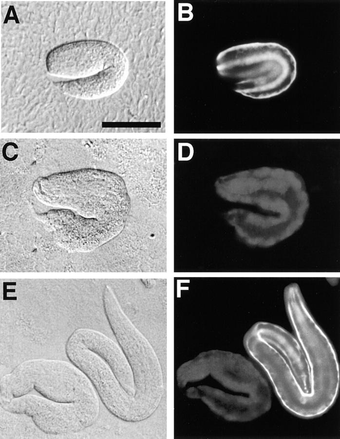
We have cloned and characterized the troponin C gene, pat-10 of the nematode Caenorhabditis elegans. At the amino acid level nematode troponin C is most similar to troponin C of Drosophila (45% identity) and cardiac troponin C of vertebrates. Expression studies demonstrate that this troponin is expressed in body wall muscle throughout the life of the animal. Later, vulval muscles and anal muscles also express this troponin C isoform. The structural gene for this troponin is pat-10 and mutations in this gene lead to animals that arrest as twofold paralyzed embryos late in development. We have sequenced two of the mutations in pat-10 and both had identical two mutations in the gene; one changes D64 to N and the other changes W153 to a termination site. The missense alteration affects a calcium-binding site and eliminates calcium binding, whereas the second mutation eliminates binding to troponin I. These combined biochemical and in vivo studies of mutant animals demonstrate that this troponin is essential for proper muscle function during development.


Figures

Figure 1
Figure 1
Map position and genome structure of the troponin C gene, pat-10. (A) Genomic map of the central part of chromosome I of the worm is shown. (B) Contig map of the region surrounding pat-10. Three YAC clones, Y37B3, Y55F5, and Y72D6 hit with cDNA fragment of the troponin C. Cosmid clones of B0490, W03G9, F54C1, C15C10 K04H3, and T21E10 were hybridized with cDNA fragment, and F54C1 and C15C10 contained the genomic fragment of pat-10. (C and D) Restriction map and intron/exon structure from sequence data of pat-10. It should be noted that only one transcript from the plasmid clone pTNC1 was matched to 674 bp of cDNA encoding 161 amino acid residues. H, HindIII; B, BamHI; E, EcoRI; P, PstI. The nucleotide sequence of pat-10 is in Fig. 2.
Figure 2
Figure 2
Nucleotide sequence of the troponin C gene pat-10. The exons have been translated using the standard one letter code. The splicing pattern of the gene is given in Fig. 1 D. Mutation sites in pat-10(st575) animal are shown on the top of the sequence at G1860A and G2179A. Restriction sites and the positions of primers for mutation sites determination are indicated. The accession number of pat-10 sequence in the GSDB, DDBJ, EMBL, and NCBI is D45895.
Figure 4
Figure 4
Western blot identification of troponin C from a worm extract and bacterial expression. Protein fractions were separated on 10–20% gels and either stained with Coomassie brilliant blue (A) or transferred to nitrocellulose for indirect probing with anti–Ascaris-troponin C antiserum (B). Protein fractions are the immunotransfers contained in a total protein extract from the wild-type N2 (lane 1), bacterial protein from the troponin I clone, pCTNI-1 (lane 2), bacterial protein from the troponin C clone, pCTNC-1 (lane 3), bacterial protein from bluescript (lane 4), respectively. Molecular size marker (M).
Figure 3
Figure 3
Comparison of amino acid sequences of troponin C-1 of C. elegans with a second troponin C of C. elegans and those of Drosophila, crayfish, and rabbits. Alignment of C. elegans troponin C, CeTNC-1 (pat-10), CeTNC-2 (cosmid ZK673.7), Drosophila troponin C Dm73F (Fyrberg et al. 1994), crayfish type (Kobayashi et al. 1989), rabbits, cardiac (Wilkinson 1980) and skeletal (Zot et al. 1987). Four calcium-binding sites are shown (I–IV). Mutation sites in the pat-10(st575) mutant are shown at 64 D to N and 153 W to termination (*), respectively. Shading indicates the residues that are identical in these six troponin C proteins.
Figure 5
Figure 5
Mobility-shift assay of the wild-type troponin C and characterization of mutant troponin C by using bacterially expressed proteins. (A) SDS-PAGE and Coomassie brilliant blue staining; (B) Western analysis using affinity-purified anti–troponin C antibody. Lane B, Total protein of E. coli JM109; lanes 1 and 2, bacterial protein from cDNA clone, pCTNC-1; lanes 3 and 4, mutant troponin C clone, pCPAT-10-m1; lanes 5 and 6, mutant troponin C clone, pCPAT-10-m2; lane N, total protein of the wild-type N2. Sample solution contained Ca2+ + (1 mM CaCl2, lanes 1, 3, and 5) and Ca2+ − (5 mM EGTA, lanes 2, 4, and 6), respectively. (C) Blotted sheet incubated with bacterially produced troponin I followed by detection with affinity-purified anti–TNI-1. For C, samples and conditions were the same as in A and B. It was noted that PAT-10-m1 did not show a band shift with and without calcium (B, lanes 3 and 4) and PAT-10-m2 did not bind troponin I (C, lanes 5 and 6). For details see the text.
Figure 6
Figure 6
Tissue-specific expression of the pat-10/lacZ genes. A fusion gene containing 5′ UTS of pat-10 and lacZ was used to study cellular expression. (A) Staining of body wall muscles, pTNCZ647; (B) pTNCZ292; (C) Expression of anterior part. It was noted that pharyngeal muscle was not stained. (D and E) pTNCZ647, side and top views of the vulva of young adult, respectively. Bar, 50 μm. (F and G) Summary of 5′ UTS of pat-10 and body wall–specific expressions. The position of regulatory sequences were shown: D, 1330/hlh-1 recognition; G, GC box; M, MEF2-binding sites, respectively.
Figure 7
Figure 7
Expression pattern of the pat-10/lacZ gene in early stages of wild-type development and the phenotypes of pat-10 animals. pTNC292 expressions at gastrulation (A), comma bean (B and C), twofold (D), and threefold (B) stages, respectively. pTNC292 expression in adult worm (E). It was noted that pat-10 started expression at comma bean stage (B and C). pat-10 heterozygotes yield both wild-type (I) and pat-10 homozygous animals that stop development at the twofold stage (F–H and I). Bars, 0.1 mm.
Figure 8
Figure 8
Immunostaing of the worm with affinity-purified anti-TnC. Staining of whole worm (A). The arrowhead indicates vulva muscles. Mail tail (B). High magnification of vulva muscles (arrowhead) and anus muscles (double arrowhead) (C). Vulva and body wall muscles are shown higher magnification (D). Immuno- and phalloidin-stained animals of the head region, respectively (E and F). These results indicate that troponin C protein locates in body wall and other minor muscles except pharyngeal muscles. Bars: (A–C and E) 0.1 mm; (D) 0.02 mm, respectively.
Figure 9
Figure 9
Nomarsky and anti–troponin C immunostaining images of wild-type and pat-10 animals. Staining of the wild-type (B) and pat-10 (st575) animals at the two hold stage (D and F), Nomarsky images of both (A, C, and E). The muscle filaments of the wild-type embryo were visible in a regular array (B and F). In contrast to wild-type, the tail portion of a mutant larva at the threefold stage has an irregular morphology (C and E). Bar, 50 μm.

Similar articles

Cited by

References

    1. Anderson P. Molecular genetics of nematode muscle. Annu. Rev. Genet. 1990;23:507–525. - PubMed
    1. Barstead R.J., Waterston R.H. The Basal component of the nematode dense-body is binculin. J. Biol. Chem. 1989;264:10177–10185. - PubMed
    1. Blumenthal T., Steward K. RNA processing and gene structure. In: Riddle D.L., editor. C. elegans II. Cold Spring Harbor Laboratory Press; Cold Spring Harbor, NY: 1997. pp. 117–145. - PubMed
    1. Brenner S. The genetics of Caenorhabditis elegans . Genetics. 1974;77:71–94. - PMC - PubMed
    1. Bullard B., Kevin L., Larkins A., Butcher G., Karlik C., Fyrberg E. Troponin of asynchronous flight muscle. J. Mol. Biol. 1988;204:621–637. - PubMed

Publication types

MeSH terms

Associated data