Comparative Study
. 1999 Jul 12;146(1):203-18.
doi: 10.1083/jcb.146.1.203.

alpha-bungarotoxin receptors contain alpha7 subunits in two different disulfide-bonded conformations

Affiliations
Comparative Study

alpha-bungarotoxin receptors contain alpha7 subunits in two different disulfide-bonded conformations

S Rakhilin et al. J Cell Biol. .

Abstract

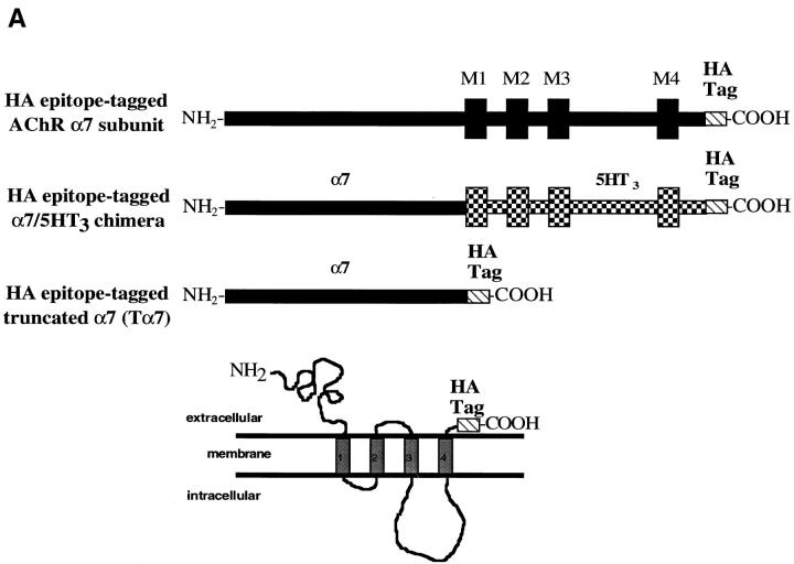
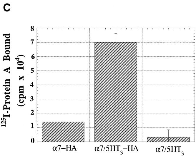
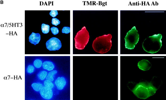
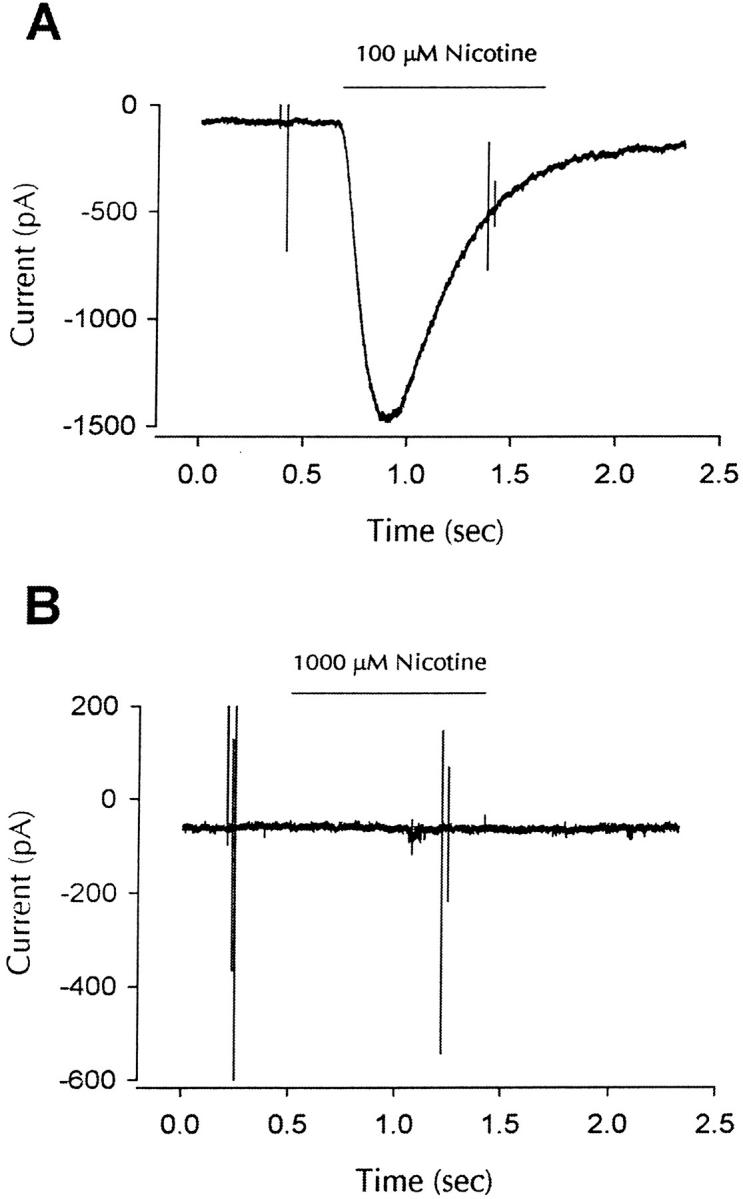
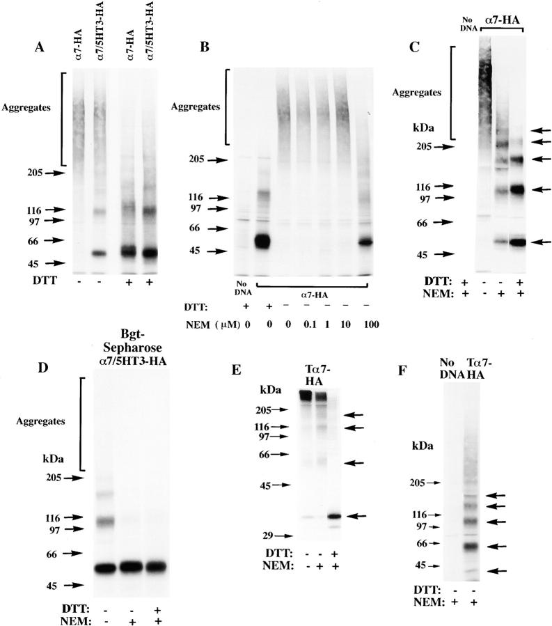
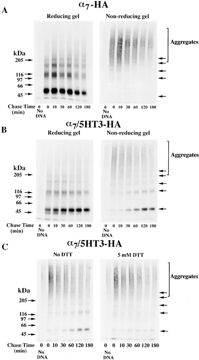
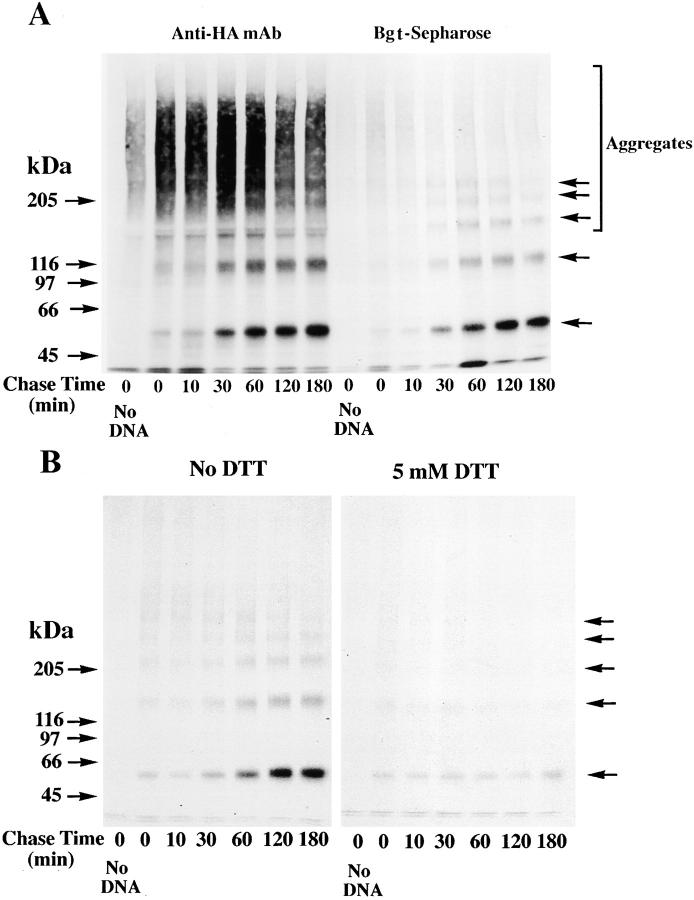
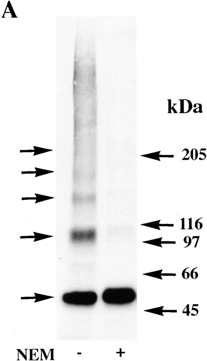
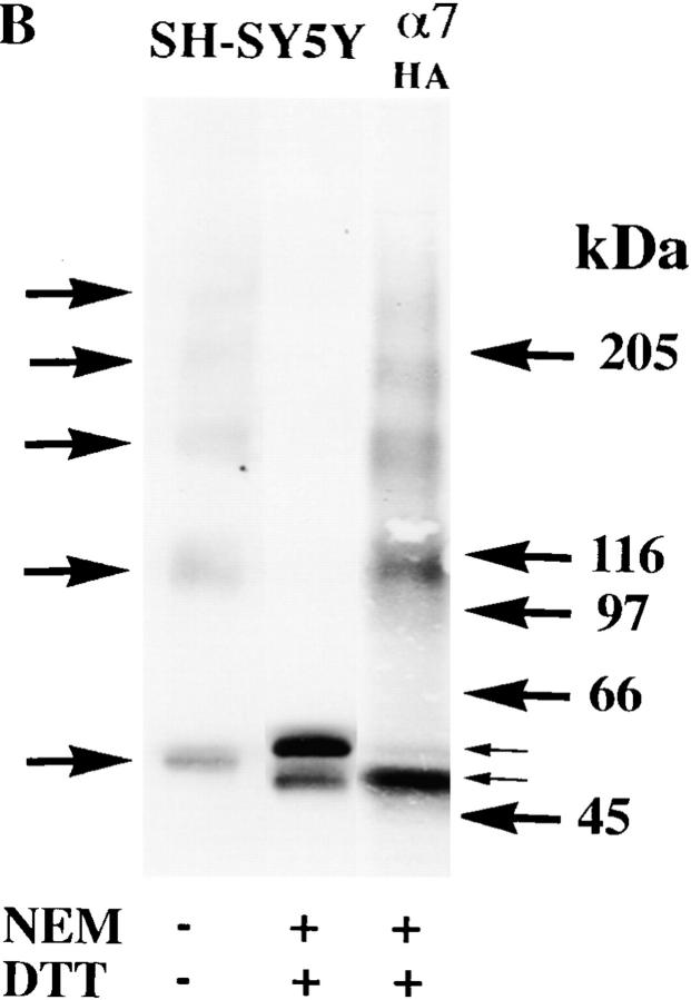
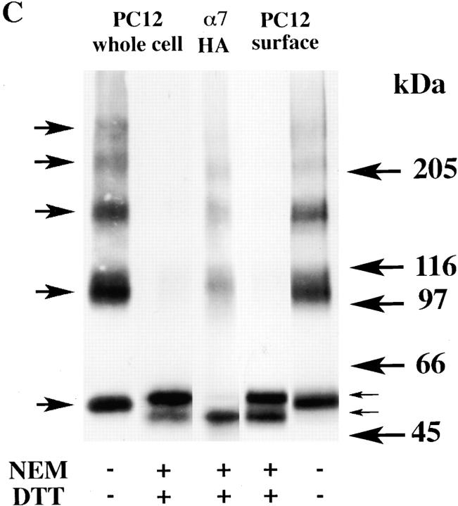
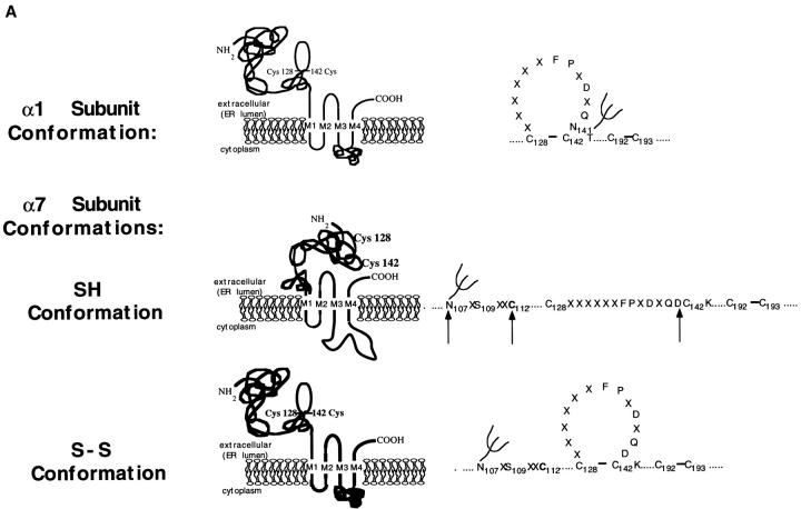
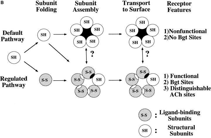
Neuronal nicotinic alpha7 subunits assemble into cell-surface complexes that neither function nor bind alpha-bungarotoxin when expressed in tsA201 cells. Functional alpha-bungarotoxin receptors are expressed if the membrane-spanning and cytoplasmic domains of the alpha7 subunit are replaced by the homologous regions of the serotonin-3 receptor subunit. Bgt-binding surface receptors assembled from chimeric alpha7/serotonin-3 subunits contain subunits in two different conformations as shown by differences in redox state and other features of the subunits. In contrast, alpha7 subunit complexes in the same cell line contain subunits in a single conformation. The appearance of a second alpha7/serotonin-3 subunit conformation coincides with the formation of alpha-bungarotoxin-binding sites and intrasubunit disulfide bonding, apparently within the alpha7 domain of the alpha7/serotonin-3 chimera. In cell lines of neuronal origin that produce functional alpha7 receptors, alpha7 subunits undergo a conformational change similar to alpha7/serotonin-3 subunits. alpha7 subunits, thus, can fold and assemble by two different pathways. Subunits in a single conformation assemble into nonfunctional receptors, or subunits expressed in specialized cells undergo additional processing to produce functional, alpha-bungarotoxin-binding receptors with two alpha7 conformations. Our results suggest that alpha7 subunit diversity can be achieved postranslationally and is required for functional homomeric receptors.


Figures

Figure 1
Figure 1
Cell-surface α7 subunits that do not bind Bgt. (A) HA epitope-tagged α7-HA and α7/5HT3-HA subunits. Diagrammed are the α7 and chimeric α7/5HT3 subunits and the location of the HA epitope. M1–M4 represent putative transmembrane domains. Based on the consensus membrane topology of the subunits shown on the bottom of the figure, the HA epitope tag on the COOH terminus of the subunits should be located within the extracellular domain of the receptors. Also diagrammed is the α7 subunit construct (Tα7-HA), which is truncated just before the first transmembrane domain (M1) and also contains the HA epitope tag at the COOH terminus. (B) Immunostaining of α7/5HT3-HA and α7-HA subunits on the cell surface. TsA201 cells, transfected with either α7/5HT3-HA or α7-HA cDNA constructs, were immobilized on slides and immunostained using an anti-HA mAb. Photographs were taken sequentially in the same field after staining with a nuclear stain, DAPI (blue), TMR-Bgt (red), and anti-HA mAb complexed with fluorescein-conjugated secondary Ab (green). DAPI staining is included to stain untransfected as well as transfected cells. Bar, 10 μm. Note the difference in scale. The cells expressing α7/5HT3-HA were photographed with a 100× objective whereas those expressing α7-HA were photographed with a 63× objective. (C) 125I-protein A binding to surface α7/5HT3-HA and α7-HA subunits. TsA201 cells were transiently transfected with either α7-HA, α7/5HT3-HA, or α7/5HT3 cDNA in 6-cm plates. To compare subunit surface expression levels 48 h after transfection, cells were incubated with saturating concentrations of anti-HA mAb and 125I-protein A, and specific binding was determined. Each bar represents the mean of three determinations ± SD.
Figure 1
Figure 1
Cell-surface α7 subunits that do not bind Bgt. (A) HA epitope-tagged α7-HA and α7/5HT3-HA subunits. Diagrammed are the α7 and chimeric α7/5HT3 subunits and the location of the HA epitope. M1–M4 represent putative transmembrane domains. Based on the consensus membrane topology of the subunits shown on the bottom of the figure, the HA epitope tag on the COOH terminus of the subunits should be located within the extracellular domain of the receptors. Also diagrammed is the α7 subunit construct (Tα7-HA), which is truncated just before the first transmembrane domain (M1) and also contains the HA epitope tag at the COOH terminus. (B) Immunostaining of α7/5HT3-HA and α7-HA subunits on the cell surface. TsA201 cells, transfected with either α7/5HT3-HA or α7-HA cDNA constructs, were immobilized on slides and immunostained using an anti-HA mAb. Photographs were taken sequentially in the same field after staining with a nuclear stain, DAPI (blue), TMR-Bgt (red), and anti-HA mAb complexed with fluorescein-conjugated secondary Ab (green). DAPI staining is included to stain untransfected as well as transfected cells. Bar, 10 μm. Note the difference in scale. The cells expressing α7/5HT3-HA were photographed with a 100× objective whereas those expressing α7-HA were photographed with a 63× objective. (C) 125I-protein A binding to surface α7/5HT3-HA and α7-HA subunits. TsA201 cells were transiently transfected with either α7-HA, α7/5HT3-HA, or α7/5HT3 cDNA in 6-cm plates. To compare subunit surface expression levels 48 h after transfection, cells were incubated with saturating concentrations of anti-HA mAb and 125I-protein A, and specific binding was determined. Each bar represents the mean of three determinations ± SD.
Figure 2
Figure 2
α7 subunit complexes are nonfunctional. (A) Whole-cell response to nicotine for cells expressing α7/5HT3-HA subunits. α7-5HT3-HA transfected cells were identified by positive staining with anti-HA mAb, and whole cell patch clamp recordings showed functional nicotine-gated currents at a potential of −70 mV (100 μM nicotine; similar responses were seen in 15 of 15 cells). No responses were seen in cells that were not positively stained with the anti-HA mAb formula image. (B) Whole-cell response to nicotine for cells expressing α7-HA subunits. Transfection with α7-HA results in positive staining of live cells, but no inward currents were seen at holding potentials of −70 mV. In total, 11 cells were exposed to a 10-fold higher nicotine concentration (1 mM). Except for their response to nicotine, cells expressing α7-HA subunits appeared to be identical to cells expressing α7/5HT3-HA subunits.
Figure 3
Figure 3
Differences in the folding of α7-HA and α7/5HT3-HA subunits. (A) A difference in α7 and α7/5HT3 subunit redox state. 6-cm cultures of tsA201 cells, transfected with α7-HA or α7/5HT3-HA cDNAs, were metabolically labeled for 1 h and chased for 1 h. The cells were solubilized in the absence of NEM and labeled subunits immunoprecipitated with anti-HA mAb. Samples, each from one 6-cm culture, were loaded on the gel with or without treatment with 10 mM DTT. α7-HA samples were loaded into lanes 1 and 3 and α7/5HT3-HA samples were loaded into lanes 2 and 4. Arrows on the left of the figure are the indicated molecular weight markers run on a separate lane. Arrows on the right of the figure indicate positions of monomer, dimer, trimer, tetramer, and pentamer subunit complexes. (B) α7 subunit alkylation prevents its aggregation. TsA201 cells were transfected with α7-HA cDNA, metabolically labeled, and precipitated as in A, except that NEM (0–100 μM) was included in the culture medium for the final 10 min of the chase. A sample from sham-transfected cells (no DNA) was run in lane 1. Arrows on the left and right of the figure are the same as in A. (C) SDS resistance of subunit multimers. TsA201 cells were transfected with α7-HA cDNA, metabolically labeled, and precipitated as in A. As indicated, NEM (2 mM) was added to the solubilization buffer and to the loading buffer after addition of DTT (1 mM). After alkylation by NEM (lane 3) and reduction by DTT before gel loading (lane 4), some subunits remained in complexes as well as migrating as monomers. Arrows on the left of the figure are the indicated molecular weight markers run on a separate lane. Arrows on the right of the figure indicate positions corresponding to monomer, dimer, trimer, tetramer, and pentamer subunit complexes. (D) Bgt-binding subunit multimers. TsA201 cells transfected with α7/5HT3-HA cDNA were metabolically labeled as in A, chased for 2 h, and subunits precipitated with Bgt-Sepharose. Subunit multimers were greatly decreased by NEM alkylation (2 mM; lane 2) and addition of DTT before gel loading (1 mM; lane 3). A sample from sham-transfected cells (no DNA) was run in lane 1. Molecular weight markers are on the left of the gel. (E and F) The truncated α7 subunit. TsA201 cells were transfected with the truncated α7 subunit cDNA (Tα7-HA; Fig. 1 A), metabolically labeled, and immunoprecipitated as in A. Labeled subunits were analyzed on 10% (E) or 4–8% gradient (F) gels. As indicated, NEM (2 mM) was added to the solubilization buffer and to the loading buffer after addition of DTT (1 mM). Truncated α7 subunits migrated as aggregates and multimers similar to full-length α7 subunits (A–C). Arrows on the left of the figures are the indicated molecular weight markers run on a separate lane. Arrows on the right of the figures indicate positions corresponding to monomer, dimer, trimer, and tetramer (E and F) plus pentamer (F) subunit complexes.
Figure 4
Figure 4
Disulfide bonding is required to effect a change in α7/5HT3 subunit redox state. (A and B) Time-dependent change in α7/5HT3-HA subunit redox state. TsA201 cells transfected with α7-HA (A) or α7/5HT3-HA (B) cDNAs were metabolically labeled for 10 min and chased for the times specified in the figure. The cells were solubilized in the absence of NEM, labeled subunits immunoprecipitated with anti-HA mAb, and samples loaded on the gels without (nonreducing) or with (reducing) 10 mM DTT. A sample from sham-transfected cells (no DNA) was run in lane 1 of each of the four gels. Arrows on the right of the figure indicate positions of putative monomer, dimer, trimer, tetramer, and pentamer α7/5HT3-HA subunit complexes. (C) Addition of 5 mM DTT to the cell medium blocks the redox state change. TsA201 cells transfected with α7/5HT3-HA cDNA were metabolically labeled for 10 min and chased for the times specified in the figure in the absence (left) or presence (right) of 5 mM DTT in the medium. Cells were solubilized in the absence of NEM and labeled subunits precipitated with anti-HA mAb. All samples were loaded on the gels without DTT, and a sample from sham-transfected cells (no DNA) was run in lane 1 (left and right). Arrows on the right of the figure indicate positions of putative monomer, dimer, trimer, tetramer, and pentamer α7/5HT3-HA subunit complexes.
Figure 5
Figure 5
Bgt-binding site formation and the change in redox state. (A) Formation of Bgt-binding site correlates with the change in redox state. TsA201 cells transfected with α7/5HT3-HA cDNA were metabolically labeled for 10 min and chased for the times specified in the figure. The cells were solubilized in the absence of NEM and labeled subunits precipitated with anti-HA mAb (lanes 2–7 on the left) or Bgt-Sepharose (lanes 2–7 on the right). Bgt-Sepharose appeared not to precipitate all of the α7/5HT3 subunit monomers precipitated by anti-HA mAb. It is possible that not all α7/5HT3 subunit monomers assemble into Bgt-binding complexes or, alternatively, Bgt-Sepharose fails to quantitatively precipitate all Bgt-binding sites. All samples were loaded on the gels without DTT, and a sample from sham-transfected cells (no DNA) was run in lane 1 (left and right). Arrows on the right of the figure indicate positions of putative monomer, dimer, trimer, tetramer, and pentamer α7/5HT3-HA subunit complexes. (B) Addition of 5 mM DTT to the cell medium blocks Bgt-binding site formation. The experiment was performed as in Fig. 4 C except that labeled subunits were precipitated with Bgt-Sepharose.
Figure 6
Figure 6
Surface α7/5HT3 and α7 subunit receptors from SH-SY5Y and PC-12 cells contain subunits in two conformations. (A) Surface Bgt-binding receptors contain subunits in different redox states. TsA201 cells, transfected with α7/5HT3-HA cDNAs, were metabolically labeled for 1 h and chased for 2 h. Cells were surface labeled with Bgt and solubilized in the absence (lane 1) or presence (lane 2) of 2 mM NEM. The Bgt-bound receptors were specifically precipitated with anti-Bgt polyclonal antiserum. Arrows on the right of the figure indicate positions of putative monomer, dimer, trimer, tetramer, and pentamer α7/5HT3-HA subunit complexes. (B) α7 subunits expressed in SH-SY5Y cells. α7 subunits stably expressed in SH-SY5Y cells were analyzed by Western blots. Cells were solubilized in the absence (lane 1) or presence (lane 2) of 2 mM NEM and subunits were precipitated using Bgt-Sepharose. The proteins were transferred to a nitrocellulose membrane and probed with the α7 subunit-specific polyclonal antiserum, C-20. Equal samples were loaded on a SDS polyacrylamide gel either directly (lane 1), or preincubated with 1 mM DTT before loading and then treated with 2 mM NEM (lane 2). For lane 3, tsA201 cells were transfected with α7-HA cDNA, metabolically labeled, and precipitated as in Fig. 3 A. The sample was loaded on the same SDS polyacrylamide gel after treatment with 1 mM DTT and then with 2 mM NEM. (C) α7 subunits expressed in PC12 cells. α7 subunits expressed endogenously in PC12 cells were analyzed by Western blots. Cells were solubilized in the absence (lanes 1 and 4) or presence (lanes 2 and 5) of 2 mM NEM. Receptors were either precipitated with Bgt-Sepharose (whole cell; lanes 1 and 2) or cell-surface, Bgt-bound receptors were selectively precipitated with anti-Bgt protein A–Sepharose (surface; lanes 4 and 5). The proteins were transferred to a nitrocellulose membrane and probed with the α7 subunit-specific polyclonal antiserum, C-20. Equal samples were loaded onto an SDS polyacrylamide gel either directly (lanes 1 and 4), or preincubated with 1 mM DTT before loading and then treated with 2 mM NEM (lanes 2 and 5). For lane 3, tsA201 cells were transfected with α7-HA cDNA, metabolically labeled, and precipitated as in Fig. 3 A. The sample was loaded on the same SDS polyacrylamide gel after preincubation with 1 mM DTT and treatment with 2 mM NEM.
Figure 6
Figure 6
Surface α7/5HT3 and α7 subunit receptors from SH-SY5Y and PC-12 cells contain subunits in two conformations. (A) Surface Bgt-binding receptors contain subunits in different redox states. TsA201 cells, transfected with α7/5HT3-HA cDNAs, were metabolically labeled for 1 h and chased for 2 h. Cells were surface labeled with Bgt and solubilized in the absence (lane 1) or presence (lane 2) of 2 mM NEM. The Bgt-bound receptors were specifically precipitated with anti-Bgt polyclonal antiserum. Arrows on the right of the figure indicate positions of putative monomer, dimer, trimer, tetramer, and pentamer α7/5HT3-HA subunit complexes. (B) α7 subunits expressed in SH-SY5Y cells. α7 subunits stably expressed in SH-SY5Y cells were analyzed by Western blots. Cells were solubilized in the absence (lane 1) or presence (lane 2) of 2 mM NEM and subunits were precipitated using Bgt-Sepharose. The proteins were transferred to a nitrocellulose membrane and probed with the α7 subunit-specific polyclonal antiserum, C-20. Equal samples were loaded on a SDS polyacrylamide gel either directly (lane 1), or preincubated with 1 mM DTT before loading and then treated with 2 mM NEM (lane 2). For lane 3, tsA201 cells were transfected with α7-HA cDNA, metabolically labeled, and precipitated as in Fig. 3 A. The sample was loaded on the same SDS polyacrylamide gel after treatment with 1 mM DTT and then with 2 mM NEM. (C) α7 subunits expressed in PC12 cells. α7 subunits expressed endogenously in PC12 cells were analyzed by Western blots. Cells were solubilized in the absence (lanes 1 and 4) or presence (lanes 2 and 5) of 2 mM NEM. Receptors were either precipitated with Bgt-Sepharose (whole cell; lanes 1 and 2) or cell-surface, Bgt-bound receptors were selectively precipitated with anti-Bgt protein A–Sepharose (surface; lanes 4 and 5). The proteins were transferred to a nitrocellulose membrane and probed with the α7 subunit-specific polyclonal antiserum, C-20. Equal samples were loaded onto an SDS polyacrylamide gel either directly (lanes 1 and 4), or preincubated with 1 mM DTT before loading and then treated with 2 mM NEM (lanes 2 and 5). For lane 3, tsA201 cells were transfected with α7-HA cDNA, metabolically labeled, and precipitated as in Fig. 3 A. The sample was loaded on the same SDS polyacrylamide gel after preincubation with 1 mM DTT and treatment with 2 mM NEM.
Figure 7
Figure 7
α7 subunits fold and assemble by two different pathways. (A) α7 subunits fold into two conformations. Based on the disulfide bonding of the AChR α1 subunit (Karlin and Akabas 1995) shown on the top of the figure, a structural interpretation of the α7 subunit redox state change is shown. In the SH conformation, we envision that the 15–amino acid cystine loop is not formed and the two cysteines at residues 128 and 142 are not oxidized. In the S-S conformation, a disulfide bond has formed between these two cysteines. The arrows mark residues in this region of the α7 subunit that are different from other AChR subunits. These residues are an aspartate at position 141 that replaces an asparagine and removes a conserved consensus site for N-linked glycosylation at residue 141, a fifth cysteine residue at position 112 not found on other AChR α subunits, and an asparagine at residue 107 that creates a consensus site for N-linked glycosylation at residue 107. Our results suggest that changes in the conformation of the NH2-terminal domain allow the disulfide bonding. The NH2-terminal domain conformational changes appear to be driven by changes in the conformation of the COOH-terminal half of the subunit, perhaps changes in the COOH-terminal half cytoplasmic domain as depicted in the figure. (B) Assembly of α7 subunit complexes by different pathways. As depicted in the figure, we have found that α7 subunits can fold and assemble into two conformations. In tsA201 cells, α7 subunits assemble by a default pathway since they remain in a single conformation, SH, and assemble into pentamers that are neither functional nor bind Bgt. In SH-SY5Y and PC-12 cells, α7 subunits fold into a second conformation, S-S, and assemble with subunits in the SH conformation into pentamers that function and bind Bgt. The question marks are included in the figure because it is unclear whether the subunits undergo the conformational change before, during, or after assembly into pentamers. If it is assumed that ACh-binding sites can form on S-S subunits at their interface with other subunits, then distinguishable sites can form at interfaces between S-S subunits (open ellipses) and between S-S and SH subunits (filled ellipses).
Figure 7
Figure 7
α7 subunits fold and assemble by two different pathways. (A) α7 subunits fold into two conformations. Based on the disulfide bonding of the AChR α1 subunit (Karlin and Akabas 1995) shown on the top of the figure, a structural interpretation of the α7 subunit redox state change is shown. In the SH conformation, we envision that the 15–amino acid cystine loop is not formed and the two cysteines at residues 128 and 142 are not oxidized. In the S-S conformation, a disulfide bond has formed between these two cysteines. The arrows mark residues in this region of the α7 subunit that are different from other AChR subunits. These residues are an aspartate at position 141 that replaces an asparagine and removes a conserved consensus site for N-linked glycosylation at residue 141, a fifth cysteine residue at position 112 not found on other AChR α subunits, and an asparagine at residue 107 that creates a consensus site for N-linked glycosylation at residue 107. Our results suggest that changes in the conformation of the NH2-terminal domain allow the disulfide bonding. The NH2-terminal domain conformational changes appear to be driven by changes in the conformation of the COOH-terminal half of the subunit, perhaps changes in the COOH-terminal half cytoplasmic domain as depicted in the figure. (B) Assembly of α7 subunit complexes by different pathways. As depicted in the figure, we have found that α7 subunits can fold and assemble into two conformations. In tsA201 cells, α7 subunits assemble by a default pathway since they remain in a single conformation, SH, and assemble into pentamers that are neither functional nor bind Bgt. In SH-SY5Y and PC-12 cells, α7 subunits fold into a second conformation, S-S, and assemble with subunits in the SH conformation into pentamers that function and bind Bgt. The question marks are included in the figure because it is unclear whether the subunits undergo the conformational change before, during, or after assembly into pentamers. If it is assumed that ACh-binding sites can form on S-S subunits at their interface with other subunits, then distinguishable sites can form at interfaces between S-S subunits (open ellipses) and between S-S and SH subunits (filled ellipses).

Similar articles

Cited by

References

    1. Alkondon M., Albuquerque E.X. Diversity of nicotinic acetylcholine receptors in rat hippocampal neurons. I. Pharmacological and functional evidence for distinct structural subtypes. J. Pharmacol. Exp. Ther. 1993;265:1455–1473. - PubMed
    1. Alkondon M., Rocha E.S., Maelicke A., Albuquerque E.X. Diversity of nicotinic acetylcholine receptors in rat brain. V. Alpha-bungarotoxin-sensitive nicotinic receptors in olfactory bulb neurons and presynaptic modulation of glutamate release. J. Pharmacol. Exp. Ther. 1996;278:1460–1471. - PubMed
    1. Blount P., Merlie J.P. Molecular basis of the two nonequivalent ligand binding sites of the muscle nicotinic acetylcholine receptor. Neuron. 1989;3:349–357. - PubMed
    1. Blumenthal E.M., Conroy W.G., Romano S.J., Kassner P.D., Berg D.K. Detection of functional nicotinic receptors blocked by alpha-bungarotoxin on PC12 cells and dependence of their expression on post-translational events. J. Neurosci. 1997;17:6094–6104. - PMC - PubMed
    1. Braakman I., Hoover L.H., Wagner K.R., Helenius A. Folding of influenza hemagglutinin in the endoplasmic reticulum. J. Cell Biol. 1991;114:401–411. - PMC - PubMed

Publication types

MeSH terms