. 1999 Jul 12;146(1):219-32.

Recycling of E-cadherin: a potential mechanism for regulating cadherin dynamics

Affiliations

Recycling of E-cadherin: a potential mechanism for regulating cadherin dynamics

T L Le et al. J Cell Biol. .

Abstract

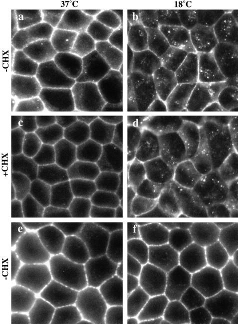
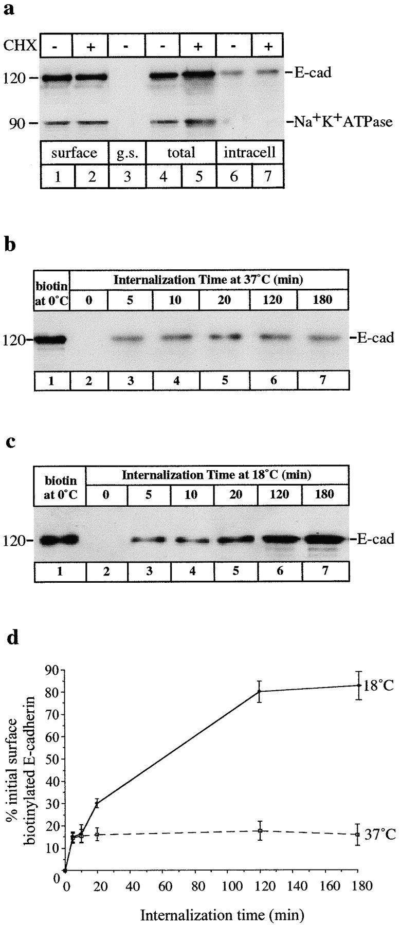
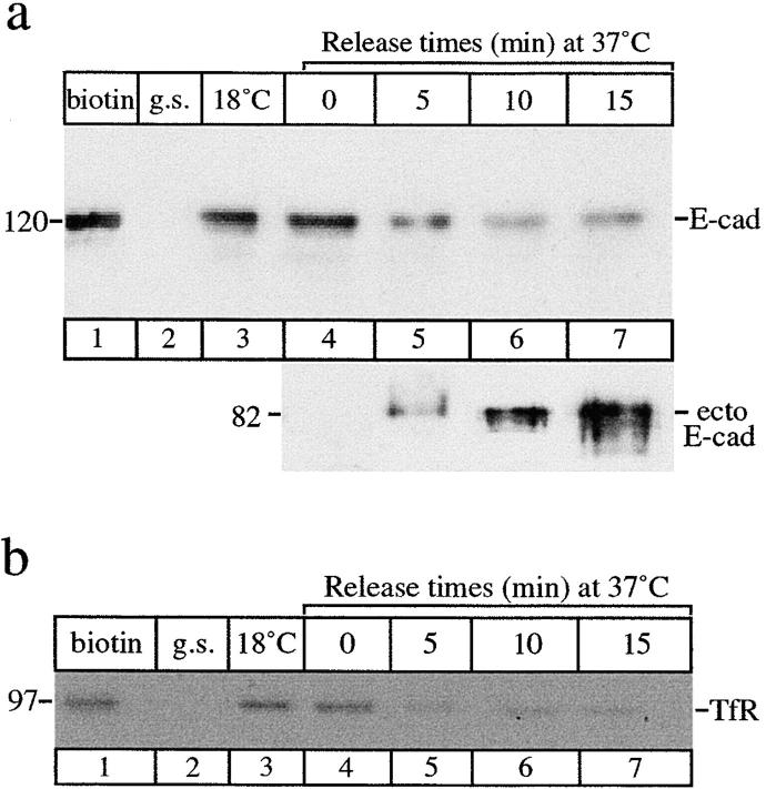
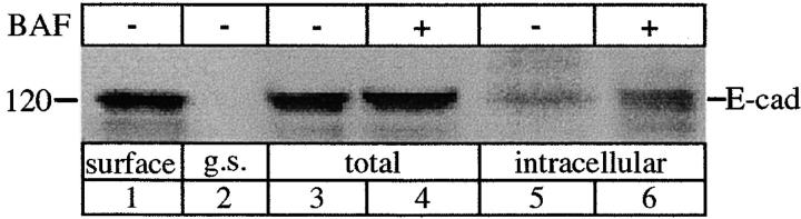
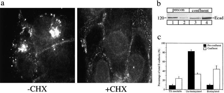
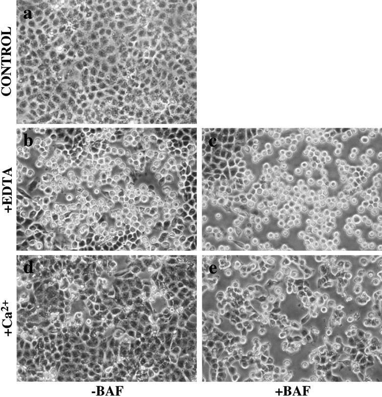
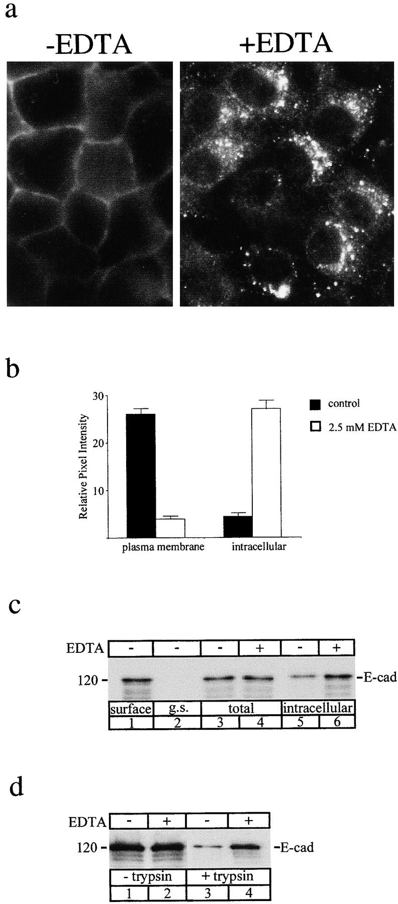
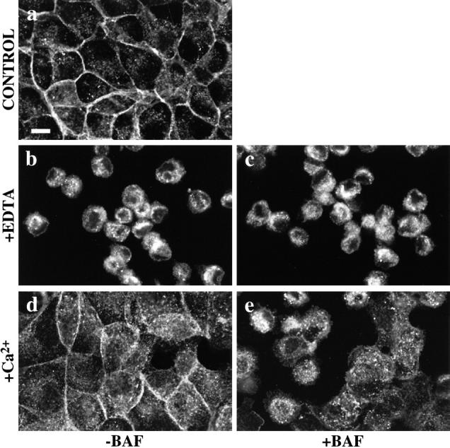
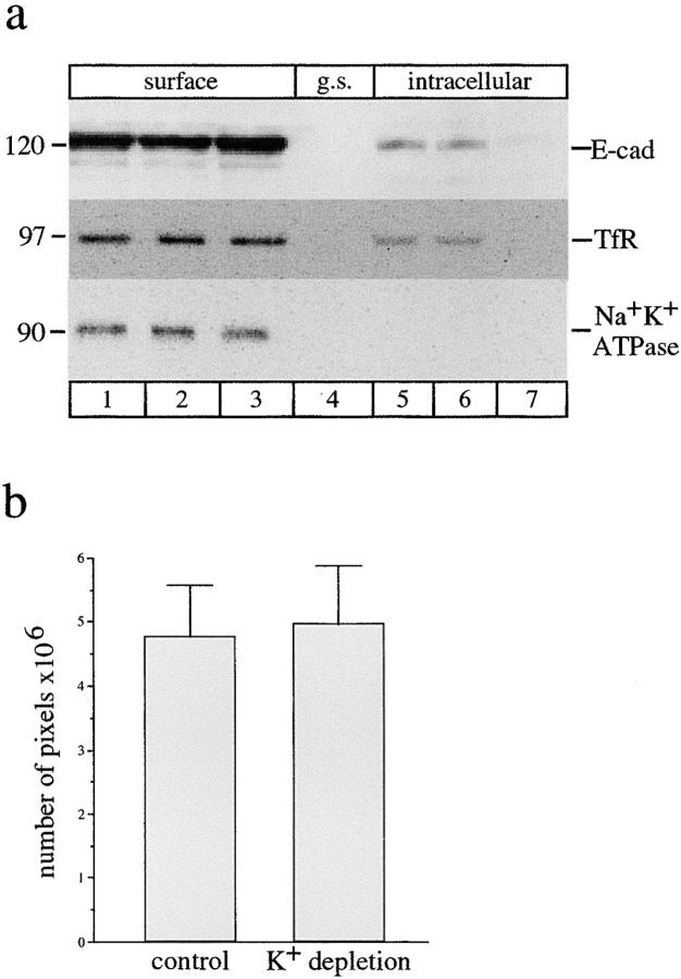
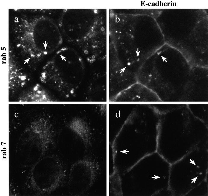
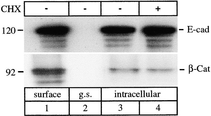
E-Cadherin plays critical roles in many aspects of cell adhesion, epithelial development, and the establishment and maintenance of epithelial polarity. The fate of E-cadherin once it is delivered to the basolateral cell surface, and the mechanisms which govern its participation in adherens junctions, are not well understood. Using surface biotinylation and recycling assays, we observed that some of the cell surface E-cadherin is actively internalized and is then recycled back to the plasma membrane. The pool of E-cadherin undergoing endocytosis and recycling was markedly increased in cells without stable cell-cell contacts, i.e., in preconfluent cells and after cell contacts were disrupted by depletion of extracellular Ca2+, suggesting that endocytic trafficking of E-cadherin is regulated by cell-cell contact. The reformation of cell junctions after replacement of Ca2+ was then found to be inhibited when recycling of endocytosed E-cadherin was disrupted by bafilomycin treatment. The endocytosis and recycling of E-cadherin and of the transferrin receptor were similarly inhibited by potassium depletion and by bafilomycin treatment, and both proteins were accumulated in intracellular compartments by an 18 degrees C temperature block, suggesting that endocytosis may occur via a clathrin-mediated pathway. We conclude that a pool of surface E-cadherin is constantly trafficked through an endocytic, recycling pathway and that this may provide a mechanism for regulating the availability of E-cadherin for junction formation in development, tissue remodeling, and tumorigenesis.


Figures

Figure 1
Figure 1
E-Cadherin distribution in confluent MDCK cells. (a) Immunofluorescence staining of E-cadherin is mostly localized to the lateral cell surfaces in confluent MDCK monolayers. Small amounts of intracellular, vesicular staining can also be seen scattered throughout the periphery of cells. (b) When cells were fixed and stained after incubation at 18°C, intracellular staining of E-cadherin was more intense. The vesicular staining relocated and was now clustered in a perinuclear position. (c and d) Preincubation of cells in cycloheximide (CHX) did not significantly alter the staining pattern; both cell surface and vesicular staining can still be seen. (e and f) Phalloidin staining of F-actin at the level of adhesion junctions shows that cell morphology was preserved in both control cultures and in monolayers incubated at 18°C. Representative fields of cells were photographed with similar exposures.
Figure 2
Figure 2
Internalization of surface-biotinylated E-cadherin. (a) Confluent MDCK cells treated with (+) or without (−) cycloheximide (CHX) were surface-biotinylated at 0°C. Detergent-soluble, surface-biotinylated proteins were recovered on streptavidin beads and analyzed by SDS-PAGE. E-Cadherin and Na+K+ATPase in these fractions were detected by immunoblotting as depicted. Biotinylated cell surface E-cadherin was recovered from the cell extracts (lanes 1 and 2). Glutathione stripping (g.s.) immediately after biotinylation at 0°C completely removed biotin from surface E-cadherin (lane 3). Surface-biotinylated cells were then incubated at 37°C and after 1 h, total biotinylated E-cadherin was recovered (lanes 4 and 5). E-Cadherin sequestered in an internal pool was recovered after glutathione stripping showing that some of the surface E-cadherin was endocytosed (lanes 6 and 7). Na+K+ATPase was biotinylated (lanes 1 and 2) but was not found in an internal pool under the same conditions (lanes 6 and 7). (b) A time course of E-cadherin uptake. CHX-treated cells were surface-biotinylated at 0°C (lane 1) and then incubated at 37°C for periods of 0–180 min (lanes 2–7). A constant amount of surface-biotinylated E-cadherin was sequestered from glutathione stripping at chase times from 5–180 min (lanes 3–7), consistent with a steady-state flux of E-cadherin into and out of this intracellular pool. (c) When the same experiments were carried out on MDCK cells incubated at 18°C, internalized E-cadherin now accumulated, resulting in increasing amounts of E-cadherin in the internal pool over 180 min (lanes 3–7). Fainter, lower molecular weight bands (lane 6 and 7) may be due to some degradation of internalized E-cadherin after prolonged incubations. (d) Relative amounts of internalized E-cadherin, quantitated by densitometry and expressed as a percentage of the total initial surface-biotinylated pool in cells incubated at 37°C or 18°C. Data are means ± SEM from three separate experiments.
Figure 3
Figure 3
Recycling of E-cadherin. Cells were surface-biotinylated on ice, then incubated at 18°C for 2 h to allow endocytosis and accumulation of E-cadherin. After glutathione stripping cells were then returned to 37°C. At each chase time (0–15 min) cells were trypsinized to release cell surface proteins. Glutathione stripping immediately after biotinylation effectively removed all surface-biotinylated proteins (g.s., lane 2). Biotinylated proteins from both the cell-associated (a, top) and trypsin-released fractions (a, bottom) were recovered on streptavidin beads and analyzed by SDS-PAGE. Intact E-cadherin (120 kD) or its trypsin-cleaved, 82-kD ecto-domain (a) and TfR (b) were detected by immunoblotting with specific antibodies. (a) The top shows that internalized E-cadherin accumulated at 18°C (lane 3) gradually disappeared from the internal pool over 15 min (lanes 4–7). The bottom shows that the 82-kD ectodomain of biotinylated E-cadherin was initially detected after 5 min at 37°C (lane 5) and maximal amounts were detected after 15 min at 37°C (lane 7) showing that the internalized E-cadherin was recycling and reappearing on the cell surface. (b) Surface-biotinylated (lane 1) TfR was also internalized (lane 4) but disappeared from the internal pool (lanes 5–7) more rapidly under the same conditions. Results are representative of four experiments.
Figure 4
Figure 4
Endocytosis of E-cadherin in the presence of bafilomycin A1 (BAF). Cells were surface-biotinylated at 0°C and then incubated at 37°C in the presence (+) or absence (−) of 1 μM BAF. Surface-biotinylated E-cadherin (lane 1) was recovered from total cell extracts (lanes 3 and 4) and from the internal pool (lanes 5 and 6) sequestered from glutathione stripping. A greater amount of internalized biotinylated E-cadherin was recovered after BAF treatment (lane 6) in comparison to the normal amount of internal E-cadherin (lane 5), consistent with BAF-induced accumulation of E-cadherin. The result is representative of three independent experiments.
Figure 5
Figure 5
Distribution of E-cadherin in preconfluent MDCK cells. (a) Immunofluorescence staining of E-cadherin in preconfluent cultures treated with or without cycloheximide (+/− CHX). At steady state (−CHX), there is a small amount of cell surface staining, prominent perinuclear staining over the Golgi complex, and bright vesicular staining throughout the cytoplasm. When protein synthesis is blocked (+CHX), the Golgi staining disappears but the vesicular staining remains. Representative fields of cells were photographed with similar exposures. (b) Surface biotinylation of E-cadherin. Preconfluent and confluent cells were surface-biotinylated; biotinylated E-cadherin in detergent-soluble cell extracts was recovered by streptavidin affinity, biotinylated and unbiotinylated (supernatant) fractions were analyzed by SDS-PAGE and immunoblotting. All of the biotinylated fraction and 20% of the total unbiotinylated fraction were loaded. Unbiotinylated E-cadherin from preconfluent cells (lane 1) and confluent cells (lane 3) was compared to the amounts of surface-biotinylated E-cadherin in preconfluent cells (lane 2) and confluent cells (lane 4). There is an increased amount of biotinylated cell surface E-cadherin in confluent cells with a concomitant decrease in the unbiotinylated fraction, which includes the intracellular pool. (c) The relative amounts of E-cadherin in detergent-soluble (biotinylated and unbiotinylated) and detergent-insoluble (TX-insoluble) fractions were compared by immunoblotting and densitometry in preconfluent and confluent cells. Detergent-insoluble E-cadherin, which is likely to represent protein incorporated into cytoskeleton-stabilized junctional complexes, increases in confluent cells. Within the detergent-soluble pool, biotinylated cell surface E-cadherin increases (from ∼10% to ∼50%) in confluent monolayers. Data are means ± SEM from three separate experiments.
Figure 6
Figure 6
Restoration of cell-cell contact after Ca2+ depletion: bafilomycin A1 (BAF) inhibits restoration of monolayer integrity following Ca2+ depletion. Phase-contrast images of confluent MDCK monolayers exposed to 2.5 mM EDTA for 45 min to chelate extracellular Ca2+ and then following restoration of Ca2+ in the presence or absence of bafilomycin A1 (BAF). Cells in untreated monolayers (a) have intact cell contacts. In the presence (c) or absence (b) of BAF (1 μM) cells are rounded and retracted from each other upon chelation of Ca2+ with EDTA. Within 1 h of restoring extracellular Ca2+ control cells had spread, covering most of the substrate (d); in contrast, cells treated with BAF (e) spread to a much lesser degree, leaving extensive regions of bare substrate exposed. Representative fields of cells show the results of three separate experiments.
Figure 7
Figure 7
Endocytosis of E-cadherin after depletion of extracellular Ca2+. Internalization of E-cadherin was compared in untreated confluent MDCK cells and in cells incubated in medium containing EDTA to chelate extracellular Ca2+. (a) Immunofluorescence localization of E-cadherin in control MDCK cells (−EDTA) and MDCK cells incubated in DMEM containing 2.5 mM EDTA (+EDTA) and cycloheximide (10 μM) for 30 min. In the presence of EDTA cells show prominent intracellular vesicular staining of E-cadherin. (b) Quantification of relative fluorescence intensities at the cell surface and inside cells measured by SOM software showed that chelation of Ca2+ resulted in a dramatic increase in intracellular staining accompanied by a concomitant decrease in plasma membrane staining, indicative of E-cadherin endocytosis stimulated by EDTA. Data are means ± SEM formula image. (c) Surface biotinylation. Cells were surface-biotinylated at 0°C (lane 1) and then incubated at 37°C in normal medium (lanes 3 and 5) or in medium containing EDTA (lanes 4 and 6) for 30 min to allow for internalization. Total biotinylated E-cadherin was unchanged in the total cell extracts under both these conditions (lanes 3 and 4). After glutathione stripping there was a significantly increased pool of internalized biotinylated E-cadherin after Ca2+ depletion (lane 6) compared to control cells (lane 5). Thus, EDTA treatment increased internalization of surface-biotinylated E-cadherin. (d) Surface trypsinization. Cells were incubated in normal media or in medium containing 2.5 mM EDTA for 30 min. Cell surface proteins were removed by trypsinization and the remaining E-cadherin in cell extracts was analyzed by SDS-PAGE and immunoblotting with a NH2 terminus antibody (3B8). Total cellular E-cadherin remained unchanged in the absence (lane 1) or presence (lane 2) of Ca2+ chelation. A small pool of internalized E-cadherin was detected after trypsin treatment in cells incubated in normal media (lane 3), but this pool was dramatically increased in the presence of EDTA (lane 4), showing increased internalization of surface E-cadherin. Results shown are representative of three independent experiments.
Figure 8
Figure 8
Bafilomycin A1 (BAF) inhibits the reaccumulation of E-cadherin in cell-cell contacts following Ca2+ depletion. Confluent MDCK monolayers were exposed to DMEM containing 2.5 mM EDTA for 45 min before restoration of normal extracellular Ca2+ concentrations before fixation and staining for E-cadherin. Before Ca2+ chelation, monolayers showed E-cadherin staining in a typical circumferential pattern of cell-cell contacts, in addition to scattered intracellular punctate deposits (a). Upon chelation of Ca2+, E-cadherin staining was concentrated in a prominent perinuclear vesicular pattern both in the presence (c) or absence (b) of BAF (1 μM). Upon restoration of extracellular Ca2+, control cells showed redistribution of E-cadherin to reforming cell-cell junctions (d). In contrast, upon restoration of extracellular Ca2+, BAF-treated cells retained significant amounts of intracellular E-cadherin in peripheral cytoplasmic vesicles as well as in the perinuclear area (e).
Figure 9
Figure 9
Endocytosis of E-cadherin is inhibited by K+ depletion. (a) Control cells (lane 1), cells pretreated with hypotonic shock alone (lane 2), or cells which were pretreated with hypotonic shock for 5 min then also incubated in K+-free medium for 15 min (lane 3) were surface-biotinylated. Biotinylated proteins were collected and analyzed by SDS-PAGE and immunoblotted to detect E-cadherin, transferrin receptor, and Na+K+ATPase. A set of duplicate cells was then treated and surface-biotinylated and then incubated at 37°C in either K+-free medium (lanes 6 and 7) or normal medium (lane 5) to allow for internalization of surface proteins. Cells were glutathione-stripped and the internal pool recovered with streptavidin beads. Biotinylated E-cadherin and TfR were both internalized in control cells (lane 5) and in cells treated with hypotonic shock alone (lane 6), but internalization of both proteins was effectively blocked in cells depleted of K+ using hypotonic shock and K+-free medium (lane 7). Na+K+ATPase did not internalize under either control or K+ depletion conditions (lanes 5–7). The results are representative of four separate experiments. (b) Clathrin-independent endocytosis of FITC-ricin. Cells were incubated in either normal media or were hypotonically shocked and incubated in K+-free media as outlined above. Cells were then surface-labeled with FITC-ricin at 4°C for 1 h and then incubated in normal or K+-free media for 30 min at 37°C to allow for internalization. Nonendocytosed FITC-ricin was removed by several washes with 0.2 M lactose in PBS. Cells were then fixed and the amount of internalized FITC-ricin was viewed and analyzed by confocal microscopy using SOM software. Similar amounts of intracellular FITC-ricin staining were obtained in both sets of cells. Inhibition of clathrin-mediated endocytosis by K+ depletion under the shown conditions in a and b, did not affect FITC-ricin uptake. Data are means ± SEM formula image.
Figure 10
Figure 10
Double labeling of E-cadherin and rab proteins. MDCK cells were incubated at 18°C, fixed, and double labeled by immunofluorescence with antibodies to E-cadherin (b and d) and rab 5 (a) or E-cadherin and rab 7 (c). Vesicular staining of rab 5 (a) is consistent with its presence on early endosomes. E-Cadherin is colocalized with rab 5 in some endosomes (a and b). Punctate staining of rab 7 on late endosomes in the perinuclear area (c) did not coincide with any of the vesicles labeled for E-cadherin (d). Representative fields of cells were photographed.
Figure 11
Figure 11
Endocytosis of β-catenin. Confluent MDCK cells treated with (+) or without (−) CHX were surface-biotinylated at 0°C, biotinylated proteins were collected with streptavidin beads, and the bound fraction was analyzed by SDS-PAGE and immunoblotting. Typically, E-cadherin was detected in this fraction, with β-catenin also present (lane 1). Glutathione stripping (g.s.) immediately after biotinylation completely removed biotin from surface E-cadherin; neither E-cadherin nor β-catenin was then collected on streptavidin beads (lane 2). Surface-biotinylated cells were then incubated at 18°C, and after 3 h E-cadherin sequestered in an internal pool after glutathione stripping (lanes 3 and 4) was recovered on streptavidin beads. A small amount of β-catenin was also recovered in the biotinylated internalized fraction suggesting that it too was endocytosed (lanes 3 and 4). The results are representative of three separate experiments.

Similar articles

Cited by

References

    1. Adams C.L., Nelson W.J., Smith S.J. Quantitative analysis of cadherin-catenin-actin reorganization during development of cell-cell adhesion. J. Cell Biol. 1996;135:1899–1911. - PMC - PubMed
    1. Adams C.L., Chen Y.T., Smith S.J., Nelson W.J. Mechanisms of epithelial cell-cell adhesion and cell compaction revealed by high-resolution tracking of E-cadherin–green fluorescent protein. J. Cell Biol. 1998;142:1105–1119. - PMC - PubMed
    1. Alexander J.S., Jackson S.A., Chaney E., Kevil C.G., Haselton F.R. The role of cadherin endocytosis in endothelial barrier regulationinvolvement of protein kinase C and actin-cadherin interactions. Inflammation. 1998;22:419–433. - PubMed
    1. Angres B., Barth A., Nelson W.J. Mechanism for transition from initial to stable cell-cell adhesionkinetic analysis of E-cadherin-mediated adhesion using a quantitative adhesion assay. J. Cell Biol. 1996;134:549–557. - PMC - PubMed
    1. Bradbury N.A., Jilling T., Berta G., Sorscher E.J., Bridges R.J., Kirk K.L. Regulation of plasma membrane recycling by CFTR. Science. 1992;256:530–532. - PubMed

Publication types

MeSH terms