. 1999 Aug;45(2):199-209.
doi: 10.1136/gut.45.2.199.

Reduced oxidative and nitrosative damage in murine experimental colitis in the absence of inducible nitric oxide synthase

Affiliations

Reduced oxidative and nitrosative damage in murine experimental colitis in the absence of inducible nitric oxide synthase

B Zingarelli et al. Gut. 1999 Aug.

Abstract

Background: Oxidative and nitrosative stress have been implicated in the pathogenesis of inflammatory bowel diseases.

Aims: To study the role of nitric oxide (NO) derived from inducible NO synthase (iNOS) in an experimental model of murine enterocolitis.

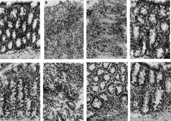
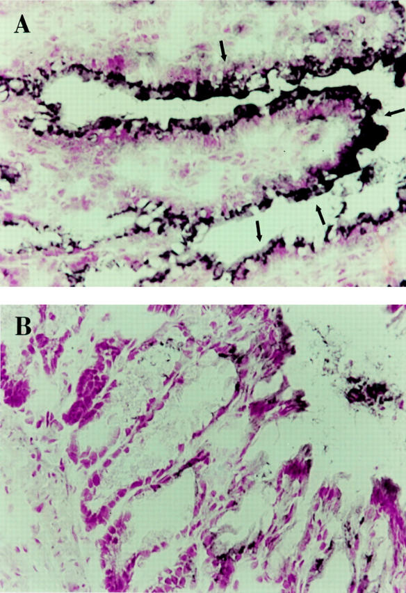
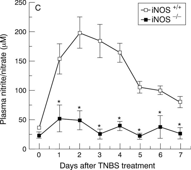
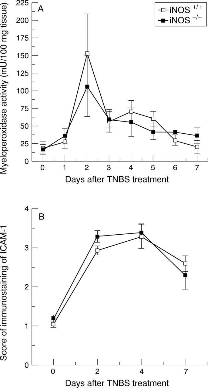
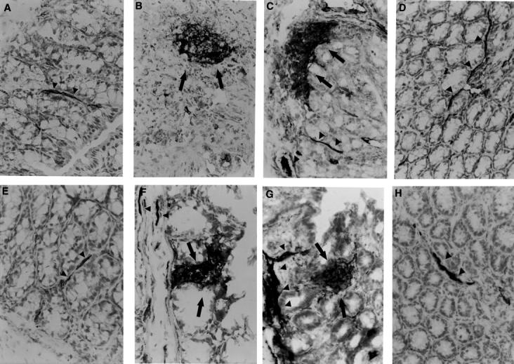
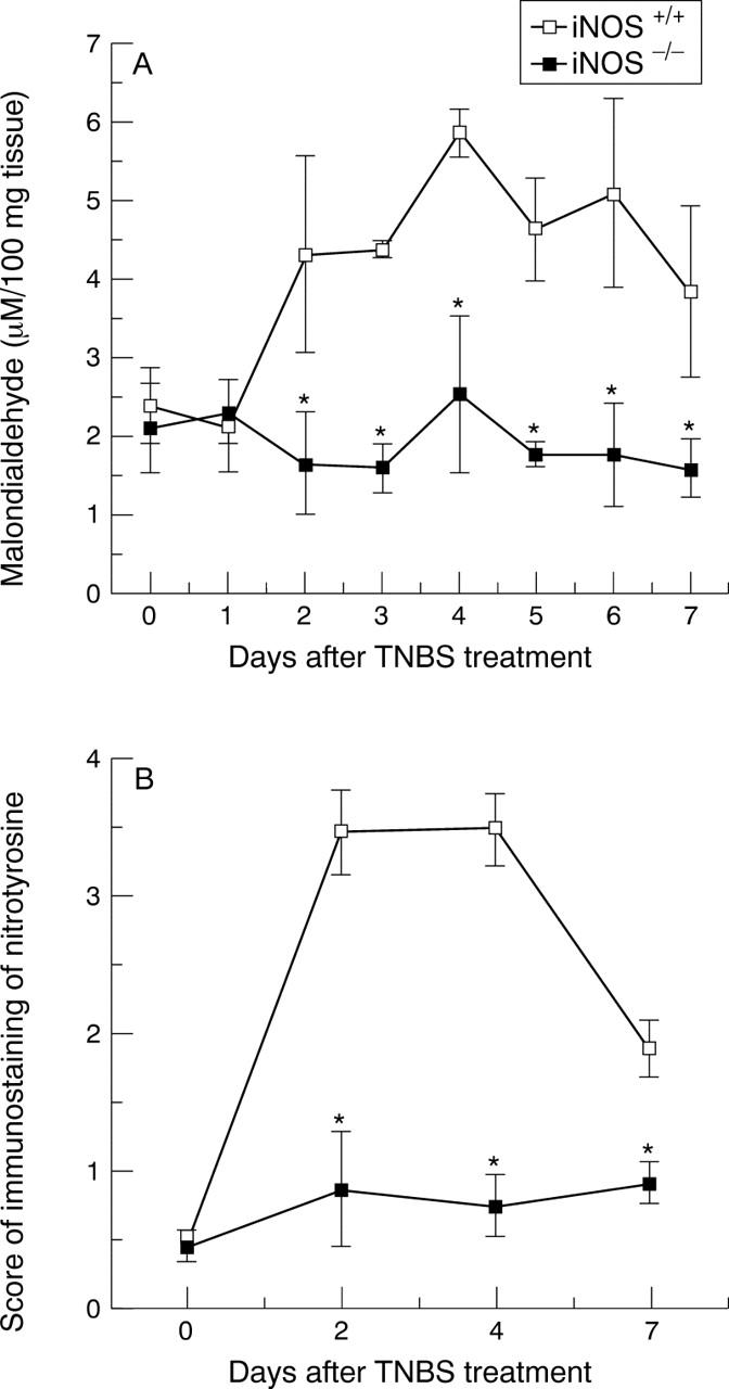
Methods: Trinitrobenzene sulphonic acid (TNBS) was instilled per rectum to induce a lethal colitis in iNOS deficient mice and in wild type controls. The distal colon was evaluated for histological evidence of inflammation, iNOS expression and activity, tyrosine nitration and malondialdehyde formation (as indexes of nitrosative and oxidative stress), myeloperoxidase activity (as index of neutrophil infiltration), and tissue localisation of intercellular adhesion molecule 1 (ICAM-1).

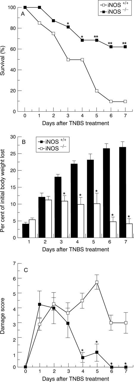
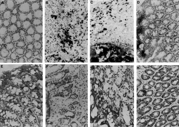
Results: TNBS administration induced a high mortality and weight loss associated with a severe colonic mucosal erosion and ulceration, increased myeloperoxidase activity, increased concentrations of malondialdehyde, and an intense staining for nitrotyrosine and ICAM-1 in wild type mice. Genetic ablation of iNOS gene conferred to mice a significant resistance to TNBS induced lethality and colonic damage, and notably reduced nitrotyrosine formation and concentrations of malondialdehyde; it did not, however, affect neutrophil infiltration and intestinal ICAM-1 expression in the injured tissue.

Conclusion: Data show that activation of iNOS is required for nitrosative and oxidative damage in experimental colitis.


Figures

Figure 1
Figure 1
Survival (A), weight loss (B), and damage score (C) in iNOS+/+ and iNOS−/− mice after TNBS intracolonic administration. Each data point is the mean (SEM) of 8-20 animals for each group. *p<0.05, **p<0.01.
Figure 2
Figure 2
Time course of changes of colonic epithelial architecture after TNBS administration. Representative colonic sections from non-treated iNOS+/+ (A) or iNOS−/− (E) mice show normal tissue structure at day 0. (B) and (C) show a notable disruption of the epithelial structure with extensive haemorrhagic necrosis and infiltration of neutrophils in a colonic section from an iNOS+/+ mouse at two and four days after TNBS administration, respectively. At day 7 (D) oedema was still present in the healing epithelium of a TNBS treated iNOS+/+ mouse. (F) shows alteration of epithelial architecture with a massive infiltration of inflammatory cells in a colonic section of an iNOS−/− mouse two days after TNBS administration. At days 4 (G) and 7 (H) inflammatory cells were still present, mainly in the submucosa, while a healing process started in the epithelium. Original magnification × 400.
Figure 3
Figure 3
Immunohistochemical localisation of iNOS (A and B) and plasma nitrite/nitrate concentrations (C). (A) shows a diffuse dark staining localised in the apical epithelium (arrows) in the inflamed colon of an iNOS+/+ mouse four days after TNBS administration. (B) shows no staining in a colonic section of an iNOS−/− mouse four days after TNBS administration. Original magnification × 400. (C) shows the time course of plasma nitrite/nitrate concentrations after TNBS administration. Each data point is the mean (SEM) of 4-8 animals for each group. *p<0.05.
Figure 3
Figure 3
Immunohistochemical localisation of iNOS (A and B) and plasma nitrite/nitrate concentrations (C). (A) shows a diffuse dark staining localised in the apical epithelium (arrows) in the inflamed colon of an iNOS+/+ mouse four days after TNBS administration. (B) shows no staining in a colonic section of an iNOS−/− mouse four days after TNBS administration. Original magnification × 400. (C) shows the time course of plasma nitrite/nitrate concentrations after TNBS administration. Each data point is the mean (SEM) of 4-8 animals for each group. *p<0.05.
Figure 4
Figure 4
Time course of myeloperoxidase activity (A) and score of immunostaining of ICAM-1 (B) in tissues from iNOS+/+ and iNOS−/− mice after induction of colitis. Each data point is the mean (SEM) of eight animals for each group.
Figure 5
Figure 5
Time course of ICAM-1 expression after TNBS induced colitis. Control tissues at day 0 from non-treated animals iNOS+/+ (A) and iNOS-/- (E) showed a dark brown staining of endothelium of blood vessels (arrow heads) indicating the presence of constitutive ICAM-1 protein. TNBS administration induced an increase in the positive staining for ICAM-1 along the endothelial vascular wall (arrow heads) in the submucosa area and in infiltrated cells (arrows) at days 2 and 4 after TNBS administration in iNOS+/+ (B and C) and iNOS−/− mice (F and G). A representative microphotograph at day 7 shows the presence of constitutive ICAM-1 staining in vessels (arrow heads) of the healing mucosa in iNOS+/+ (D) and iNOS−/− mice (H). Original magnification × 400.
Figure 6
Figure 6
Time course of colonic concentrations of malondialdehyde (A) and score of immunostaining of nitrotyrosine (B) in tissues from TNBS treated iNOS+/+ and iNOS−/− mice. Each data point is the mean (SEM) of eight animals for each group. *p<0.05.
Figure 7
Figure 7
Immunohistochemistry for nitrotyrosine. In colonic sections from non-treated mice (day 0) no staining for nitrotyrosine was found in iNOS+/+ (A) and iNOS−/− mice (E). At days 2 (B) and 4 (C) after TNBS administration a diffuse dark staining was localised in infiltrated inflammatory cells and in the necrotic epithelium of an iNOS+/+mouse. At day 7 (D) nitrotyrosine staining was still present in the apical epithelium in the area of healing mucosa of an iNOS+/+mouse. Nitrotyrosine staining was absent at days 2 (F), 4 (G), and 7 (H) after TNBS administration in the mucosa of an iNOS−/− mouse. Original magnification × 400.

Similar articles

Cited by

References

    1. Anal Biochem. 1979 Jun;95(2):351-8 - PubMed
    1. New Horiz. 1995 Feb;3(1):33-45 - PubMed
    1. Gastroenterology. 1988 Dec;95(6):1528-34 - PubMed
    1. Gastroenterology. 1989 Mar;96(3):795-803 - PubMed
    1. Life Sci. 1989;45(20):1869-76 - PubMed