

Strictures in Crohn's disease are characterised by an accumulation of mast cells colocalised with laminin but not with fibronectin or vitronectin
- PMID: 10403732
- PMCID: PMC1727598
- DOI: 10.1136/gut.45.2.210
Strictures in Crohn's disease are characterised by an accumulation of mast cells colocalised with laminin but not with fibronectin or vitronectin
Abstract
Background/aims: Intestinal fibrosis and stricture formation is an unresolved problem in Crohn's disease. The aim of this study was to investigate whether mast cells accumulate in these tissues and whether their localisation is associated with extracellular matrix components.
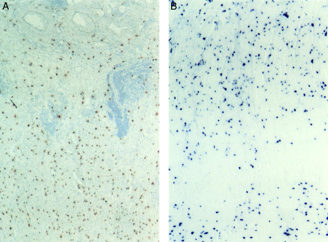
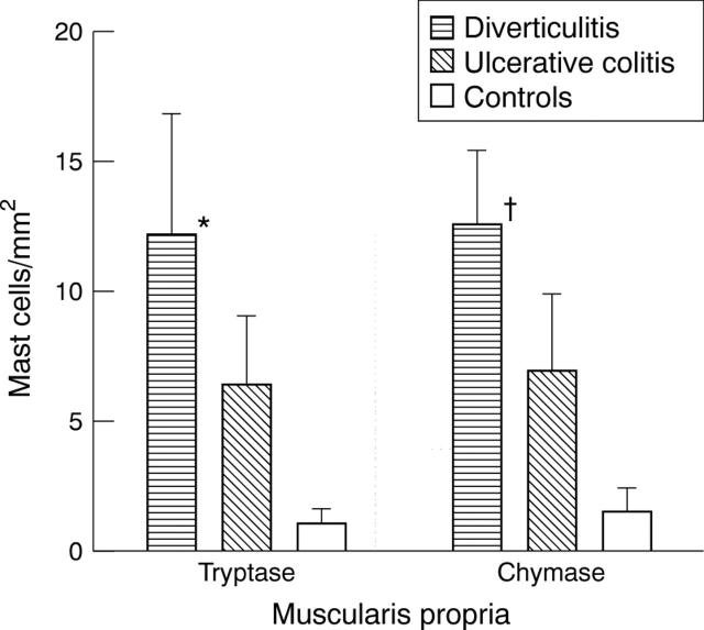
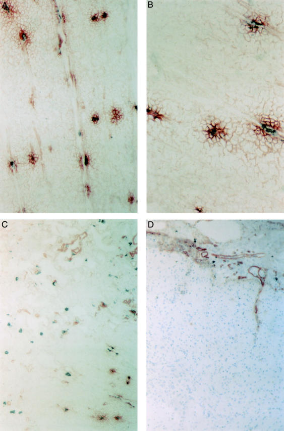
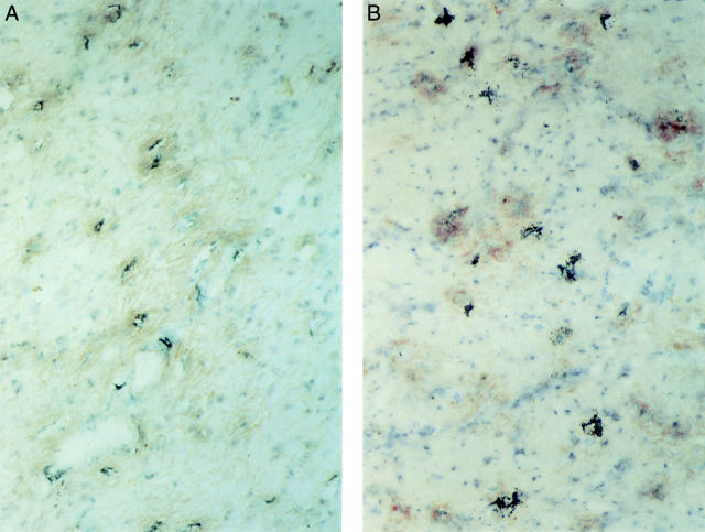
Methods: Mast cells were visualised by immunohistochemical staining of the mast cell specific proteases chymase and tryptase. Their localisation in relation to extracellular matrix components was shown by immunohistochemical double labelling.
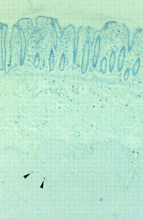
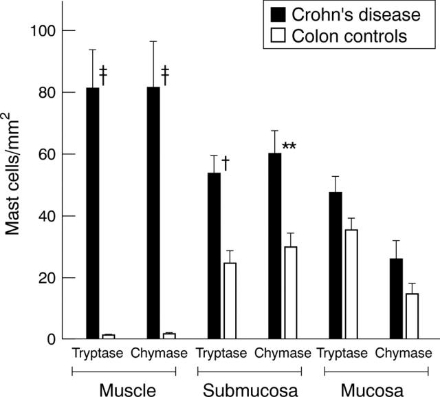
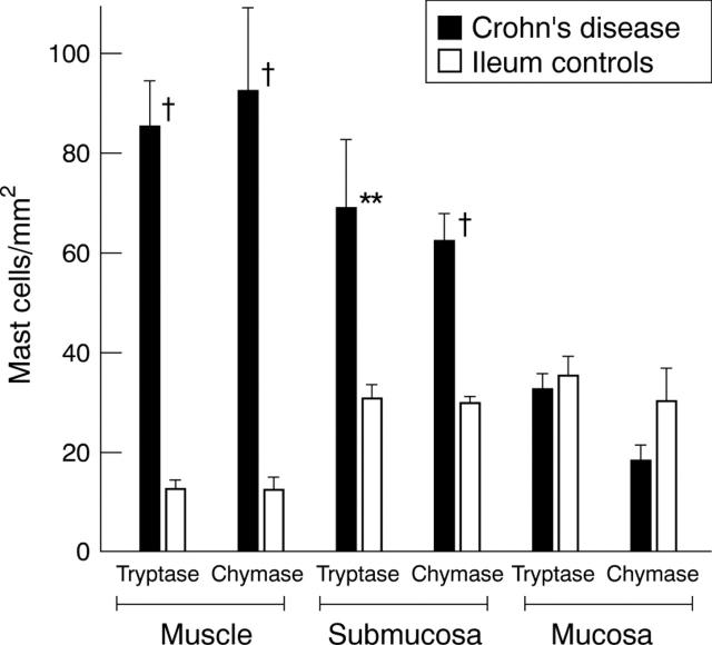
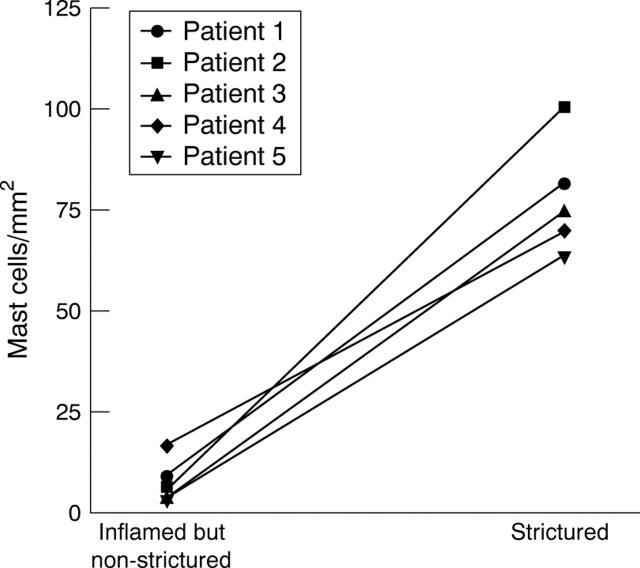
Results: In strictures in Crohn's disease, a striking accumulation of mast cells was seen particularly in the hypertrophied and fibrotic muscularis propria, with a mean (SEM) mast cell number of 81.3 (14.9) v 1.5 (0.9)/mm(2) in normal bowel (p<0.0005). All mast cells in the muscularis propria were colocalised with patches of laminin. In contrast, in the submucosa, laminin was exclusively found in the basal lamina of blood vessels where many adherent mast cells were seen. No colocalisation of mast cells was found with fibronectin or vitronectin.
Conclusions: The large accumulation of mast cells in the muscle layer of strictured bowel suggests a functional role for these cells in the hypertrophic and fibrotic response of the smooth muscle cells. The colocalisation with laminin indicates a mechanism of interaction between smooth muscle cells and mast cells that may be important in the role of mast cells in the process of fibrosis.
Figures
Similar articles
-
Different distribution of mast cells and macrophages in colonic mucosa of patients with collagenous colitis and inflammatory bowel disease.Hepatogastroenterology. 2002 May-Jun;49(45):678-82. Hepatogastroenterology. 2002. PMID: 12063968
-
Collagen content and types in the intestinal strictures of Crohn's disease.Gastroenterology. 1988 Feb;94(2):257-65. doi: 10.1016/0016-5085(88)90411-8. Gastroenterology. 1988. PMID: 3335305
-
Immunohistochemical study of chymase-positive mast cells in inflammatory bowel disease.Oncol Rep. 2006 Jul;16(1):103-7. Oncol Rep. 2006. PMID: 16786130
-
Mechanisms of initiation and progression of intestinal fibrosis in IBD.Scand J Gastroenterol. 2015 Jan;50(1):53-65. doi: 10.3109/00365521.2014.968863. Scand J Gastroenterol. 2015. PMID: 25523556 Review.
-
Contribution of morphology for the comprehension of mechanisms of fibrosis in inflammatory enterocolitis.Acta Gastroenterol Belg. 2000 Oct-Dec;63(4):371-6. Acta Gastroenterol Belg. 2000. PMID: 11233520 Review.
Cited by
-
The role of mast cells in bacterial enteritis.Am J Pathol. 2007 Aug;171(2):399-401. doi: 10.2353/ajpath.2007.070501. Epub 2007 Jun 14. Am J Pathol. 2007. PMID: 17569775 Free PMC article. No abstract available.
-
Mast cells control neutrophil recruitment during T cell-mediated delayed-type hypersensitivity reactions through tumor necrosis factor and macrophage inflammatory protein 2.J Exp Med. 2000 Nov 20;192(10):1441-52. doi: 10.1084/jem.192.10.1441. J Exp Med. 2000. PMID: 11085746 Free PMC article.
-
Effect of Dietary Fiber and Metabolites on Mast Cell Activation and Mast Cell-Associated Diseases.Front Immunol. 2018 May 29;9:1067. doi: 10.3389/fimmu.2018.01067. eCollection 2018. Front Immunol. 2018. PMID: 29910798 Free PMC article. Review.
-
Modulatory effect of nitric oxide on mast cells during induction of dextran sulfate sodium colitis.Dig Dis Sci. 2007 Jan;52(1):45-51. doi: 10.1007/s10620-006-9409-5. Epub 2006 Dec 8. Dig Dis Sci. 2007. PMID: 17160477
-
Essential role for mast cell tryptase in acute experimental colitis.Proc Natl Acad Sci U S A. 2011 Jan 4;108(1):290-5. doi: 10.1073/pnas.1005758108. Epub 2010 Dec 20. Proc Natl Acad Sci U S A. 2011. PMID: 21173247 Free PMC article.
References
Publication types
MeSH terms
Substances
LinkOut - more resources
Full Text Sources
Other Literature Sources
Medical