

Impaired expression of MHC class II molecules in response to interferon-gamma (IFN-gamma) on human thymoma neoplastic epithelial cells
- PMID: 10403908
- PMCID: PMC1905478
- DOI: 10.1046/j.1365-2249.1999.00933.x
Impaired expression of MHC class II molecules in response to interferon-gamma (IFN-gamma) on human thymoma neoplastic epithelial cells
Abstract
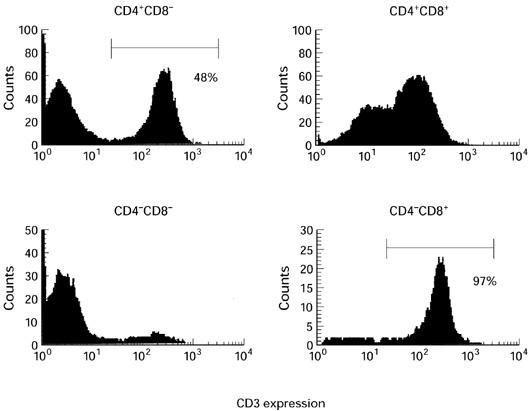
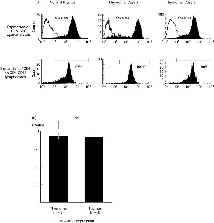
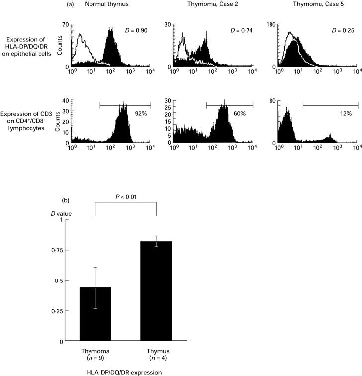
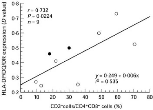
A human thymoma is a neoplasm derived from the thymic epithelial cell, and is well known for its association with autoimmune diseases, especially myasthenia gravis. The neoplastic epithelial cells of thymoma clearly retain thymic epithelial functions, but the development of T cells in thymoma is somewhat impaired. In this study, we quantified by flow cytometry the in vitro expression of MHC molecules on neoplastic epithelial cells precultured with IFN-gamma. While MHC class I expression was comparable with that on normal thymic epithelial cells, the level of MHC class II molecules on neoplastic epithelial cells was lower than in controls, and also varied greatly from case to case. Additionally, there was a significant positive correlation between the expression level of MHC class II and the proportion of mature CD3+ cells in the CD4+CD8- subset. Thus, accumulation of CD3-CD4+CD8- cells in thymoma may result from impaired expression of the MHC class II molecules, suggesting that the function of the neoplastic epithelial cells might determine the maturation and the positively selected repertoire of T cells in thymomas.
Figures
Similar articles
-
Immunological function of thymoma and pathogenesis of paraneoplastic myasthenia gravis.Gen Thorac Cardiovasc Surg. 2008 Apr;56(4):143-50. doi: 10.1007/s11748-007-0185-8. Epub 2008 Apr 10. Gen Thorac Cardiovasc Surg. 2008. PMID: 18401674 Review.
-
Altered T cell development in human thymoma is related to impairment of MHC class II transactivator expression induced by interferon-gamma (IFN-gamma).Clin Exp Immunol. 2000 Jul;121(1):59-68. doi: 10.1046/j.1365-2249.2000.01256.x. Clin Exp Immunol. 2000. PMID: 10886240 Free PMC article.
-
T-cell development in human thymoma.Pathol Res Pract. 1999;195(8):541-7. doi: 10.1016/S0344-0338(99)80003-X. Pathol Res Pract. 1999. PMID: 10483584
-
Two-color flow cytometric analysis of thymic lymphocytes from patients with myasthenia gravis and/or thymoma.Clin Immunol Immunopathol. 1992 Jan;62(1 Pt 1):91-6. doi: 10.1016/0090-1229(92)90027-l. Clin Immunol Immunopathol. 1992. PMID: 1370261
-
Alterations of the immune system in thymic malignancies.J Thorac Oncol. 2014 Sep;9(9 Suppl 2):S137-42. doi: 10.1097/JTO.0000000000000299. J Thorac Oncol. 2014. PMID: 25396311 Review.
Cited by
-
Chromosome 6 suffers frequent and multiple aberrations in thymoma.Am J Pathol. 2002 Oct;161(4):1507-13. doi: 10.1016/S0002-9440(10)64426-4. Am J Pathol. 2002. PMID: 12368223 Free PMC article.
-
Current immunotherapy for thymic epithelial tumors: a narrative review.Mediastinum. 2024 Oct 11;8:47. doi: 10.21037/med-24-24. eCollection 2024. Mediastinum. 2024. PMID: 39781199 Free PMC article. Review.
-
Immunological function of thymoma and pathogenesis of paraneoplastic myasthenia gravis.Gen Thorac Cardiovasc Surg. 2008 Apr;56(4):143-50. doi: 10.1007/s11748-007-0185-8. Epub 2008 Apr 10. Gen Thorac Cardiovasc Surg. 2008. PMID: 18401674 Review.
-
Characterization of T cell maturity in thymic epithelial cell tumors from BUF/Mna spontaneous thymoma rats and BUF/Mna-Rnu/+ rats showing delayed thymomagenesis.Int J Clin Exp Pathol. 2010 Jun 30;3(6):587-92. Int J Clin Exp Pathol. 2010. PMID: 20661406 Free PMC article.
-
Glucocorticoids induce G1 cell cycle arrest in human neoplastic thymic epithelial cells.J Cancer Res Clin Oncol. 2005 May;131(5):314-22. doi: 10.1007/s00432-004-0646-8. Epub 2005 Feb 10. J Cancer Res Clin Oncol. 2005. PMID: 15703942 Free PMC article.
References
-
- Rosai J, Levine GD. Atlas of tumor pathology. Washington DC: Armed Forces Institute of Pathology; 1976. Tumors of the thymus.
-
- Bernatz PE, Harrison EG, Clagett OT. Thymoma: a clinicopathologic study. J Thorac Cardiovasc Surg. 1961;42:424–44. - PubMed
-
- Salyer WR, Eggleston JC. Thymoma: a clinical and pathological study of 65 cases. Cancer. 1976;37:229–49. - PubMed
-
- Pedraza MA. Thymoma immunological and ultrastructural characterization. Cancer. 1977;39:1455–61. - PubMed
Publication types
MeSH terms
Substances
LinkOut - more resources
Full Text Sources
Medical
Research Materials
