

Depletion of peritoneal CD5+ B cells has no effect on the course of Leishmania major infection in susceptible and resistant mice
- PMID: 10403925
- PMCID: PMC1905474
- DOI: 10.1046/j.1365-2249.1999.00953.x
Depletion of peritoneal CD5+ B cells has no effect on the course of Leishmania major infection in susceptible and resistant mice
Abstract
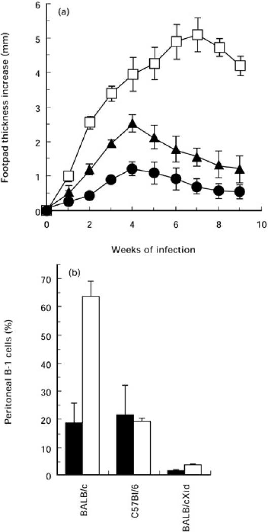
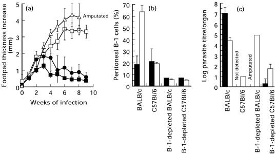
The mouse peritoneal cavity contains a unique self-renewing population of B cells (B-1) derived from fetal liver precursors and mainly producing polyreactive antibodies. Since B-1 cells are a potential source of IL-10, it has been suggested that these cells may contribute to the susceptibility of BALB/c mice to Leishmania major infection by skewing the T helper cell network towards a Th2 phenotype. Accordingly, L. major infection of B cell-defective BALB/c Xid mice (lacking B-1 cells) induces less severe disease compared with controls. However, in addition to the lack of B-1 cells, the Xid immune deficiency is characterized by high endogenous interferon-gamma (IFN-gamma) production. In the present study, the role of B-1 cells during L. major infection was investigated in mice experimentally depleted of peritoneal B-1 cells. Six weeks old C57Bl/6 and BALB/c mice were lethally irradiated and reconstituted with autologous bone marrow which allows systemic depletion of B-1 cells. Untreated BALB/c, C57Bl/6 as well as BALB/c Xid mice were used as controls. After reconstitution, mice were injected with L. major amastigotes and progression was followed using clinical, parasitological and immunological criteria. As previously reported, BALB/c Xid mice showed a significant reduction in disease progression. In contrast, despite the dramatic reduction of B-1 cells, B-1-depleted BALB/c mice showed similar or even worse disease progression compared with control BALB/c mice. No differences were found between B-1-depleted or control C57Bl/6 mice. Our data suggest that the B-1 cells do not contribute to the susceptibility of BALB/c mice to L. major infection.
Figures

Similar articles
-
How to B(e)-1 Important Cell During Leishmania Infection.Front Cell Infect Microbiol. 2020 Jan 14;9:424. doi: 10.3389/fcimb.2019.00424. eCollection 2019. Front Cell Infect Microbiol. 2020. PMID: 31993374 Free PMC article. Review.
-
The Xid defect determines an improved clinical course of murine leishmaniasis in susceptible mice.Int Immunol. 1994 Aug;6(8):1117-24. doi: 10.1093/intimm/6.8.1117. Int Immunol. 1994. PMID: 7981141
-
The early IL-4 response to Leishmania major and the resulting Th2 cell maturation steering progressive disease in BALB/c mice are subject to the control of regulatory CD4+CD25+ T cells.J Immunol. 2002 Sep 15;169(6):3232-41. doi: 10.4049/jimmunol.169.6.3232. J Immunol. 2002. PMID: 12218142
-
B-cell outgrowth and ligand-specific production of IL-10 correlate with Th2 dominance in certain parasitic diseases.Exp Parasitol. 1996 Nov;84(2):168-77. doi: 10.1006/expr.1996.0102. Exp Parasitol. 1996. PMID: 8932766
-
Activation of B-cells by sIgM cross-linking induces accumulation of CD5 mRNA.Curr Top Microbiol Immunol. 1995;194:219-28. doi: 10.1007/978-3-642-79275-5_26. Curr Top Microbiol Immunol. 1995. PMID: 7534670 Review.
Cited by
-
How to B(e)-1 Important Cell During Leishmania Infection.Front Cell Infect Microbiol. 2020 Jan 14;9:424. doi: 10.3389/fcimb.2019.00424. eCollection 2019. Front Cell Infect Microbiol. 2020. PMID: 31993374 Free PMC article. Review.
-
Uptake of Leishmania major by dendritic cells is mediated by Fcgamma receptors and facilitates acquisition of protective immunity.J Exp Med. 2006 Jan 23;203(1):177-88. doi: 10.1084/jem.20052288. Epub 2006 Jan 17. J Exp Med. 2006. PMID: 16418399 Free PMC article.
-
B-1 cell response in immunity against parasites.Parasitol Res. 2019 May;118(5):1343-1352. doi: 10.1007/s00436-019-06211-2. Epub 2019 Apr 2. Parasitol Res. 2019. PMID: 30941496 Review.
-
Dependency of B-1 Cells in the Maintenance of Splenic Interleukin-10 Producing Cells and Impairment of Macrophage Resistance in Visceral Leishmaniasis.Front Microbiol. 2017 Jun 2;8:978. doi: 10.3389/fmicb.2017.00978. eCollection 2017. Front Microbiol. 2017. PMID: 28626451 Free PMC article.
-
Igh-6(-/-) (B-cell-deficient) mice fail to mount solid acquired resistance to oral challenge with virulent Salmonella enterica serovar typhimurium and show impaired Th1 T-cell responses to Salmonella antigens.Infect Immun. 2000 Jan;68(1):46-53. doi: 10.1128/IAI.68.1.46-53.2000. Infect Immun. 2000. PMID: 10603367 Free PMC article.
References
-
- Müller I, Garcia-Sanz J, Titus R, Behin R, Louis J. Analysis of the cellular parameters of the immune responses contributing to resistance and susceptibility of mice to infection with the intracellular parasite Leishmania major. Immunol Rev. 1989;112:95–113. - PubMed
-
- Reiner SL, Locksley RM. The regulation of immunity to Leishmania major. Annu Rev Immunol. 1995;13:151–77. - PubMed
-
- Locksley RM, Heinzel FP, Holaday BJ, Reiner SS, Sadick MD. Induction of Th1 and Th2, CD4+ subsets during murine Leishmania major infection. Res Immunol. 1991;142:28–32. - PubMed
-
- Belosevic M, Finbloom DS, Van Der Meide PH. Administration of monoclonal anti-IFN-gamma antibodies in vivo abrogates natural resistance of C3H/HeN mice to infection with Leishmania major. J Immunol. 1989;143:266–74. - PubMed
Publication types
MeSH terms
Substances
LinkOut - more resources
Full Text Sources
Molecular Biology Databases
Research Materials
