

Presence of circulating abnormal CD34+ progenitors in adult Langerhans cell histiocytosis
- PMID: 10403933
- PMCID: PMC1905468
- DOI: 10.1046/j.1365-2249.1999.00950.x
Presence of circulating abnormal CD34+ progenitors in adult Langerhans cell histiocytosis
Abstract
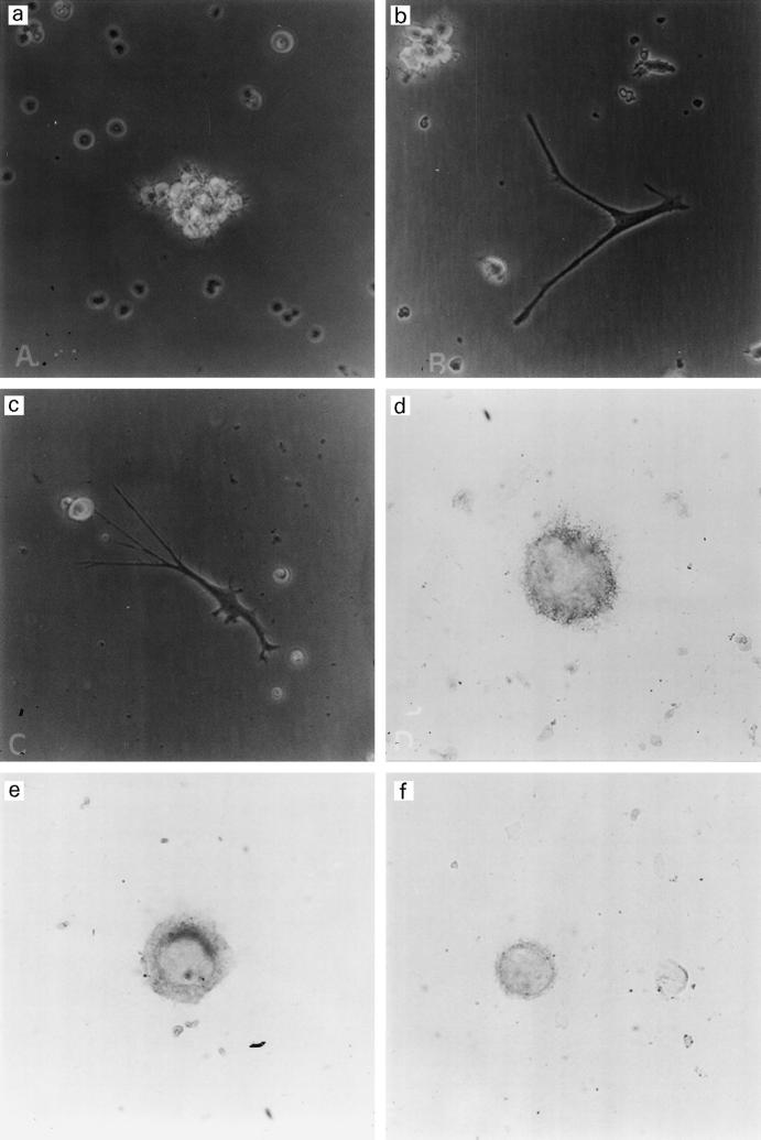
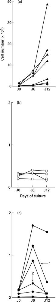
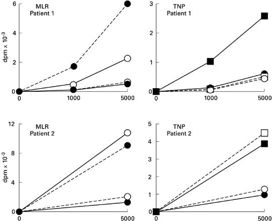
Langerhans cell histiocytosis (LCH) is related to the proliferation of cells, which are similar to Langerhans cells (LC) but possess many abnormal characteristics. Lesions are widespread and this fact suggests that LCH cells or their precursors are present in the blood of patients. In five adult patients, we have isolated and cultured CD34+ blood progenitors of dendritic cells. We studied their phenotype by flow cytometry and their functional properties in mixed culture with heterologous lymphocytes and with autologous lymphocytes in the presence of tri-nitro-phenyl antigen (TNP). The amount of CD34+ precursors was dramatically higher than controls but a high mortality occurred during the in vitro differentiation. The phenotype of surviving cells was similar to LC phenotype (CD1a+, CD83+, Lag+) but some of them expressed CD2. These cells were able to induce T cell proliferation in mixed culture. They could not initiate primary response to TNP, except in a patient treated with thalidomide. In our hands, these CD34+ cells may be precursors of LCH cells.
Figures
References
-
- Cline MJ. Histiocytes and histiocytosis. Blood. 1995;84:2840–53. - PubMed
-
- Lichtenstein L. Integration of eosinophilic granuloma of bone, Letterer–Siwe disease and Schüller–Christian disease as related manifestations of a single nosologic entity. Arch Pathol. 1953;56:84–102. - PubMed
-
- Misery L, Lyonnet S, Cambazard F, Faure M. Encyclopidie médico-chirurgicale-dermatologie. 12-798-A-10. Paris: Editions Techniques; 1993. Histiocytose X (histiocytose langerhansienne) p. 5.
-
- Malpas JS, Norton AJ. Langerhans cell histiocytosis in the adult. Med Ped Oncol. 1996;27:540–6. - PubMed
-
- Cambazard F, Misery L, Kanitakis J, Archimbaud E, Hemier C. Acute monoblastic leukemia and histiocytosis X: a case report and review of the literature. Eur J Dermatol. 1991;1:11–17.
Publication types
MeSH terms
Substances
LinkOut - more resources
Full Text Sources
Medical
