

Detection of bovine herpesvirus type 1 in blood from naturally infected cattle by using a sensitive PCR that discriminates between wild-type virus and virus lacking glycoprotein E
- PMID: 10405392
- PMCID: PMC85268
- DOI: 10.1128/JCM.37.8.2498-2507.1999
Detection of bovine herpesvirus type 1 in blood from naturally infected cattle by using a sensitive PCR that discriminates between wild-type virus and virus lacking glycoprotein E
Abstract
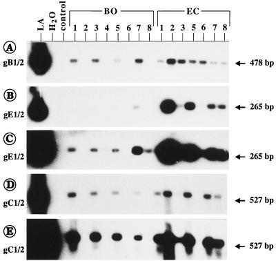
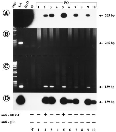
In the present study, we report for the first time on the detection of bovine herpesvirus type 1 (BHV-1) in whole-blood samples derived from naturally infected cattle. Sensitive PCR assays specific for glycoprotein B (gB), gC, and gE of BHV-1 allow the detection of one BHV-1 DNA copy in 10(5) to 10(7) peripheral blood leukocytes (PBLs). The incidence of BHV-1-positive PBLs in naturally infected cattle appears to be quite high (92.2% positive PBLs among all samples tested), although in most cases only between 10(-5) and 10(-7) positive leukocytes were present. The results demonstrate that the viral DNA is detectable not only in the peripheral blood of acutely infected animals but, more importantly, also in the peripheral blood of subclinically infected cattle. The gE-specific PCR described in the report allows discrimination between wild-type (WT) virus-infected and vaccinated animals, which is of importance for control programs that use the recently introduced vaccination strategy with a gE-negative virus. The results further show that doubtful serological results can be verified or falsified and that individual animals can be monitored for the presence or absence of WT BHV-1 or gE-negative virus in cattle herds. The PCR protocols allow the detection of BHV-1 prior to seroconversion or in BHV-1-seronegative cattle. Finally, the results indicate the simultaneous presence of WT and gE-negative vaccine virus in the PBLs of several cattle. Therefore, investigations of viremia in naturally and experimentally infected cattle and on the identification of infected cell types of bovine PBLs can be now performed.
Figures
Similar articles
-
Isolation of a glycoprotein E-deleted bovine herpesvirus type 1 strain in the field.Vet Rec. 2003 Aug 16;153(7):209-12. doi: 10.1136/vr.153.7.209. Vet Rec. 2003. PMID: 12956298 Clinical Trial.
-
A glycoprotein E deletion mutant of bovine herpesvirus 1 infects the same limited number of tissues in calves as wild-type virus, but for a shorter period.J Gen Virol. 1995 Sep;76 ( Pt 9):2387-92. doi: 10.1099/0022-1317-76-9-2387. J Gen Virol. 1995. PMID: 7561782
-
Genomic and pathogenic studies on a glycoprotein E variant field isolate of bovine herpesvirus 1.Vet Res Commun. 2000 Sep;24(6):423-31. doi: 10.1023/a:1006430402043. Vet Res Commun. 2000. PMID: 11014611
-
[Diagnostic markers in the prevention of bovine herpesvirus type 1: possibilities and limitations].Berl Munch Tierarztl Wochenschr. 2003 May-Jun;116(5-6):183-91. Berl Munch Tierarztl Wochenschr. 2003. PMID: 12784550 Review. German.
-
A review of the biology of bovine herpesvirus type 1 (BHV-1), its role as a cofactor in the bovine respiratory disease complex and development of improved vaccines.Anim Health Res Rev. 2007 Dec;8(2):187-205. doi: 10.1017/S146625230700134X. Anim Health Res Rev. 2007. PMID: 18218160 Review.
Cited by
-
Regulation of the latency-reactivation cycle by products encoded by the bovine herpesvirus 1 (BHV-1) latency-related gene.J Neurovirol. 2011 Dec;17(6):535-45. doi: 10.1007/s13365-011-0060-3. Epub 2011 Dec 3. J Neurovirol. 2011. PMID: 22139602 Review.
-
Production of bovine herpesvirus type 1-seronegative latent carriers by administration of a live-attenuated vaccine in passively immunized calves.J Clin Microbiol. 2000 Nov;38(11):4233-8. doi: 10.1128/JCM.38.11.4233-4238.2000. J Clin Microbiol. 2000. PMID: 11060097 Free PMC article.
-
Genome sequence of bubaline herpesvirus-1 associated with pustular vulvovaginitis in Indian water buffalo.Microbiol Resour Announc. 2024 Mar 12;13(3):e0088923. doi: 10.1128/mra.00889-23. Epub 2024 Feb 20. Microbiol Resour Announc. 2024. PMID: 38376342 Free PMC article.
-
Detection and molecular characterisation of bovine corona and toroviruses from Croatian cattle.BMC Vet Res. 2015 Aug 13;11:202. doi: 10.1186/s12917-015-0511-9. BMC Vet Res. 2015. PMID: 26268320 Free PMC article.
-
Comparison between DNA Detection in Trigeminal Nerve Ganglia and Serology to Detect Cattle Infected with Bovine Herpesviruses Types 1 and 5.PLoS One. 2016 May 25;11(5):e0155941. doi: 10.1371/journal.pone.0155941. eCollection 2016. PLoS One. 2016. PMID: 27224314 Free PMC article.
References
-
- Ayoub I A, Yang T J. Age-dependent changes in peripheral blood lymphocyte subpopulations in cattle: a longitudinal study. Dev Comp Immunol. 1996;20:353–363. - PubMed
-
- Babiuk L A, van Drunen Little-van den Hurk S, Tikoo S K. Immunology of bovine herpesvirus 1 infection. Vet Microbiol. 1996;53:31–42. - PubMed
-
- Bosch J C, DeJong M C M, Franken P, Frankena K, Hage J J, Kaashoek M J, Maris-Veldhuis M A, Noordhuizen J P T M, Van der Poel W H M, Verhoeff J, Weerdmeester K, Zimmer G M, Van Oirschot J T. An inactivated gE-negative marker vaccine and an experimental gD-subunit vaccine reduce the incidence of bovine herpesvirus 1 infection in the field. Vaccine. 1998;16:265–271. - PubMed
-
- Brenner J, Ungar-Waron H, Elad D, Abraham A. Clinical and serological follow up of young calves experimentally infected with virulent bovine herpesvirus (BHV-1) Isr J Vet Med. 1989;45:21–25.
-
- Castrucci G, Ferrari M, Traldi V, Tartaglione E. Effects in calves of mixed infections with bovine viral diarrhea virus and several other bovine viruses. Comp Immunol Microbiol Infect Dis. 1992;15:261–270. - PubMed
Publication types
MeSH terms
Substances
Associated data
- Actions
LinkOut - more resources
Full Text Sources
Other Literature Sources
Miscellaneous
